Paroxysmal supraventricular tachycardia, abbreviated as PVT, is a type of arrhythmia. Accompanied by a heart rate (HR) from 140 to 220 or more beats per minute. It arises spontaneously and ends unexpectedly. The duration of PNT is at least three cardiac cycles, while maintaining a regular heart rhythm. The attack itself can last from a few seconds to several days.
PNT occurs due to disruption of the nervous regulation of cardiac activity or damage to other organs.
In the first case, excessive nerve stimulation leads to an increase in heart rate.
PNT can also occur in healthy people under the influence of certain factors:
- prolonged physical activity;
- stress;
- consumption of energy and stimulant drinks;
- bad habits.
In the case of organic damage, paroxysmal supraventricular tachycardia can be caused by:
- damage to the heart muscle due to ischemia, inflammation, intoxication;
- myocardial infarction;
- heart disease;
- pathology of the cardiac conduction system.
In addition to heart disease, other organs can affect paroxysmal supraventricular tachycardia. It is worth checking the functioning of the kidneys, lungs and gastrointestinal tract, especially if there are chronic or acute diseases.
Symptoms of paroxysmal supraventricular tachycardia
Paroxysmal tachycardia always has a sudden, distinct onset. The patient feels a push or compression, and sometimes a prick, in the area of the heart.
The attack may be accompanied by symptoms:
- dizziness;
- noise in the head;
- lightheadedness or fainting;
- sweating;
- nausea;
- trembling in the body.
To correctly diagnose tachycardia, it is important to conduct a thorough interview and examination of the patient. Highly qualified doctors of the Cardiology Center of the Federal Scientific and Clinical Center of the Federal Medical and Biological Agency will be able to quickly identify the disease.
What is paroxysmal tachycardia?
As a rule, attacks of paroxysmal tachycardia begin and end suddenly and have different durations.
They make the heart work uneconomically, reduce the efficiency of blood circulation, and lead to its failure. According to the mechanism of development and etiology, paroxysmal tachycardia resembles extrasystole. Therefore, cardiologists regard successive extrasystoles as a short attack of tachycardia.
Classification of paroxysmal tachycardia
1. Based on the location of ectopic impulses, doctors distinguish:
- atrial paroxysmal tachycardia;
- atrioventricular (atrioventricular) paroxysmal tachycardia;
- ventricular paroxysmal tachycardia.
In turn, atrioventricular and atrial tachycardia are combined into a supraventricular or supraventricular form.
2. Taking into account the nature of the course, the following types of paroxysmal tachycardia are distinguished:
- chronic (constantly recurrent);
- spicy;
- continuously relapsing (lasts for years, can cause circulatory failure and dilated, arrhythmogenic cardiomyopathy).
3. According to the characteristics of the development mechanism, paroxysmal supraventricular tachycardia is classified into:
- focal (ectopic);
- reciprocal (associated with the re-entry mechanism in the sinus node);
- multifocal (multifocal).
In children, paroxysmal tachycardia manifests itself in the same way as in adults.
Diagnostics
Tachycardia can be sinus and paroxysmal; they differ from the location of the electrical impulse that causes the heart to contract. To make a diagnosis, the doctor of the Federal Scientific and Clinical Center of the Federal Medical and Biological Agency performs a physical examination, collects anamnesis and prescribes tests.
Physical examination includes:
- external examination of the patient;
- heart rate measurement;
- blood pressure measurement.
Instrumental research:
- ECG;
- ECG with additional load on the body;
- daily ECG monitoring;
- ECHO;
- stress echocardiography;
- MRI;
- CT cardiography.
Diagnosis of paroxysmal tachycardia
Paroxysmal tachycardia is recognized by the typicality of the attack (sudden onset and sudden end) and data obtained from a cardiac examination. The supraventricular and ventricular forms differ in the degree of increase in heart rate:
- with ventricular tachycardia, the heart rate is up to 180 beats/min, tests with excitation of the vagus nerve are negative;
- with supraventricular tachycardia, the heart rate is about 220-250 beats/min, the attack is easily stopped by excitation of the vagus nerve.
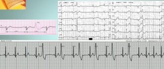
An ECG with paroxysmal tachycardia shows a change in the polarity and shape of the P wave, as well as a violation of its location in relation to the ventricular QRS complex. These signs allow us to judge what form of the disease we are talking about.
Thus, an ECG with atrial paroxysmal tachycardia shows the location of the positive/negative P wave in front of the QRS complex. If the paroxysm comes from the atrioventricular junction, a negative P wave is recorded, located behind/merging with the QRS complex. An ECG with ventricular paroxysmal tachycardia registers expansion and deformation of the QRS complex, and an unchanged P wave can also be recorded.
Paroxysmal tachycardia cannot always be detected using an ECG. Then cardiologists resort to daily ECG monitoring, which allows them to observe short episodes of attacks that are not subjectively felt by the patient.
In order to exclude organic cardiac pathology, the following are additionally carried out:
- MRI;
- Ultrasound of the heart;
- MSCT of the heart (layer-by-layer scanning of cardiac structures).
In some cases, an endocardial electrocardiogram is recorded by inserting electrodes into the heart.
Prevention
Prevention lies in early diagnosis. Often, success directly depends on the patient and his attitude towards his health. Basic recommendations that should be followed during prevention:
- moderate regular physical activity;
- proper nutrition;
- rejection of bad habits;
- avoidance of situations that lead to stress and cause anxiety;
- compliance with the instructions of the attending physician.
The main thing is to diagnose tachycardia in a timely manner in order to move on to prevention in time. You can get acquainted with the programs of our center by following the link.
Treatment of paroxysmal tachycardia
The treatment regimen for paroxysmal tachycardia depends on:
- forms of arrhythmia (ventricular, atrial, atrioventricular);
- reasons that caused the disease;
- duration and frequency of paroxysms;
- presence/absence of complications.
Attacks of paroxysmal tachycardia require emergency hospitalization (with the exception of idiopathic variants with a benign course, which are easily stopped by administering an antiarrhythmic drug to someone).
For the treatment of the supraventricular form, complicated by cardiovascular or heart failure, patients are hospitalized in the cardiology department. Planned hospitalization is carried out if attacks recur more often than 2-3 times a month. Then the question of the relevance of surgical intervention is decided.
Emergency care for paroxysmal tachycardia includes intravenous administration of antiarrhythmic drugs (Propranolol, Aymalin, Rytmodan, Quinidine, Ethmozin, Cordarone, Isoptin, etc.). If the attack is prolonged and cannot be controlled with medications, electropulse therapy is performed. Then the patient undergoes outpatient cardiological treatment, during which effective antiarrhythmic drugs are selected.
To relieve paroxysmal tachycardia, doctors often use vagal maneuvers - techniques that have a pronounced mechanical effect on the vagus nerve. These include:
- straining;
- Aschner's test (moderate and uniform pressure on the inner upper corner of the eyeball);
- Valsalva maneuver (vigorous exhalation with the mouth and nasal cavity closed);
- irritating the root of the tongue to trigger a gag reflex;
- Chermak-Hering test (uniform pressure on the carotid sinuses in the area of the carotid artery).
If attacks of paroxysmal tachycardia occur several times a month, the patient is prescribed long-term anti-relapse therapy:
- quinidine preparations (Disopyramide, Kinylentine, Amiodarone, Verapomil, etc.);
- cardiac glycosides (Celanide, Digoxin).
The selection of an effective dosage is carried out taking into account the patient’s well-being and ECG results.
Surgical treatment of paroxysmal tachycardia is used only if the disease is severe and cannot be controlled with medications. Among the most common surgical methods are:
- mechanical, laser, electrical, cryogenic, chemical destruction of additional impulse pathways or ectopic foci of automatism;
- RFA;
- implantation of a pacemaker;
- implantation of an electrical defibrillator.
If a patient wants to treat paroxysmal tachycardia using traditional methods, he should first consult with a cardiologist, since failure to take prescribed antiarrhythmic drugs can lead to serious complications.
How to treat paroxysmal supraventricular tachycardia
There are several treatment options at the FMBA center. Depending on the severity of the illness, the attending physician may place you in the therapeutic department. You will be under the constant supervision of professionals and receive timely medical care. This is usually drug therapy. If the symptoms of standard therapy are not enough, then you may be offered catheter ablation of the arrhythmia. At the same time, by puncture access under local anesthesia, under X-ray control using a catheter, an accurate diagnosis of the source of arrhythmia is made and, by precisely influencing it with a radiofrequency pulse or cold, the cause of paroxysmal tachycardia is eliminated.
Causes of paroxysmal tachycardia
Supraventricular paroxysmal tachycardia occurs as a result of increased activation of the sympathetic nervous system. Ventricular is a consequence of necrotic, inflammatory, sclerotic or dystrophic damage to the heart muscle.
In the ventricular form, the focus of ectopic excitation is located in the His bundle, its legs and Purkinje fibers, that is, in the ventricular sections of the conductive nervous system. Most often, the pathology is diagnosed in elderly men who have suffered a myocardial infarction, coronary heart disease, or suffer from hypertension, myocarditis, or heart defects.
The main causes of the development of paroxysmal tachycardia also include:
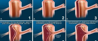
1. The presence of additional innate impulse pathways in the myocardium. That is:
- Kent's bundle between the atria and ventricles, bypassing the atrioventricular node;
- Macheim fibers between the atrioventricular node and the ventricles.
2. The presence in the myocardium of additional impulse pathways resulting from myocardial damage (infarction, myocarditis, cardiomyopathy).
3. Additional pathways cause pathological movement of excitation throughout the myocardium.
Cardiology knows cases when longitudinal dissociation appears in the atrioventricular node, causing uncoordinated functioning of the fibers of the atrioventricular connection. Then some of the fibers function normally, while the other, on the contrary, conducts excitation in the retrograde (opposite) direction and acts as the basis for the circular circulation of impulses from the atria to the ventricles and along the opposite fibers back to the atria.
In young children, essential (idiopathic) paroxysmal tachycardia of unknown etiology is sometimes diagnosed.
Tachycardia with wide QRS complexes (differential diagnosis, therapeutic tactics)
Download Slideshow
Author: Lebedev D.S.
Tachycardias with wide QRS complexes are often encountered in the practice of cardiologists, resuscitators, and emergency physicians and require an accurate differential diagnosis to correctly determine treatment tactics [8,11]. Difficulties in the differential diagnosis of broad complex tachycardias are due to the insufficient sensitivity of numerous criteria.
Tachycardia with wide QRS complexes is considered to be tachycardia with a heart rate (HR) of more than 150 beats/min and a QRS complex duration of more than 120 ms in one of the unipolar chest leads (V1-V6) or tachycardia with a QRS duration of 110 to 120 ms. with a morphology characteristic of blockade of one or both bundle branches.
Tachycardias with wide QRS complexes include:
- Paroxysmal ventricular tachycardia.
- Paroxysmal supraventricular tachycardia:
- paroxysms of antidromic reciprocal atrioventricular (AV) tachycardias in WPW syndrome.
- paroxysms of reciprocal AV tachycardias (RAVT) in the presence of abnormal conduction pathways with tachydependent functional blockades of the bundle branches.
- Paroxysms of flutter (PTP) or atrial fibrillation (AAF) with tachydependent functional blockade of the bundle branches.
- Severe sinus tachycardia in patients with organic blockade of the bundle branch or branches.
Various types of tachycardias with wide QRS complexes have different clinical significance and require a certain set of emergency measures. Therefore, it is important to quickly identify variants of tachycardias with wide QRS complexes.
For the differential diagnosis of tachycardias with wide QRS complexes, the following can be used: standard ECG, transesophageal electrocardiography (TE ECG), surface ECG mapping, dynamic ECG (DECG), programmed transesophageal pacing (TEP pacing), endocardial electrogram and intracardiac electrophysiological study (IC). EFI).
It is obvious that the listed methods differ in the speed of their use, the degree of difficulty in interpreting the results, and the complexity of the equipment. The sensitivity and specificity of the listed methods are also different for different types of tachycardias with wide QRS complexes.
Among the different types of tachycardias with wide QRS complexes, ventricular tachycardia (VT) is of particular importance in determining treatment tactics. Therefore, the clinical picture during paroxysm of tachycardia is important. Hemodynamic disturbances, collapse, acute cardiac and/or coronary failure, severe electrical instability of the myocardium do not leave the doctor time for a thorough examination using the listed methods and require urgent emergency measures.
A full range of examinations is possible only in cases where there is no threat to life and the patient’s condition is sufficiently stable. It turned out that in these cases the clinical picture for determining the mechanism of tachycardia is not of great importance in the differential diagnosis of tachycardias with wide QRS complexes. However, in some cases, studying the clinical picture during an attack of VT may be even more important than a careful study of the standard ECG at this moment.
Thus, a history of previous or current myocardial infarction (MI) is of great importance, which may indicate in favor of VT [13, 28]. The patient’s age, congenital anomalies of valves and large vessels, information about pre-existing bundle branch block, manifesting WPW syndrome can also facilitate the diagnosis [18, 22]. In this report, special attention is paid to changes in the standard ECG in the diagnosis of tachycardias with wide QRS complexes.
Electrocardiographic criteria for tachycardias with wide QRS complexes.
In the literature of recent years, one can find many studies devoted to the development of criteria for the differential diagnosis of tachycardias with wide QRS complexes [9,36,37]. HJWelens in 1978 [48] proposed four criteria for the differential diagnosis of VT: AV dissociation, deviation of the electrical axis of the heart, an increase in the duration of the QRS complex and the configuration of the QRS complex in unipolar chest leads V1 and V6.
The use of these criteria by the authors [49, 50] made it possible to correctly diagnose VT in 82-92% of cases. In the works of P. Brugada [15] and M. J. Griffith [27], the use of additional ECG criteria made it possible to increase the sensitivity of the ECG method to 96% and 98.7%, with a specificity of 64% and 96.5%, respectively. In the works known to us, there is no comparison of omission errors and false alarms when using the previously listed methods and assessing all syndromes that have two main features: tachycardia and a wide QRS complex.
Therefore, the assumption that using modern ECG criteria for differential diagnosis can correctly determine the mechanism of tachycardia in 90% of cases based on surface ECG data raises certain doubts.
The duration of the QRS complex is determined by the time of depolarization of the ventricular myocardium. It should be taken into account that there are significant methodological difficulties in determining the end of the QRS complex. The normal sequence of conduction of excitation through the AV node and the His-Purkinje system (HPS) leads to an orderly depolarization of the interventricular septum, right and left ventricles, resulting in the formation of the QRS complex, the duration of which normally in standard leads ranges from 80 to 110 ms., and in the chest leads up to 120 ms.
Disruption of the sequence of depolarization of the ventricular myocardium during a rhythm that has a source of heterotopic activity in the SGP leads to the formation of a wide QRS complex (>120 ms). However, a wide QRS complex also occurs with organic or functional tachydependent blockade of one or both branches of the His bundle, as well as with antidromic tachycardia, when each QRS complex is confluent, since the myocardium is excited both through the regular AV node and through the additional AV node. connection - Kent beam. Therefore, for differential diagnosis, the features of the morphology of the QRS complex in each of these cases should be considered:
- ventricular tachycardia,
- NVT with aberrant conduction,
- atrial flutter,
- AV tachycardia (orthodromic tachycardia with the participation of DAVS),
- nodal AV tachycardia.
- NVT with eccentric conduction,
- atrial tachycardia/flutter,
- AV tachycardia (antidromic tachycardia with the participation of DAVS),
- nodal tachycardia conducted via DAVS,
- tachycardia type Maheim (nodoventricular tract).
According to [44], VT is the cause of wide complex tachycardia in 80% of the total number of cases and in 95% of patients requiring emergency care. These data are questionable, since the number of patients examined did not include cases of arborization block, organic pre-existing bundle branch block and antidromic reciprocal tachycardia.
With aberrant conduction of excitation, it is delayed below the AV node (in the legs or branches of the His bundle, Purkinje fibers). The causes of aberrant conduction are:
- functional blockade of the bundle branch, when excitation is carried out with incompletely restored conductivity, for example, with early extrasystoles (ESp, ESzh);
- functional frequency-dependent blockade of the bundle branch, when with a significant increase in heart rate, arborization blockade occurs more often than the right bundle branch;
- pre-existing bundle branch block, occurring before the paroxysm of tachycardia.
Functional bundle branch block is rarely the cause of aberrant conduction in sustained tachycardia with wide QRS complexes; it is observed in 4-8% of cases and is unstable. Frequency-dependent bundle branch block (usually the right one - 90%) occurs at high heart rates due to the fact that at high frequencies the corresponding parts of the cardiac conduction system (CCS) do not have time to fully recover.
At a high heart rate, this phenomenon is also characteristic of normal SGP. In contrast to functional conduction aberration, with frequency-dependent blockade, normal conduction of excitation can be restored when the rhythm decreases below a critical level [46].
Pre-existing bundle branch block is detected in normal sinus rhythm (SR). Against the background of tachycardia, it is possible to change the degree of blockade (more often it increases up to the transition from incomplete to complete blockade of the leg). Up to 15% of patients with wide QRS complex tachycardia have pre-existing bundle branch block [51].
The conduction of excitation along the Kent bundle is characterized by a shortening of the PQ interval (up to 100-120 ms), a delta wave, which is actually the cause of the widening of the QRS complex, and secondary repolarization disorders, the degree of which may vary depending on which part of the ventricular myocardium is excited by additional abnormal pathways (APP). As a rule (up to 90%), with WPW syndrome, orthodromic reciprocal tachycardias (ROART) occur, less often PMA or a combination of PROART with PMA. According to our data, PMA was observed in 20% of patients with DPP.
Antidromic tachycardia itself, when anterograde conduction is carried out through the APP, and retrograde conduction through the ventriculoatrial (VA) connection or paraseptal APP, is rarely observed (about 5% of patients with WPW syndrome). These tachycardias are characterized by the most pronounced delta wave, a long interval R-P' and R-P'>P'-R, where P' is a retrogradely conducted atrial P wave, which can rarely be detected on a standard ECG.
Electrocardiographic criteria for the differential diagnosis of tachycardias with wide QRS complexes.
There are a number of criteria that allow, with varying degrees of reliability, a differential diagnosis of VT and other variants of tachycardias with wide QRS complexes. The most reliable criteria for VT are the presence of AV dissociation with an independent rhythm of the atria and ventricles, ventricular captures, which on the ECG are characterized by normal QRS complexes that interrupt the chain of ventricular QRS complexes, as well as drain complexes due to partial capture of the ventricular myocardium by conducting sinus impulses along the AV connection and partial capture of the myocardium by an ectopic impulse from the His-Purkinje system.
However, this phenomenon can rarely be detected on a standard ECG. It is easier to detect in lead V1. Emergency ECG helps to increase the frequency of detection of drainage complexes. The sensitivity of the criterion is 24%, and the specificity is 100% [19]. At the same time, in 30% of cases of VT, retrograde VA conduction is observed [7], when a retrogradely conducted P wave is detected after the QRS complex or coincides with it. The remaining ECG signs are of relative value.
Some authors believe that the duration of the QRS complex is more than 160 ms. characteristic of VT [19]. The sensitivity of this criterion reaches 65%, and the specificity is 97%. The specificity of the criterion increases with such an expansion of the complex in more than 2 leads [19]. When the duration of the QRS complex is up to 140 ms. the specificity of the complex duration criterion in diagnosing VT decreases to 69%.
When assessing this criterion, it is necessary to take into account the presence of pre-existing bundle branch block, previous MI or other cicatricial changes caused by other organic myocardial diseases, as well as the use of antiarrhythmic drugs (AAP), which can lead to an increase in QRS duration and reduce the diagnostic significance of this criterion. Its use is also limited for tachycardias with a frequency of more than 190 beats/min, when it is difficult to determine the beginning and end of the ventricular complex.
Extreme degrees of deviation of the electrical axis of the QRS in the frontal plane are also of diagnostic significance (Table 1). The criteria for diagnosing VT are: high right (from -90 to +180) position of the axis, its deviation to the left (from -60 to -90) with tachycardia with QRS morphology such as right bundle branch block. Deviation of the electrical axis of the heart to the right (from +120 to +180) with tachycardia with QRS morphology such as left bundle branch block.
Table 1. The significance of the electrical axis criterion in the diagnosis of VT (according to Drew, Scheinman [3]).
| Electric axis quadrant | NVT (n=35) | ZhT (n=98) | Sensitivity | Specificity |
| Upper right (- 90° — +180° ) | 1 | 22 | 22% | 97% |
| Left (- 60° — — 90° ) morphology of right bundle branch block | 1 | 23 | 38% | 95% |
| Right (+120° - +180) morphology of left bundle branch block | 1 | 11 | 30% | 94% |
| Total | 3 | 56 | 57% | 91% |
Absence of complexes such as RS or QS>100 ms. in the precordial leads, as a criterion for VT was proposed by P. Brugada et al.[15]. To determine this criterion, clear recording of the six chest leads is required. The sensitivity of the criterion is 12% and is significantly reduced due to artifacts, myogram, and poor skin contact of the electrodes. The specificity of the trait is 100% [19]. Monophasic Q or R, biphasic QR and triphasic QRS complexes are found in patients with VT in leads V1 - V6.
If an RS type complex is present in one of these leads, then the interval from the beginning of the R wave to the top of the S wave, the so-called internal deviation time, is in favor of VT, exceeding 100 ms. This criterion is difficult to use when the heart rate is high and the QRS complexes overlap each other. The use of the criterion may also lead to diagnostic error in fascicular VT [12,16].
With the morphology of the QRS complex in lead V1 and tachycardia with a form of right bundle branch block, the criteria for diagnosing VT are split complexes of the Rr' and QRr' type with a higher and thinner first peak, as well as biphasic RS and QR. At the same time, supraventricular tachycardia with aberrant conduction of excitation is characterized by three-phase rsR' or biphasic rR' (Table 2).
The criteria for VT with left bundle branch block morphology are any of the following:
- R wave duration is at least 40 ms;
- the presence of a “notch” on the descending limb of the S wave;
- delayed peak of the S wave (from the beginning of the QRS to the apex of the S wave more than 60 ms).
And supraventricular tachycardia with QRS aberration is indicated by the absence of these criteria (Table 3) [6, 19, 40]. The morphology of the QRS complex in lead V6 is represented by one of three types:
- monophasic QS;
- morphology of rS (R/S < 1) in tachycardia with the type of right bundle branch block;
- triphasic qRs (R/S > 1) with tachycardia with a type of right bundle branch block.
It is believed that the first two criteria correspond to VT, and the last one to supraventricular tachycardia with QRS aberration (Tables 4, 5).
In the literature you can find descriptions of such criteria as tachycardia frequency, T wave discordance in relation to the QRS complex. However, these criteria are not significant. However, a heart rate over 180 beats/min is typical for PMA, PROAVT with tachydependent blockade of the SGP and antidromic tachycardia. High heart rate is used to differentiate P' in PROAVT and retrogradely conducted P in ventricular tachycardia.
Esophageal electrocardiogram
The literature does not sufficiently reflect the importance of the esophageal electrocardiogram (PE ECG) method in the differential diagnosis of tachycardias with wide QRS complexes. However, having our own experience, we consider it necessary to include this technique in the diagnostic program for patients with wide QRS complexes, since the emergency ECG increases the frequency of detection of AV dissociation - a reliable sign of VT [7,10], allows us to determine the mechanism of tachycardia (antidromic tachycardia, nodal reciprocal AV tachycardia, atrial flutter) [10, 26, 39]. The value of the method also increases due to the possibility of stopping tachycardia (or converting atrial flutter into atrial fibrillation) by electrical stimulation [5].
Dynamic electrocardiography
Daily ECG monitoring is of great importance in the differential diagnosis of ventricular arrhythmias. First of all, it makes it possible to identify high grade ES, which often precedes VT. DECG also makes it possible to detect short paroxysms of unsustained VT and facilitates the identification of confluent QRS complexes and “captures” in the chain of complexes of ventricular origin. In addition, DECG allows one to determine the onset of an attack of tachycardia and thereby facilitate the identification of previous blockade of the bundle branches and tachydependent functional blockades, since often the first QRS complexes when tachycardia occurs have a normal duration. The presence of manifesting WPW syndrome before an attack makes the recognition of antidromic tachycardia reliable.
Transesophageal electrophysiological study.
Transesophageal electrophysiological study significantly facilitates the differential diagnosis of tachycardias with wide QRS complexes, primarily because, like the emergency ECG, it allows one to recognize AV dissociation due to the reliable determination of the P wave. Based on the assessment of the R-P' and P'-R intervals, it allows one to establish nodal reciprocal tachycardia, and due to the correspondence of P' and R, determine retrogradely conducted atrial waves in VT.
Emergency EPI allows, with programmed atrial pacemaker, to stop up to 98% of attacks of reciprocal AV tachycardia. Consequently, the diagnosis of antidromic tachycardia, RAVT with tachydependent blockades and previous bundle branch blocks is significantly simplified. It is also easier to carry out tests with ATP or adenosine. The indicated advantages of emergency EPI allow emergency treatment measures to be carried out, in particular, the relief of PRVT and the conversion of a regular form of atrial flutter using frequent and ultra-frequent stimulation into atrial fibrillation.
Endocardial electrophysiological study.
Endocardial electrophysiological study (EndoEPI) occupies a special place in the diagnostic program of patients with tachycardias with wide QRS complexes. This diagnostic procedure, being the final stage of diagnosis, can at the same time be combined with a radical therapeutic effect - catheter ablation. A huge number of works are devoted to endoEPI and catheter ablation in patients with tachyarrhythmias [2, 9, 14, 21, 35, 43, 47, etc.]. Let's give just a few numbers.
The effectiveness of catheter ablation for WPW syndrome, nodal AV tachycardias, idiopathic VT, and atrial flutter is 90-95% [20, 33, 34, 51]. In accordance with the American College of Cardiology Guidelines, endo-EPI in patients with wide-complex tachycardias should be performed in all cases of persistent clinically significant tachyarrhythmia [31].
Thus, the feasibility of performing this procedure in patients with tachycardias with wide QRS complexes, both for diagnostic and therapeutic purposes, is obvious.
Determining the correct treatment tactics is the main task of the differential diagnosis of tachycardias with wide QRS complexes. In the case of an established mechanism of tachycardia, the doctor’s tactics cause less difficulty. However, in a significant proportion of cases, especially in emergency situations, the mechanism of tachycardia remains unclear or requires too much time to clarify it [45].
When determining treatment tactics, the doctor faces a number of questions, and the time the doctor has to solve them is determined by the severity of the clinical picture and the prognosis. Among the questions are the following:
- What is the mechanism of tachycardia with wide QRS complexes?
- Which antiarrhythmic drug should be chosen to stop tachycardia?
- Is electrical impulse therapy necessary?
- Is it possible to use electrostimulation methods of cupping?
The importance of determining an algorithm for providing care is evidenced at least by studies devoted to severe complications observed with the administration of isoptin against the background of tachycardia: increased heart rate, collapse, transformation into ventricular fibrillation during VT and antidromic tachycardias [6, 23, 32, 46].
Recently, a significant number of works have been devoted to the use of ATP or adenosine in the diagnosis and termination of tachycardias with wide QRS complexes [17, 24, 27, 42]. The use of adenine nucleosides helps in diagnosing the mechanism of tachycardias due to their high stopping effect in reciprocal AV tachycardias, due to the ability to cause short-term disturbances in AV conduction of excitation.
At the same time, the activity of adenosine and ATP against VT and antidromic tachycardias is low. Only with idiopathic VT of the right ventricular outflow tract can one expect a relief effect when using adenine nucleosides. At the same time, adenosine and ATP are well tolerated both by patients with VT, even in cases of severe myocardial damage, and by patients with tachycardia due to WPW syndrome.
The sensitivity of bolus administration of ATP in doses from 10 to 30 mg, according to our data, is 98% for PRVT. Sensitivity when using adenosine in cases of supraventricular tachycardia is 90%, and specificity is 93% [25]. The administration of adenosine was used in the diagnostic and treatment algorithm proposed by AJCamm, CJGarratt [26] (Table 6).
In general, when diagnosing tachycardias with wide QRS complexes, one of two approaches is proposed. In the first case, it is considered appropriate to construct diagnostic algorithms by excluding the diagnosis of VT according to special criteria [1, 3, 4]. However, in recent publications, most authors consider a different approach necessary, using the diagnosis of VT as a “default diagnosis” and gradually excluding supraventricular tachycardias step by step [29, 38]. In this case, if there are difficulties in diagnosing supraventricular tachycardias, the diagnosis of VT remains as a “working” diagnosis. With this approach, there is some possible overdiagnosis of VT, which is considered quite justified [25, 41].
The main goal of differential diagnosis of tachycardias with wide QRS complexes is the correct determination of treatment tactics for a given patient. Since there are no absolute diagnostic criteria, and the implementation of diagnostic algorithms takes quite a lot of time, there is an opinion that it is inappropriate to perform complex algorithms in unstable hemodynamics, acute heart failure and other emergency conditions [19].
Thus, the American guidelines for the provision of resuscitation care [30] contain recommendations not to use electrocardiographic criteria to differentiate VT from tachycardia with aberrant conduction and “treat the patient, not the ECG curves,” and if indicated, the implementation of (EIT) should not be delayed .
Apparently, one should distinguish between diagnostic efforts in emergency situations, when the patient’s condition requires rapid effective assistance, and in planned situations, when there are no indications for emergency care or after stopping tachyarrhythmias. Using the diagnosis of ventricular tachycardia in diagnostically unclear cases allows us to take more active therapeutic measures to stabilize hemodynamics in these patients. An attempt to implement such an approach is Table. 6.
A minimum of examinations is justified, including registration of emergency ECG, use of pacemaker and intravenous administration of ATP or adenosine both for differential diagnostic purposes and for stopping paroxysm of tachycardia. In a planned manner, it is necessary to resolve the issue of indications for endocardial EPI, to achieve an accurate diagnosis and resolve the issue of preventive drug treatment or catheter ablation in patients with clinically significant ventricular tachycardia.
Literature
1. Andreev N.A., Pichkur K.K. Cardiac arrhythmias: diagnosis. - Riga, Zinatne, 1985. - 239 p. 2. Grigorov S.S., Smirnov B.V., Kozlov V.L. Electrical stimulation of the heart for ventricular tachycardia. Determination of the mechanism of tachycardia.// Ter. archive, 1980.- N.10.-p.22-25. 3. Doshitsyn V.L. Clinical analysis of the electrocardiogram.-M., Medicine, 1982, -207 p. 4. Isakov I.I., Kushakovsky M.S., Zhuravleva N.B. Clinical electrocardiography. Heart rhythm and conduction disorders. - L., Medicine. -1984. — 272 p. 5. Kirkutis A.A., Rimsha E.D., Nevyarauskas Yu.V. Methods of using transesophageal electrical stimulation of the heart. Kaunas, 1990, pp. 32-33. 6. Kushakovsky M.S. Life-threatening arrhythmias and heart block // Guide for emergency physicians, ed. Mikhailovich V.A.-L., Medicine -1989-p.335-348. 7. Kushakovsky M.S. Cardiac arrhythmias: A guide for doctors. - Publishing house "Hippocrates", St. Petersburg - 1992. - 544 p. 8. Mazur N.A. Paroxysmal tachycardia. M., Medicine, 1984.-208 p. 9. Mikhailova G.A., Golitsyn S.P. Ventricular heart rhythm disturbances: issues of diagnosis and treatment.//Cardiology-1988.-T. 28. -N.2.-p.111-118 10. Chazov E.I. edited by Emergency conditions and emergency medical care. - Moscow, Medicine, -1988.-p. 16-18. 11. Akhtar M., Shenasa M., Jazayeri M., et al. Wide QRS complex tachycardia. Reappraisal of a common clinical problem. Ann Intern Med 1988.-V.109.-P. 905 -912 12. Andrade FR, Ealsmi M., Elias J. et al. Diagnostic Clues from the Surface ECG to Identify Idiopathic (Fascicular) Ventricular Tachycardia: Correlaton with Electrophysiologic Findings. J. Cardiovascular. Electrophysiol. 1996.-V.7.-P 2-8. 13. Baerman JM, Morady F, Di-Carlo LA, et al. Differentiation of ventricular tachycardia with aberration: Value of the clinical history. Ann. Emerg Med. 1987.-V.16.-P.40-43. 14. Brugada P., Green M., Abdollah H., et al. Significance of ventricular arrhythmias initiated by programmed ventricular stimulation: The importance of the type of ventricular arrhythmias induced and the number of premature stimuli required. Circulation 1984.-V.69.-P.87-92 15. Brugada P., Brugada J., Mont L., et al. A New Approach to the Differential Diagnosis of a Regular Tachycardia with a Wide QRS Complex. Circulation 1991.-V.83.-P.1649-1659. 16. Cohen HC, Gozo EG, Pick A. Ventricular tachycardia with narrow QRS complex (left posterior fascicular tachycardia). Circulation 1972.-V.15. -P.1035-1043 17. Conti JB, Belardinelli L., Curtis AB Usefulness of adenosine in the diagnosis of tachyarrhythmias. Am. J. Cardiol. 1995.-V.75.-P.952-955. 18. Dongas J, Lehmann MH, Mahmud R, et al. Value of preexisting bundle block in the electrocardiographic differentiation of supraventricular from ventricular origin of wide QRS tachycardia. Am. J. cardiol. 1985.-V.55. -P.717-721. 19. Drew BJ, Scheinman MM ECG Criteria to Distinguish Between Aberrantly Conducted Supraventricular Tachycardia and Ventricular Tachycardia: Practical Aspects for the Immediate Care Seetting. PACE 1995.- V.18.- P. 2194-2208. 20. Evans GT, Scheinman MM, Zipes DP, et al. The Percutaneous Cardiac Mapping and Ablation Registry: final summary of results. PACE 1988.-V.11. -P.1621-1626. 21. Ferrick KJ, Maher M, Roth JA, et al. Reproducibility of electrophysiological testing during antiarrhythmic therapy for ventricular arrhythmias unrelated to coronary artery disease. PACE 1995.-V.18.-P.1395- -1400. 22. Fitzpatrick AP The ECG in Wolff-Parkinson-White syndrome. PACE 1995. -V.18.-P.1469-1473. 23. Garratt C., Antoniou A., Ward E., et al. Misuse of verapamil in pre-excited atrial fibrillation. Lancet.-1989.-V.1.-P.367-369. 24. Garratt CJ, Griffith MJ, O'Nunain S, et al. Effect of intravenous adenosine on antegrade refractoriness of accessory atrioventricular connections. Circulation-1991.-V.84.-P.1962-1968. 25. Garratt CJ, Griffith MJ Clinical Approaches to Tachyarrhythmias // Ed. by Camm AJV2. Electrocardiographic Diagnosis of Tachycardias. NY, 1994, pp. 22-46 26. German L., Parker D., Bardy G., et al. Ventricular tachycardia induced by atrial stimulation in patients without symptomatic cardiac disease. Am. J. Cardiol. 1983.-V.52.-P.1202-1207. 27. Griffith MJ, Linker NJ, Ward DE, et al. Adenosine in the diagnosis of broad complex tachycardia. Lancet 1988.-V.1.-P.672-675. 28. Griffith MJ, de Belder MA, Linder NJ, et al. Multivariate analysis to simplify the differential diagnosis of broad complex tachycardia. Br. Heart J. 1991.-V.66.-P.166-174. 29. Griffith MJ, Barratt CJ, Mounsey P. et al. Ventricular Tachycardia as Default Diagnosis in Broad Complex Tachycardia. Lancet 1994.-V343.-P.386 -388. 30. Guadelines for cardiopulmonary resuscitation and emergency cardiac care. Emergency Cardiac Care Committee and Subcommittee, American Heart Association. Part III. Adult advanced cardiac life support. JAMA 1992. - V. 268.- P.2199- -2241. 31. Guidelines for clinical intracardiac electrophysiologic studies: a report of the ACC/AHA task force on assessment of diagnostic and therapeutic cardiovascular procedures. J. Cardiovasc. Electrophysiol. 1995.-V.6.-N.8. — P.652-679. 32. Gulamhusein S, Ko P, Klein GJ et al. Ventricular fibrillation following verapamil in the Wolff-Parkinson-White syndrome. Am. Heart J.- 1982. -V.106.-P.145-147 33. Kuck K.-H., Schluter M., Geiger M., et al. Radiofrequency current catheter ablation of accessory atrioventricular pathways. Lancet 1991.-V. 337.-P.1557-1561. 34. Lesh MD, van Hare GF, Epstein LM, et al. Radiofrequency catheter ablation of atrial arrhythmias. Results and mechanisms. Circulation 1994. -V.89.-P. 1074-1089. 35. Li HG, Thakur RK, Yee R. et al. The value of electrophysiologic testing in patients resuscitated from documented ventricular fibrillation. J. Cardiovasc. Electrophysiol. 1994.-V.5.-P.805-809. 36. Marriot HJL, Sandler IA Criteria, old and new, for differentiating between ectopic ventricular beats and aberrant ventricular conduction in the presence of atrial fibrillation. Prog. Cardiovasc. Dis. 1966.-V.9.-P. 18-28 37. Morady F., Kadish A., Calkins H. et al. Diagnosis and immediate cure of paroxysmal supraventricular tachycardia. Circulation 1990.-V.82.-P. 689 38. Morady F, Bareman JM, Di-Carlo LA et al. A common misconception regarding wide complex tachycardias. JAMA, 1985 V.254.-P.2790-2792 39. Nallasivan M., Appel RA, Welch WJ et al. Ventricular tachycardia due to bundle branch reentry: induction by spontaneous atrial premature beat. Am. Heart J. 1988.-V.116.-P.552-555. 40. Sandler A., Marriot HJL The differential morphology of anomalous ventricular complex of RBBB type in lead V1 - Ventricular ectopy nersus aberration. Circulation 1965.-V.31.-P.551-556. 41. Serge Barold S. Bedside diagnosis of wide QRS complex tachycardia. PACE 1995.-V.18.-P.2109-2111. 42. Sharma AD, Klein GJ, Yee R. Intravenous adenosine triphosphate during wide QRS complex tachycardia: Safety, therapeutic efficacy and diagnostic utility. Am. J. Med. 1990.-V.88.-P.337-343. 43. Simons GR, Sorrentino RA, Zimerman LI et al. Bundle branch reentry tachycardia and possible sustainednterfascicular reentry tachycardia with a shared unusual induction pattern. J. Cardiovasc. Electrophysiol. 1996.-V.7. -P.44-50 44. Steinman RT, Herrara C., Schuger C. et al. Wide QRS tachycardia in the conscious adult. Ventricular tachycardia is the most frequent cause. JAMA, 1989.-V.261.-P.1013-1016. 45. Steward RB, Baray GH, Greene HL Wide complex tachycardia: misdiagnosis and outcome after emergent therapy. Ann. Intern. Med. 1986. -V.104.-P.771-776. 46. Tchou PJ, Jazayeri M, Avitall B et al. Wide QRS tachycardias: Machanisms differential diagnosis and acute management. // Ed by GV Naccarelli. Cardiac Arrhythmias: A Practical Approach. Mount Kisco, NY, Futura Publishing, 1991.-P.217-241 47. Wang PJ, Friedman PL “Clockwise” and “counterclockwise” bundle branch reentry as machanism for sustained ventricular tachycardia masquerading as supraventricular tachycardia. PACE 1989.-V.12.-P.1426-1432. 48. Wellens HJ, Bar FW, Lie KL The Value of the Electrocardiogram in the Differential Diagnosis of a Tachycardia with a Widened QRS Complex. Am. J. Med. 1978. - V.64.- 27-33. 49. Wellens HJJ, Brugada P. Diagnosis of Ventricular Tachycardia from the 12-lead Electrocardiogram. Cardiol. Clin. 1987.-V.5.-P.511-525. 50. Wellens HJJ The Wide QRS Tachycardia. Ann. Intern. Med. 1986. -V.104.-P.879. 51. Zipes DP ed. by. Catheter ablation of Arrhythmias. NY, 1994,-P. 61-81.
Diet for paroxysmal tachycardia
Patients with paroxysmal tachycardia should monitor blood sugar and cholesterol levels and maintain normal body weight. It is recommended that they take food in small portions 4-5 times a day. This is explained by the fact that overeating leads to irritation of the nerve receptors that are responsible for the functioning of the heart, and this is fraught with attacks of tachycardia.
You can't eat late at night. The last meal should be no later than 2-3 hours before going to bed. Exclude from the diet:
- coffee Tea;
- products containing large amounts of sugar, starch;
- sweets, baked goods;
- fatty foods (lard, mayonnaise, fatty meat, butter).