Angioma is a benign vascular formation. As a rule, the neoplasm does not cause discomfort to its owner. Unless it causes deformity if it is located on the face, neck, or other open parts of the body. But sometimes small red spots are dangerous. Pathology can degenerate and spread. Therefore, vascular anomalies always require observation, and in some cases, treatment.
Let's figure out what angiomas are, why they appear, and how to remove vascular pathology. Frequently asked questions from patients are answered by doctors at the Lasersvit Center for the Diagnosis of Moles and Vascular Pathologies. We conduct consultations in Kharkov, offering children and adults ultra-precise diagnosis, treatment and prevention of skin and cutaneous-vascular abnormalities.
If you have an angioma on your skin, we suggest you make an appointment with a dermatologist. We will not only conduct a thorough diagnosis of the skin, but also tell you which skin tumors pose a hidden threat. A comprehensive study allows you to accurately determine the need for treatment of skin abnormalities.
Make an appointment with a dermatologist
Important : Angioma is often compared to a red mole. However, the vascular formation is not a mole. Red moles are formed by melanocyte cells, and skin angioma is a bundle of dilated lymphatic or blood vessels. In the first case it is a lymphangioma, and in the second it is a hemangioma.
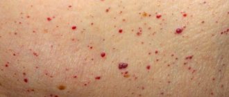
Vascular angiomas appear in various parts of the body. They can be seen on the face, torso, and scalp. Medical statistics show that 80% of people over 50 have these formations. Sometimes the tumors disappear on their own. This happens with infantile hemangiomas. But often their number only increases with age. There is even such a thing as multiple senile angiomas. They appear due to the fragility of capillaries and their loss of elasticity. The vessels dilate and are unable to regain their previous diameter.
Forecast
Possible consequences of the disease
Hemangiomas can cause necrosis (death) of tissue, so the lesion becomes an entry point for infection. The addition of a purulent process can cause sepsis. With hemangioma of bones, their destruction is possible. In addition, the process can contribute to blood clotting disorders and the formation of blood clots. Malignancy is possible - degeneration of hemangioma into cancer.
With timely consultation with a doctor, the neoplasm is successfully treated in 100% of cases. Source: What a pediatrician needs to know about infant hemangiomas. Zakharova I.N., Kotlukova N.P., Roginsky V.V., Sokolov Yu.Yu., Zaitseva O.V., Maykova I.D., Idrisova G.R., Pshenichnikov I.I.: Medical Council , 2021.
Angiomas: treatment
Angioma in adults and children is easily diagnosed. The doctor performs a dermatoscopy to examine the formation. A digital dermatoscope repeatedly magnifies the skin, allowing you to determine the depth, nature, and boundaries of the pathology. In some cases, angiography, blood flow analysis, and histology may be prescribed.
Important: Many patients are interested in why angiomas are removed if the formation is benign and non-aggressive. The main reasons have already been mentioned above. A large angioma on the face disfigures the appearance and there is a risk of complications. In this case, it is enough to observe congenital small spots, since there is a high probability of their disappearance without a trace.
When an angioma on the body or face is perceived as a serious aesthetic defect, if the risk of complications is high, in case of bleeding the formation must be removed. Today, several methods of treating vascular formations are practiced: radio wave therapy, cold treatment (cryotherapy), electrocoagulation, surgery and laser treatment. Each method has its own advantages. But there are also disadvantages. For example, removing part of the skin with a scalpel leaves scars and does not allow you to work with pathology accurately. Cold treatment can negatively affect adjacent tissues. And electrocoagulation is not suitable for neoplasms of significant depth and large area.
The most effective treatment for pathology of any complexity is laser removal of angioma. The laser beam removes abnormally dilated capillaries without affecting adjacent tissue. The doctor controls the intensity and depth of the impact, so he works with high precision. It is important that the treatment is bloodless, painless, and safe. The beam also cauterizes blood vessels, preventing infection. There are no scars left on the skin.
Important: Many people try to get rid of senile moles and red spots on their legs, arms, and face with dubious means at home. Aggressive agents and various ointments, including those containing hormones, are used. Doctors strongly do not recommend doing this, since you most likely will not be able to get rid of the pathology. But serious harm to health can be caused. Moreover, it is not difficult to treat skin lesions professionally today.
In each case, the dermatologist practices an individual approach to the patient. This allows you to get optimal results.
Important: If the formation does not bother you and is located in an inconspicuous place, you can replace treatment with a wait-and-see approach. Because there is a high chance that it will disappear.
Types of hemangiomas
There are 2 types of hemangiomas - congenital - formed during intrauterine development - and acquired; acquired in early childhood are often called infantile. By structure, single and multiple hemangiomas (hemangiomatosis) are distinguished; by structure - capillary, arterial and venous.
By education they are local and segmental. Local hemangiomas grow from one point, are distinguished by smooth edges and relatively small sizes. Segmental - large, with a “ragged” edge, often develop as part of combined pathologies of the chest, aorta, pelvis and sacrum, heart defects, etc.
Hemangiomas are classified according to their location:
- simple – formed on the surface of the skin;
- cavernous – grows under the skin;
- combined – the process involves the superficial and deep layers of the dermis;
- mixed - not only dermal, but also nervous and muscle tissues are involved in the process.
Advantages of treating angiomas at the Lazersvit clinic
Our specialists have extensive experience in treating skin pathologies and vascular formations of the skin. Highly qualified dermatologists, the use of international protocols, and the use of the latest laser equipment in treatment ensure high cosmetic and therapeutic results. In the vast majority of cases, we completely remove vascular anomalies without consequences or complications. In the case of large and deep pathology, several sessions may be required at intervals of 2-3 weeks.
Important: Laser removal of angiomas does not require complex preparation and does not require a long rehabilitation period. The procedure is performed on an outpatient basis. In one session you can completely get rid of a small formation. Immediately after the intervention, the patient returns to his usual lifestyle.
You can learn more about the work of the Mole Diagnostic Center and directly about the treatment of angiomas during a face-to-face consultation with a doctor. You can make an appointment by phone.
Why do hemangiomas appear?
The exact causes of hemangiomas have not yet been identified; It is generally accepted that their appearance can provoke disruption of intrauterine development of the fetus due to a viral illness of the mother or oxygen starvation. Additional risk factors for the formation of congenital hemangioma:
- multiple and/or late pregnancy;
- increased amount of estrogen in the mother;
- her sedentary lifestyle;
- unbalanced diet;
- alcohol or nicotine intoxication of the fetus;
- his low birth weight.
In adults, the cause of neoplasm can be a hereditary predisposition, diseases of the cardiovascular system, excessive ultraviolet radiation, and increased levels of the female hormones estrogen.
Angiomas: causes of appearance
Until now, scientists cannot name the exact reasons for the appearance of vascular formations. Doctors agree that vascular pathology begins during embryonic development. Infectious diseases suffered by the mother can affect this. Toxicosis, anemia of a woman during pregnancy, and serious hormonal fluctuations also contribute to the appearance of abnormalities. Acquired angiomas occur due to sunburn, liver disease, skin injuries, and increased fragility of blood vessels.
Signs of hemangioma and pathogenesis
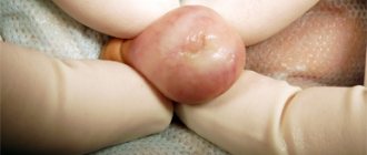
Hemangioma is a red spot, in the center of which there is a point, and from it comes a network of small vessels. Usually the neoplasm is smooth and can protrude 1-2 cm above the skin. When pressed, it turns pale, but quickly returns to its normal appearance. A characteristic symptom is temperature unevenness: upon palpation, the hemangioma is noticeably warmer than the surrounding areas of the body. It may not manifest itself clinically and may be discovered by chance during examination.
The disease occurs in phases: growth, stabilization, and optionally spontaneous regression. Complications are possible during the growth and stabilization phases of hemangioma. If a neoplasm grows near a functional organ, its function may deteriorate - for example, in the periorbital zone it can contribute to decreased vision, and on the laryngeal mucosa - obstruction of breathing.
If the hemangioma is localized near the nerve ending, as it grows, weakness and numbness of the limbs, disruption of the bladder and gastrointestinal tract are possible. Large spinal hemangiomas can cause pain and increase the risk of compression fractures. New growths are easily injured, so they can cause bleeding.
Types of angiomas
According to the etiology and pathogenesis, formations caused by the dilation of small vessels are divided into hemangiomas, formed from a cluster of capillaries, and lymphangiomas, consisting of small lymphatic vessels. Formations of the lymphatic system are less common. Their peculiarity is that the skin with this pathology does not change color. Flesh-colored nodules simply appear on the skin. Angiomas on the body are another matter. This disease always causes red spots to appear. And according to the type of structure, such formations come in several types.
Simple angiomas
Vascular formations of this type are, as a rule, congenital pathologies. They can be smooth or protruding above the surface of the skin. The color of the new growths can be scarlet, burgundy, sometimes with a bluish tint. Vascular angioma grows on any part of the body, but mainly on the upper part of the body. The formation of dilated capillaries reaches a size of 10 cm in diameter. The anomaly is not dangerous, but its presence on the skin can lead to emotional imbalance. After all, the stain greatly spoils the appearance and becomes the reason for the close attention of others and their ridicule. Capillary hemangioma can especially ruin life when it is located on the face, neck, or arms.
Cavernous angiomas
A benign formation of this type looks like a pulsating purple spot. The structure of the pathology is characteristic. Blood enters the cavernous chambers through narrow arteries and is drained from the formation through wide venous channels. The spot is soft on palpation, and after squeezing it quickly restores its shape and appearance.
Branched angioma
The pathology often affects the limbs, less often appears on the scalp. The structure of the formation looks like intertwined dilated vessels. The skin over such a pathology is often affected by simple angioma. This is why capillary hemangioma must be deeply investigated. After all, a branched angioma may be hidden under it.
Intraosseous angiomas
These formations develop on the bones and are detected when they spread to nearby tissues. Bleeding becomes a symptom of the pathology. Accurate diagnosis is possible after obtaining radiographic images. The photo clearly shows the boundaries of the pathology.
Of all the listed types of angiomas, it is simple capillary formations that affect the skin.
How are hemangiomas treated?
The main method of treatment is surgical. In addition to cosmetic reasons, there are medical indications for this:
- rapid growth and threat of malignancy of the tumor;
- disruption of the normal functioning of organs - for example, if there is a tumor on the eyelid or tongue;
- impaired blood circulation – if the tumor is on a large vessel;
- infection and bleeding – for example, when on the genitals.
The priority is gentle, minimally invasive methods for removing hemangioma or vascular surgery methods to reduce blood flow in it. The excised tissue is sent for histological analysis.
If the neoplasm is small, it is possible to use electrocoagulation, radio wave or laser surgery, or cryodestruction with liquid nitrogen. For some forms of hemangiomas, sclerotherapy is effective - “gluing” damaged capillaries by injecting special solutions. Source: Hemangiomas and vascular malformations. Modern theories and therapeutic tactics. Goncharova Y.A.: Child’s health, 2013.
For slow-growing tumors with a low risk of malignancy, conservative drug therapy with drugs from the group of beta-adrenergic receptor blockers is possible. Source: Wong A., Hardy K., Kitajewsky A. Propranolol causes functional changes in hemangioma stem cells and hemangioma endothelial cells // Abstract book. ISSVA the 19th International Workshop on Vascular Anomalies. — 2012. – p. 245.. In some cases (if the tumor does not grow, does not bother, or there is a tendency towards its reverse development), they resort to wait-and-see tactics.
To ensure the effectiveness of treatment, after its completion, a control study is prescribed - ultrasound, tomography or dermatoscopy. To consult with a specialized specialist in St. Petersburg, fill out the online form.
Sources:
- What a pediatrician needs to know about infantile hemangiomas. Zakharova I.N., Kotlukova N.P., Roginsky V.V., Sokolov Yu.Yu., Zaitseva O.V., Maykova I.D., Idrisova G.R., Pshenichnikov I.I.: Medical Council , 2016
- Infantile hemangioma: classification, clinical picture and methods of correction. Sheptiy O.V., Kruglova L.S.: Russian Journal of Skin and Venereal Diseases, 2021.
- Hemangiomas and vascular malformations. Modern theories and therapeutic tactics. Goncharova Y.A.: Child’s health, 2013.
- Sires V. Systemic corticosteroid use in orbital lymphangioma /V. Sires, C. Goins, R. Anderson // Ophthal. Plast.Reconstr.Surg. — 2001. Mar. - Vol. 17(2). — P. 85 — 90.
- Wong A., Hardy K., Kitajewsky A. Propranolol causes functional changes in hemangioma stem cells and hemangioma endothelial cells // Abstract book. ISSVA the 19th International Workshop on Vascular Anomalies. — 2012. – p. 245.
The information in this article is provided for reference purposes and does not replace advice from a qualified professional. Don't self-medicate! At the first signs of illness, you should consult a doctor.
Angiomas in children
Angioma in children is usually diagnosed at birth or during the first weeks of life. Girls get sick twice as often as boys. Angioma in a child goes through several stages. In the first few months, the formation actively grows, the size of the spot increases. At the next stage, growth stops. By the age of 9, in 90% of cases, self-healing occurs - spontaneous involution of the pathology. The stain disappears, leaving no traces in its place.
Unfortunately, in some cases, benign formation is accompanied by the following complications:
- ulceration and bleeding of the skin as a result of trauma, for example, due to rubbing with a diaper when the formation is located on the leg or buttock;
- formation of scars at the site of healing ulcers after ulceration of the skin at the site of formation;
- dysfunction of internal organs due to the germination of the formation or compression of them by its boundaries;
- When the formation is located on the forehead, ophthalmological diseases develop.
Important: Very often, angioma in a child is located on the face, neck, or chest. This leads to a pronounced cosmetic defect.
It is recommended to observe uncomplicated disease in children. To do this, during the appointment, photographs of the formation are taken using a dermatoscope. At subsequent appointments, images can be compared to analyze the behavior of the formation.
Prevention of angioma
It is impossible to prevent the occurrence of congenital vascular pathologies, so effective prevention does not exist. There are several ways to reduce the risk of abnormalities such as red spots. Women are advised to carefully plan their pregnancy, and while carrying a child, follow all the recommendations of the obstetrician-gynecologist.
As for senile vascular anomalies, their occurrence can be prevented by avoiding prolonged exposure to the sun and cold, and by building a healthy diet rich in minerals and vitamins. It is also recommended that older people lead a healthy lifestyle and do not ignore physical activity agreed upon with their family doctor.