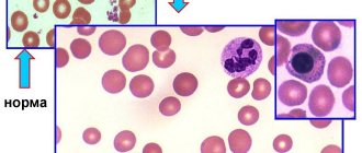
While carrying a child, the female body needs a large amount of useful substances that participate in the processes that occur during pregnancy and ensure the growth and development of the fetus. One of the most important microelements is iron. Unfortunately, 40% of pregnant women develop iron deficiency - this condition is called iron deficiency anemia. It is caused by a decrease in the level of hemoglobin, a complex iron-containing protein that binds oxygen and delivers it to tissues and organs. As a result of anemia, cells do not receive enough oxygen, which is dangerous for the health of both the woman and the child.
Why does iron deficiency occur during pregnancy, what are its signs and consequences?
Causes of iron deficiency in the female body during pregnancy
During the period of bearing a child, the volume of blood in the female body increases by 50%. At the same time, the amount of fluid increases, causing the blood to “thin out.” Therefore, a decrease in hemoglobin levels is natural: the lower limit of hemoglobin in the blood of pregnant women is set at 110 g/l. For comparison: the norm for non-pregnant women is from 120 to 140 g/l. It is worth considering that women often enter pregnancy with a deficient or borderline level of iron in the blood. Iron levels may be low due to heavy menstruation lasting more than 5 days, poor nutrition, problems with the gastrointestinal tract, in which this trace element cannot be fully absorbed in the body.
Iron deficiency anemia during pregnancy is indicated when the hemoglobin level drops below 110 g/l. There are three degrees of anemia:
- Mild severity (hemoglobin level - from 90 to 110 g/l).
- Moderately expressed (from 70 to 89 g/l).
- Heavy (from 40 to 69 g/l).
Risk group for anemia in pregnant women:
- Pregnancy in adolescence.
- Pregnancy and lactation during the previous 3 years, many births (more than 3 with a short interval).
- Excessive menstrual blood loss (monthly blood loss more than 100 ml).
- Gynecological diseases, especially uterine fibroids and endometrial diseases, inflammatory diseases of the pelvic organs.
- Diets low in animal protein (vegetarianism).
- Regular blood donation.
- Diseases of the gastrointestinal tract.
- Low socio-economic status.
- Obesity.
At the same time, WHO recommends that all menstruating women be considered at risk, provided that the prevalence of anemia in non-pregnant women in the region exceeds 20%.
Symptoms of anemia during pregnancy
With iron deficiency during pregnancy, the following symptoms are observed:
- Constant weakness, possible fainting.
- Dizziness.
- Pale, dry, flaky skin.
- Pale mucous membranes (lips, eyelids, etc.).
- Depressed mood, irritability, drowsiness.
- Tachycardia (heart beats quickly - more than 90 beats per minute).
- Brittle nails with white spots.
- Brittle, dull hair with split ends.
- Non-standard taste preferences (for example, when you want to eat chalk or sand).
If the hemoglobin level decreases gradually, then the expectant mother may feel well for a long time, although the problem still exists. Therefore, it is important to regularly take all the tests prescribed during pregnancy. Anemia will be detected by a complete blood test.
How to prevent anemia?
Treatment of anemia is based on the use of iron supplements and a balanced diet. Here are data on the iron content of some foods (mg iron per 100 g of food):
- Liver, oatmeal, beans, beans, soybeans - 5–15 mg.
- Eggs, meat, fish, spinach, prunes, apricots, bread - 1–5 mg.
- Milk, potatoes, carrots, beets, apples - less than 1 mg.
But I repeat, the maximum amount of iron that we can get from food is 2.5 mg/day! Therefore, during pregnancy you need to additionally take vitamin complexes with iron or iron supplements according to indications. The course of treatment is usually 1–1.5 months.
Do not be ill!
What are the dangers of iron deficiency during pregnancy?
Anemia increases the risk of pregnancy complications. Women with iron deficiency are more likely to experience toxicosis. In 40% of pregnant women with anemia, gestosis is diagnosed, in which blood pressure rises, swelling appears, and protein is found in the urine. 15–42% of women with anemia experience miscarriages and labor begins prematurely.
Acute iron deficiency can lead to serious consequences:
- Premature birth.
- Weak labor.
- Premature placental abruption.
- Intrauterine growth retardation and fetal development.
- Bleeding after childbirth.
- Increased risk of inflammation after childbirth.
- Reduced milk supply during breastfeeding.
Children whose mothers experienced iron deficiency during pregnancy also often develop anemia by the first year of life. In addition, such babies are more susceptible to viral diseases. The fact is that iron is required for the proper formation of the immune system. If there was little iron during intrauterine development, then the child’s immunity will be weak.
Treatment of anemia:
Treatment of anemia consists of proper nutrition and taking iron supplements with vitamins for a long time. Your doctor will prescribe medications for you, but you need to know how to eat properly during pregnancy.
The female body needs 1.5-1.7 mg of iron per day, during pregnancy - up to 6-7 mg. Throughout pregnancy you need 2000 mg of iron. With normal nutrition during pregnancy, a woman receives 700-800 mg and another 400-500 mg is consumed from the depot.
What if the iron supply in the depot is reduced (ferritin below 30 mcg/l)? 12 - 15% of women in the reproductive period of the Russian Federation suffer from iron deficiency anemia, and 60% from hidden iron deficiency. Therefore, it is so necessary to create an iron reserve before pregnancy. How often do women say at a doctor’s appointment: “But my hemoglobin is always low and I live.” But this is dangerous for pregnancy, as it poses a risk not only for the mother, but also for her child. It is right for such women to be examined and treated before pregnancy.
Anemia is one of the most common complications of pregnancy. The leading sign of anemia in pregnant women is a decrease in hemoglobin (Hb) level less than 110 g/l [1].
In 90% of cases, anemia in pregnant women is iron deficiency. Such anemia is characterized by a violation of Hb synthesis due to iron deficiency developing as a result of various physiological and pathological processes. According to WHO, the incidence of iron deficiency anemia in pregnant women ranges from 21 to 80%. The presence of iron deficiency anemia leads to impaired quality of life, reduces performance, and causes functional disorders of many organs and systems. Iron deficiency in pregnant women increases the risk of complications during childbirth, and in the absence of timely and adequate therapy, iron deficiency may also occur in the fetus [1].
Iron is one of the vital elements; the human body contains about 4 g, 75% of iron is in Hb. Iron is most fully absorbed from animal products (meat), and much worse from plant foods. The release of iron from foods is reduced when they are cooked, frozen, or stored for a long time.
Iron is excreted from a woman’s body in an amount of 2-3 mg/day through the intestines, bile, urine, through the skin epithelium, during lactation and menstruation.
Outside of pregnancy, the iron requirement is 1.5 mg/day. During pregnancy, this figure increases steadily: in the first trimester by 1 mg/day, in the second trimester by 2 mg/day, in the third trimester by 3-5 mg/day. Iron loss is most pronounced in 16-20 weeks of pregnancy, which coincides with the period of the onset of hematopoiesis in the fetus and an increase in blood volume in the pregnant woman (Table 1). During childbirth (with physiological blood loss), 200 to 700 mg of iron is lost, and subsequently, during lactation, about another 200 mg. Thus, about 800-950 mg of iron is consumed from the maternal depot during pregnancy and the postpartum period. The body is able to restore iron reserves within 4-5 years. If a woman plans a pregnancy before this period, she will inevitably develop anemia [2].
Table 1. Some peripheral blood parameters in different trimesters of pregnancy
The following processes lead to the development of anemia during pregnancy:
- metabolic changes occurring in the patient’s body during pregnancy;
- decrease in the concentration of a number of vitamins and microelements - cobalt, manganese, zinc, nickel;
- changes in hormonal balance during pregnancy, in particular, an increase in the amount of estradiol, which causes inhibition of erythropoiesis;
- deficiency in the body of a pregnant woman of vitamin B12, folic acid and protein;
- lack of oxygen, which disrupts redox processes in the body;
- immunological changes in the body of a pregnant woman, occurring due to constant antigenic stimulation of the maternal body from the tissues of the developing fetus;
- consumption of iron from the mother’s body depot, necessary for the proper development of the fetus.
During pregnancy, so-called physiological, or “false” anemia may occur. The occurrence of this form is due to an uneven increase in individual blood components. The fact is that during pregnancy, as a compensatory reaction, the mother’s blood volume increases by 30-50%, but mainly due to plasma. Accordingly, the ratio of the volume of formed elements of blood and plasma shifts towards the latter. This form of anemia does not require treatment [3].
Pathological forms of anemia during pregnancy are the following:
1. Hypochromic anemia, which may be a consequence of concomitant pathology with infectious diseases (sepsis) and parasitic infestations (helminthiases, malaria) during pregnancy. Hypochromic anemia during pregnancy can occur as a result of liver disease, stomach disease, and nutritional dystrophies.
2. Megaloblastic anemia associated with folic acid (FA) deficiency accounts for 1% of all anemias in pregnant women and most often develops in the third trimester of pregnancy, before childbirth and in the early postpartum period. FA plays an important role in many physiological processes, participates in the synthesis of a number of amino acids, and plays a key role in cell division processes. Tissues with a high rate of cell division (bone marrow, intestinal mucosa) are characterized by an increased need for F.K. Deficiency of F.K. in the body occurs due to insufficient content of it in the diet, increased need for it (pregnancy, prematurity, hemolysis, cancer); impaired absorption and increased excretion from the body (some skin diseases, liver diseases). The daily requirement of a pregnant woman's body for FA increases to 400 mcg, and by the time of delivery - up to 800 mcg; the need for FA during lactation is 300 mcg. Folic acid deficiency occurs in approximately 30% of pregnant women and adversely affects the course of pregnancy and fetal development. With such a deficiency, the formation of neural tube defects (anencephaly, encephalocele, spina bifida) is possible. Another important fact confirming the role of folic acid during pregnancy is the close relationship between the level of folic acid in the mother’s body and the birth weight of the child. Several weeks before birth, the fetus uses the mother's FA to increase its own weight and replenish its folate reserves. As a result, in women with FA deficiency, the likelihood of having a child with malnutrition and a reduced FA reserve increases significantly. The main sources of FA in food are raw green vegetables and fruits, beef liver, cheese, and egg yolk.
3. Essential (cryptogenic) malignant anemia of Birmer-Ehrlich (synonyms: megaloblastic anemia, pernicious anemia, Addison-Birmer anemia). This anemia is rare during pregnancy. This form of anemia is associated with a deficiency of vitamin B12 or FK. The development of this anemia is facilitated by past infections, insufficient intake of vitamins from food, diseases of the stomach and duodenum, the use of medications (acyclovir, anticonvulsants, nitrofurans, oral contraceptives) and Crohn's disease.
4. Hypo- or aplastic anemia, in which there is a sharp suppression of bone marrow hematopoiesis. The causes of this form of anemia are most often ionizing radiation; taking medications (chloramphenicol, aminazine, butadione, cytostatics); entry into the body of chemicals (benzene, arsenic) that have a myelotoxic effect; chronic infectious diseases (viral hepatitis, pyelonephritis); autoimmune processes. The diagnosis is made based on the results of a bone marrow puncture, which reveals the complete disappearance of bone marrow elements and their replacement with adipose tissue and hemorrhages. The mortality rate for a pregnant woman with this form of anemia reaches 45%. Therefore, it is advisable to terminate pregnancy before 12 weeks followed by splenectomy. If hypoplastic anemia is diagnosed in late pregnancy, surgical delivery by cesarean section in combination with splenectomy is recommended.
5. Hemolytic anemia is a large group of diseases, the main distinguishing feature of which is a shortening of the lifespan of red blood cells due to their hemolysis. There are hereditary and acquired hemolytic anemias.
— Microspherocytic hemolytic anemia is hereditary. It develops due to a defect in the structure of the erythrocyte membrane, which leads to the penetration of excess sodium into the cell and the accumulation of water. The red blood cells become spherical and are destroyed in the spleen.
— Autoimmune hemolytic anemia, in which the formation of antibodies to one’s own red blood cells occurs. There are symptomatic and idiopathic autoimmune hemolytic anemia. The symptomatic form includes anemia, which develops against the background of hemoblastosis, systemic lupus erythematosus, ulcerative colitis, and chronic hepatitis. The idiopathic form includes those cases of anemia in which it is impossible to establish the underlying disease. Rarely occurs during pregnancy. The prognosis for the mother is favorable. Delivery is carried out through the natural birth canal.
6. True iron deficiency anemia (IDA) occurs most often during pregnancy. A characteristic feature of this form is either an absolute decrease in the number or functional deficiency of red blood cells. Clinical manifestations of IDA are caused, on the one hand, by the presence of anemic syndrome, on the other, by iron deficiency [4].
Pregnancy is contraindicated in the following forms of diseases of the blood and hematopoietic system:
— chronic IDA of III-IV degree;
- hemolytic anemia;
— bone marrow hypo- and aplasia;
- leukemia;
- Werlhof's disease with frequent exacerbations.
If pregnancy occurs due to these diseases, it is advisable to terminate it before 12 weeks.
Clinical manifestations of anemia.
Anemia is manifested by a complex of nonspecific symptoms and is caused by insufficient oxygen supply to tissues. The main clinical manifestations of this pathology are general weakness, increased fatigue, anxiety, dizziness, tinnitus, floaters before the eyes, tachycardia, shortness of breath during exercise, fainting, insomnia, morning headache, forgetfulness and decreased performance.
The consequence of iron deficiency is dry skin, the formation of cracks on it; violation of the integrity of the epidermis; the appearance of ulcerations and cracks in the corners of the mouth with inflammation of surrounding tissues; changes in nails (fragility, layering, transverse striations; nails become flat, take on a concave spoon-shaped shape); hair damage (hair splits, ends split).
Due to iron deficiency, patients experience a burning sensation in the tongue; taste is distorted (desire to eat chalk, toothpaste, ashes, clay, sand, raw cereals); there is an unhealthy addiction to certain odors (acetone, gasoline, kerosene, naphthalene), difficulty swallowing dry and solid food; there is a feeling of heaviness and abdominal pain, as with gastritis, urinary incontinence when coughing and laughing, nocturnal enuresis, muscle weakness, pale skin; possible arterial hypotension; low-grade fever. In severe forms of IDA, anemic myocardial dystrophy develops.
The course of pregnancy with iron deficiency anemia.
Due to the fact that during pregnancy, oxygen consumption increases by 15-33%, pregnant women with IDA are characterized by severe tissue hypoxia with the subsequent development of secondary metabolic disorders, which may be accompanied by the appearance of dystrophic changes in the myocardium, as well as in the uterus and placenta, which lead to to the formation of placental insufficiency and fetal development retardation.
IDA is characterized by disturbances in protein metabolism with the occurrence of protein deficiency in the body, which leads to the development of edema in a pregnant woman.
The main complications of pregnancy with IDA are the following:
- threat of miscarriage (20-42%),
- gestosis (40%),
- arterial hypotension (40%),
— premature placental abruption (25-35%),
- delayed fetal development (25%),
- premature birth (11-42%).
Childbirth is often complicated by bleeding (48%). In the postpartum period, various inflammatory complications can occur (12%) [5].
Diagnosis of anemia.
In addition to assessing standard indicators in a clinical blood test (Hb, erythrocytes, hematocrit, ESR), the diagnosis of IDA is based on the assessment of such indicators as color, average hemoglobin content in an erythrocyte, morphological picture of erythrocytes, serum iron, ferritin, transferrin, total iron-binding capacity of serum blood.
The “gold standard” for diagnosing anemia remains the determination of ferritin (Table 2).
Table 2. Dynamics of ferritin levels during pregnancy (according to V.A. Demikhov) [6]
The dynamics of ferritin and hemoglobin levels during pregnancy are as follows:
- in the first trimester, ferritin level is about 50 ng/ml, Hb - 130 g/l,
- in the second trimester, ferritin is about 30 ng/ml, Hb is about 118-120 g/l,
- in the third trimester, ferritin is at least 12 ng/ml, Hb - 109-110 g/l.
After childbirth, ferritin levels increase.
Therefore, for early detection of iron deficiency after pregnancy, examination is necessary after childbirth, after cessation of lactation. If the ferritin level decreases to less than 30 ng/ml, it is necessary to carry out preventive courses of treatment with iron supplements for 2-3 months in combination with multivitamin complexes and probiotics. If a decrease in ferritin of less than 20 ng/ml is detected, then the duration of therapy is increased until normal ferritin concentrations are obtained (50-70 ng/ml, Hb - 130-135 g/l).
Prevention of anemia should primarily be carried out among pregnant women who are at high risk of developing it.
These pregnant women experience the following:
1. Reduced intake of iron into the body from food (vegetarianism, anorexia).
2. Chronic diseases of internal organs (rheumatism, heart defects, pyelonephritis, hepatitis). In liver diseases, the processes of iron accumulation in the body and its transportation are disrupted. Gastrointestinal bleeding due to gastric and duodenal ulcers, hemorrhoids, intestinal diverticulosis, ulcerative colitis, helminthic infestation.
3. The presence of diseases manifested by chronic nosebleeds (thrombocytopathies, thrombocytopenic purpura).
4. Gynecological diseases accompanied by heavy menstruation or uterine bleeding (endometriosis, uterine fibroids, hyperplastic processes in the endometrium).
5. Complicated obstetric history: multiparous women, history of spontaneous miscarriages, bleeding during childbirth.
6. Complicated course of this pregnancy: multiple pregnancy, vomiting of pregnant women, young age of the pregnant woman (under 17 years), primiparas over 30 years old, arterial hypotension, exacerbation of chronic infectious diseases during pregnancy, preeclampsia, placenta previa, premature placental abruption.
Given the high incidence of anemia in pregnant women, preventive measures are necessary. Prevention of IDA in pregnant women at risk of developing this pathology consists of prescribing small doses of iron supplements or iron-containing multivitamin complexes for 4-6 months, starting from 14-16 weeks of pregnancy, in courses of 2-3 weeks, with breaks of 14-21 days, only 3-5 courses per pregnancy.
At the same time, it is necessary to change the diet in favor of increasing the consumption of foods containing large amounts of easily digestible iron. However, with food, a pregnant woman cannot receive the necessary dose of iron and synergistic vitamins necessary to improve the absorption of iron. The maximum amount of iron that can be absorbed from food is 2.5 mg/day.
To prevent anemia and treat mild forms of anemia, it is recommended to prescribe medications containing a physiological dosage of iron - 60 mg. WHO experts recommend the use of oral Fe2+ rather than Fe3+. Such drugs include the drug Vitrum Prenatal Forte. It contains the recommended dosage of iron 60 mg, in the recommended form Fe2+.
According to WHO recommendations, all women should take medications containing iron and its synergists to prevent and treat anemia throughout pregnancy and in the first 6 months of lactation [7].
Treatment
of anemia.
Pregnant women with IDA, in addition to drug treatment, are prescribed a special diet. The greatest amount of iron is found in meat products. The iron they contain is absorbed by the human body by 25-30%. The absorption of iron from other products of animal origin (eggs, fish) is 10-15%, from plant products - only 3-5%.
The largest amount of iron (in mg per 100 g of product) is found in liver (19.0 mg), cocoa (12.5 mg), egg yolk (7.2 mg), heart (6.2 mg), veal liver (5 .4 mg), stale bread (4.7 mg), apricots (4.9 mg), almonds (4.4 mg), turkey meat (3.8 mg), spinach (3.1 mg) and veal (2 .9 mg).
It is important to remember that 2.5 mg of iron per day is absorbed from food, while 15-20 times more from medications! Therefore, drug therapy for anemia during pregnancy is necessary!
Treatment with iron supplements should be long-term. The Hb content increases only at the end of the 3rd week of IDA therapy. Normalization of blood counts occurs after 5-8 weeks of treatment, but this does not indicate restoration of iron reserves in the body. For this purpose, WHO experts recommend that after 2-3 months of treatment and elimination of anemia, do not stop therapy, but only halve the dose of the drug used for treatment. This course of treatment continues for 3 months. Even with complete restoration of iron reserves in the body, it is advisable to take small doses of iron-containing drugs for six months. In this case, it is recommended to prescribe the drug Vitrum Prenatal Forte.
It is most preferable to take iron supplements orally rather than by injection, since in the latter case various side effects may more often occur [8, 9].
In addition to iron, drugs for the treatment of IDA contain various components that enhance the absorption of iron (copper, manganese, vitamin B12, ascorbic acid, FA, etc.). For better tolerability, iron supplements should be taken with meals. It must be taken into account that under the influence of certain substances contained in food (phosphoric acid, phytin, tannin, calcium salts), as well as with the simultaneous use of a number of medications (tetracycline antibiotics, almagel), the absorption of iron in the body decreases.
The reasons for the ineffectiveness of treatment for patients with anemia during pregnancy are the following:
1. Insufficient volume of therapy, stopping medications when Hb levels improve or because of fear of taking medications for a long time (2, 3, 6 months).
2. The cause of anemia has not been eliminated (continuing blood loss due to heavy menstruation, hemorrhoidal bleeding, long-term vegetarian diet, etc.).
3. Insufficient consideration of the balance of vitamins and microelements involved in iron metabolism.
4. The presence of dysbacteriosis, in which the synthesis of transport proteins (metal protectors, transferrin) is disrupted.
Iron deficiency is a diselementosis. The body needs a sufficient amount of various biologically active substances that make up the so-called biological chain: iron, calcium, copper, zinc, manganese, etc., as well as vitamins - FA, ascorbic acid, B1, B12, B2, B6, biotin and others [10, 11].
Each of these micronutrients has its own important biological function.
Copper is one of the main essential trace elements; it is involved in the process of respiration and erythropoiesis, the maturation of reticulocytes and in the process of granulocytopoiesis. Copper deficiency can block the activity of superoxide dismutase, an enzyme responsible for inhibiting lipid peroxidation of cell membranes. Copper is closely related to iron deficiency. Decreased serum iron levels may result in decreased serum cerulloplasmin and copper levels.
Manganese is an essential trace element. Manganese is a cofactor in many multienzyme systems and is involved in the synthesis of nucleic acids and the metabolism of various hormones. Manganese is an essential part of superoxide dismutase, which is responsible for inhibiting lipid peroxidation and protecting cell membranes. With a lack of manganese in the blood, the Hb level also decreases. It has been noted that the therapeutic combination of iron and manganese better satisfies the need for these two elements than isolated treatment with iron.
FA (vitamin B9) takes part in protein metabolism, ensuring their synthesis, as well as the synthesis of purine and pyrimidine bases, choline, formic acid, biotin, and the amino acid tryptophan. The absorption of folate, like iron, occurs in the duodenum and in the initial part of the small intestine and depends on the presence of vitamins A and C. A decrease in the content of vitamin A leads to a lack of vitamin C, without which F.K. is not absorbed. A decrease in the vitamin-like substance biotin also leads to a decrease in FA levels in the body. Further and final absorption occurs with the obligatory participation of Escherichia coli, which in the body is most concentrated in the ascending colon [12].
Vitamin B12 is involved in the process of hematopoiesis and, like FA, is synthesized by normal intestinal bacteria. When a deficiency of these vitamins occurs, bacteria usually lose their ability to synthesize vitamins.
The physiological effect of vitamin C is diverse and consists of the following: the formation of the main substance of connective tissue - hydroxyproline (with hypovitaminosis C, the ligamentous apparatus becomes weak and too extensible); vitamin C is part of polysaccharides, including cartilage tissue, includes sulfur in chondroitin sulfate; converts ferric iron into divalent iron, which ensures its absorption, divalent copper into monovalent (active), binds to calcium and ensures its bioavailability.
Vitamin C is also involved in the formation of corticosteroids, adrenocorticotropic hormone, which affects the catabolism of cholesterol; regulates the formation of glycogen in the liver and ensures the entry of glucose into the erythrocyte, ensuring glucose tolerance; regulates the production of enzymes: hepatic and pancreatic esterase, cathepsin, inhibits the enzyme urease; is a regulator of many immune reactions: promotes the production of antibodies, increases phagocytosis, activates complement along with the amino acid cysteine, has antioxidant, radioprotective and desensitizing properties; improves hematopoiesis, adaptive and regenerative processes, accelerates the healing of fractures.
There are many factors that contribute to increased consumption of vitamin C in everyday life: cooling, burns, overheating, bleeding, intoxication (including alcohol), oxygen deprivation, ether anesthesia, poisoning with salts of heavy metals, deficiency of other vitamins, especially A and E.
Vitamin C is absorbed in the small intestine without the participation of microflora, however, varieties of pathogenic bacteria have been isolated that destroy vitamin C. The process of increased destruction of the vitamin occurs if the pathogenic flora enters from the large intestine, where there are a lot of bacteria, into the small intestine, where bacteria are normal much less or should not be at all. With insufficiency of ascorbic acid, reflux from the large intestine to the small intestine appears, and the bauhinium valve becomes functionally defective. Colonization of the small intestine by bacteria leads to its inflammation and blockade of the absorption of many useful substances [13].
Vitamin C has a bactericidal and bacteriostatic effect against some pathogenic bacteria; in other words, the vitamin selectively acts as an antibiotic.
In the presented table. Table 3 shows the norms for consumption of essential microelements and vitamins for pregnant women. Thus, for the treatment of iron deficiency conditions in pregnant women, not only iron supplements are needed, but also a sufficient set of vitamins and minerals, as well as correction of intestinal dysbiosis [13].
Table 3. Norms of physiological needs* of vitamins and microelements for pregnant and postpartum women Note. * State Research Institute of Nutrition of the Russian Academy of Medical Sciences, 2008.
Iron deficiency anemia in pregnant women is an important problem related to both the health of the mother and her fetus, therefore, prevention of IDA in pregnant women helps to create higher iron stores in newborns, preventing the development of iron deficiency and anemia in infants.
The use of medications with a balanced content of iron and its synergists in one tablet (Vitrum Prenatal Forte) allows one to achieve good results in the treatment of mild IDA, and their prophylactic use throughout pregnancy, lactation and in preparation for pregnancy prevents the development of severe complications in the mother and fetus