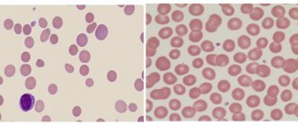
Erythremia is a tumor clonal disease of the hematopoietic system, in which there is proliferation of erythroid, granulocytic and megakaryocytic lineages of hematopoiesis with predominant activation of erythropoiesis. At the same time, an increase in the level of red blood cells and hemoglobin, thrombocytosis and leukocytosis is noted in the blood. Almost all patients with erythremia are carriers of the JAK2V617F mutation.
- How does erythremia develop?
- Symptoms of erythremia
- Stages of erythremia
- Diagnosis of erythremia
- Treatment of erythremia
- Prognosis for erythremia
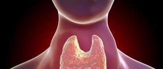
general description
Erythremia is a chronic leukemia characterized by tumor proliferation of all hematopoietic germs, especially the erythroid germ, which is accompanied by an increase in the number of red blood cells (in some cases, leukocytes and platelets), hemoglobin, mass, viscosity and blood coagulability.
Erythremia occurs mainly in old age. The incidence ranges from 0.6 to 1.6 per 100,000 population. Familial cases of the disease are observed.
There are 3 stages of the disease: initial, advanced (erythremic) and terminal.
Erythremia
In stage I, which lasts 5 years or more, there is a moderate increase in circulating blood, the spleen cannot be palpated. In the blood at this stage, moderate formation of red blood cells predominates. In the bone marrow, there is an increase in all hematopoietic germs. Vascular and visceral complications at this time are possible, but not frequent.
The identification of the initial (I) stage of erythremia is conditional. Essentially, this is a stage with few symptoms, more typical for older patients. The spleen is usually not palpable, but examination of it often reveals a slight enlargement. Thrombotic complications are also possible at this stage of the disease.
Stage IIA of the process - erythremic - is extensive, and myeloid transformation of the spleen is uncharacteristic for it. The duration of this stage is 10-15 years or more. The volume of circulating blood is increased, the spleen is enlarged, and a little earlier the liver may become enlarged. Thrombosis of arterial and venous vessels, hemorrhagic complications are observed more often at this stage. A blood test indicates “pure” erythrocythemia or erythrocythemia and thrombocytosis or panmyelosis and neutrophilosis with a band shift, an increase in the number of basophils. In the bone marrow, total three-line hyperplasia with pronounced megakaryocytosis is observed; reticulin and focal collagen myelofibrosis are possible.
Stage IIB also includes an erythremic, extensive process, but with myeloid metaplasia of the spleen. The increase in blood volume can be expressed to a greater or lesser extent, and an enlargement of the liver and spleen is observed. In the blood at this stage there is an increase in red blood cells, platelets with leukocytosis above 15 H 103 in 1 μl and a shift in the leukocyte formula to myelocytes, single erythrokaryocytes. In the bone marrow, as in stage IIA, an increase in granulocytic lineage may predominate, and reticulin and focal collagen myelofibrosis is possible.
Allergic complications and urate diathesis are often the leading ones in the clinical picture.
At this stage, the patient may be exhausted, worsening thrombotic complications and bleeding.
Stage III of erythremia is called anemic. Myelofibrosis may be expressed in the bone marrow; myelopoiesis is preserved in some cases and reduced in others. Myeloid transformation is observed in the enlarged spleen and liver. The outcome of erythremia at this stage can be acute leukemia, chronic myeloid leukemia, hypoplastic state of hematopoiesis, and hematological changes that are difficult to classify.
Arterial hypertension, which occurs with erythremia in 35-50% of cases, is caused by an increase in peripheral resistance in response to increased blood viscosity, the development of urate diathesis, chronic pyelonephritis, circulatory disorders in the renal parenchyma, thrombosis and sclerosis of the renal arteries.
Skin itching associated with washing, specific to erythremia, is observed in 50-55% of patients. For many patients, it becomes the main complaint; it arises not only from contact with water, but also spontaneously, affecting performance.
Frequent complications of the advanced stage of the disease are microcirculatory disorders with clinical manifestations of erythromelalgia, transient disorders of cerebral and coronary circulation and hemorrhagic edema of the legs, as well as thrombosis of venous and arterial vessels and bleeding. Already at this stage there may be hemostasis disorders, which often look like a latent thrombogenic danger, detected only in the laboratory and without clinical manifestations. At the same time, hemostasis disorders can be more pronounced, leading to local intravascular coagulation such as microthrombosis or to disseminated intravascular coagulation - DIC syndrome.
Diagnosis of erythremia
- Complete blood count: increased levels of red blood cells, hemoglobin, platelets, leukocytes.
- Sternal puncture.
- Ultrasound of the abdominal organs: enlarged liver, spleen.
Criteria for diagnosing erythremia:
- Increase in the mass of circulating red blood cells: for men - more than 36 ml/kg, for women - more than 32 ml/kg.
- Normal arterial blood oxygen saturation (more than 92%).
- Enlarged spleen.
- Leukocytosis is more than 12x109 in 1 μl (in the absence of infections and intoxications).
- Thrombocytosis is more than 400x109 in 1 µl (in the absence of bleeding).
- Increased content of neutrophil alkaline phosphatase (in the absence of infections and intoxications).
- Increase in unsaturated B12-binding capacity of blood serum.
The diagnosis is reliable when any 3 signs are confirmed.
How does erythremia develop?
The causes of erythremia are still unknown. It is believed that this is a multi-stage disease in which, under the influence of external factors, the genome of a normal cell is damaged, which leads to its malignant transformation and the formation of a tumor cell clone that replaces normal hematopoiesis.
Almost all patients have a mutation in the JAK2 gene. Exon 14 is usually affected; 90-96% of patients have the V617F mutation. 2% of patients have mutations in exon 12. Damage to other genes, in particular MPL, CALR, is very rarely detected. All of these genetic pathologies are specific to erythremia, so their determination is necessary to confirm the diagnosis.
So, molecular genetic disorders cause activation of the JAK-STAT signaling pathway, which leads to increased proliferation of hematopoietic sprouts and an increase in the number of blood cells.
Monocytes and megakaryocytes (platelet precursors) produce a variety of cytokines - biologically active molecules that stimulate fibrotic changes, the formation of new blood vessels, which ultimately leads to osteosclerosis and bone marrow fibrosis. In addition, massive production of cytokines contributes to the development of tumor intoxication, which aggravates the general condition of patients.
There is also a disruption in the connection of stem cells with the microenvironment. This provokes the formation of foci of hematopoiesis outside the bone marrow. The liver and spleen are primarily affected.
Treatment of erythremia
- Bloodletting (frequency and volume depend on the stage of the disease).
- Cytostatic drugs for severe disease.
- Anticoagulants.
- Antiplatelet agents (Aspirin, Clopidogrel).
- Antihypertensive therapy.
- Transfusion of fresh frozen plasma in cases of increased bleeding.
Essential drugs
THERE ARE CONTRAINDICATIONS. CONSULT YOUR DOCTOR.
- Aspirin (antiplatelet agent). Dosage regimen: orally, after meals, with a sufficient amount of water at a dose of 50-250 mg/day. for 3 doses.
- Hydroxyurea (antitumor agent, antimetabolite). Dosage regimen: orally, in the morning on an empty stomach at an initial dose of 0.5-2.0 g/day. once until blood counts normalize. After achieving remission (temporary weakening or disappearance of the manifestations of the disease), maintenance therapy of 0.5 g of the drug daily continuously.
- Reaferon (antitumor, immunomodulatory agent). Dosage regimen: IM, SC 15-30x106 IU 2-3 times a week.
- Anagrelide (antiplatelet agent). Dosage regimen: orally, 1-10 mg/day, maintenance therapy 1.5-2.5 mg/day.
Prognosis for erythremia
In general, the prognosis for erythremia is relatively favorable. The overall 10-year survival rate is about 75%. But the disease can transform into acute myeloid leukemia (5% risk) or myelofibrosis (slightly less than 10% risk).
Symptoms of erythremia (headaches, bone pain, paresthesia) can worsen the quality of life of patients. The main cause of death in patients is thrombosis, bleeding and severe infections, the risk of which increases when the pathology transforms into acute myeloid leukemia or myelofibrosis.
Treatment of erythremia at the Euroonko clinic is carried out using modern treatment protocols. In difficult cases, the decision is made collegiately by a council of specialists. We attach great importance to monitoring the patient during the period of remission. This also makes it possible to achieve an increase in the effectiveness of the treatment.
Book a consultation 24 hours a day
+7+7+78
Incidence (per 100,000 people)
| Men | Women | |||||||||||||
| Age, years | 0-1 | 1-3 | 3-14 | 14-25 | 25-40 | 40-60 | 60 + | 0-1 | 1-3 | 3-14 | 14-25 | 25-40 | 40-60 | 60 + |
| Number of sick people | 0 | 0 | 0.1 | 0.4 | 0.4 | 2.68 | 7.8 | 0 | 0 | 0.1 | 0.3 | 0.3 | 1.1 | 7.6 |
Causes
The exact causes of the disease are still unknown. Scientists suggest that hereditary predisposition, exposure to ionizing radiation and toxic substances play a significant role in the development of pathological changes.
In 20-30% of patients with erythremia, chromosomal abnormalities are detected ( https://cyberleninka.ru/article/n/klinicheskie-rekomendatsii-po-diagnostike-i-terapii-ph-negativnyh-mieloproliferativnyh-zabolevaniy-istinnaya-politsitemiya-1/viewer ).
The likelihood of developing erythremia increases in funeral home employees who embalm bodies, in people who are forced to frequently come into contact with gasoline and other petroleum products, and in those exposed to low doses of radiation. In people who survived the atomic bombing of Nagasaki and Hiroshima, a high risk of developing IP was not observed.
Symptoms
| Occurrence (how often a symptom occurs in a given disease) | |
| Constant or periodic shortness of breath without connection with body position or stress | 70% |
| Itchy skin from contact with hot water | 50% |
| Bleeding gums | 50% |
| Heaviness, pain in the left hypochondrium | 50% |
| Burning pain in the fingertips | 30% |
| Monotonous dull pain of varying intensity in the right hypochondrium | 10% |
| Increased blood pressure (high blood pressure, hypertension, arterial hypertension) | 10% |
| Monotonous dull pain of varying intensity in the epigastric region | 5% |
Symptoms of erythremia
Clinically, erythremia manifests itself in two syndromes:
- Plethora (plethora). This syndrome is characterized by an increase in the number of circulating red blood cells. Symptomatically manifested by headaches, dizziness, attacks of rapid heartbeat, itchy skin and visual disturbances. The skin and mucous membranes have a bluish tint. Vascular complications are also possible: thrombosis, erythromelalgia (redness of the fingers, pain and burning sensation in them).
- Myeloproliferative syndrome - develops as a result of hyperplasia of hematopoietic germs. Symptomatically manifested by weakness, fever, sweating, itchy skin, bone pain. With the breakdown of granulocytes, a disturbance in urate metabolism is observed, which leads to the development of gout, kidney stones and urate diathesis.
Symptoms of the disease
Clinical manifestations of IP are divided into 2 groups:
- Plethoric (“plethora” - plethora) syndrome - the mass of red blood cells increases, due to which:
- headaches and dizziness occur;
- pressure increases;
- the skin and mucous membranes acquire a bluish or red-cherry tint;
- disturbing skin itching;
- the conjunctiva turns red;
- vision deteriorates, spots appear before the eyes;
- erythromelalgia develops - small vessels periodically dilate, which provokes redness of the fingers and toes, unbearable burning pain and burning;
- thrombosis and cerebrovascular insufficiency occur;
- angina attacks are observed.
2. Myeloproliferative syndrome is caused by hyperplasia of three hematopoietic lineages: erythroid, granulocytic, megakaryocyte
With polycythemia vera, myeloid metaplasia of the spleen and splenomegaly often develop (the size of the spleen increases). The most common complications are thrombosis (occurring in 39% of patients), thromboembolism and bleeding (gingival, nasal, gastrointestinal, menorrhagia).
The risk of transformation of polycythemia vera into acute myeloid leukemia is 5%, and into myelofibrosis - less than 10% ( https://cyberleninka.ru/article/n/istinnaya-politsitemiya-sovremennye-predstavleniya-o-pathogeneze-diagnostike-lechenii/viewer ) .
With polycythemia vera symptoms
depend on the stage:
- Stage I (asymptomatic). The main signs (plethora, erythrocytosis, skin hyperemia) are moderate, the number of leukocytes and platelets, the size of the spleen is normal, there are no vascular complications or manifestations of fibrosis. Blood pressure increases and blood circulation is disrupted. Duration – 5 years or more.
- Stage IIA (erythraemic). There is no myeloid metaplasia of the spleen. Skin hyperemia, hepatosplenomegaly, and scleral injection are evident. A general blood test shows thrombocytosis, erythrocytosis, neutrophilia, and bone marrow examination shows focal myelofibrosis, total three-line hyperplasia, and often reticulin fibrosis. Thromboembolic complications are common.
- Stage IIB (erythraemic). Myeloid metaplasia is present. Hepatosplenomegaly develops - the liver and spleen simultaneously increase in size. A general blood test reveals leukocytosis, shifted to myelocytes, and a possible decrease in the number of platelets and red blood cells. When diagnosing bone marrow, focal myelofibrosis and panmyelosis are noticeable.
- Stage III (posterythremic, anemic) – terminal. The concentration of blood cells and hemoglobin sharply decreases. Symptoms of tumor intoxication become noticeable, and hemorrhagic complications occur. Myelofibrosis develops and blast crisis occurs.