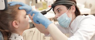
30% of patients of ENT doctors come to clinics with complaints of diseases of the nose and paranasal sinuses. As a rule, these are sinusitis, sinusitis and other viral infections. Against the background of these diseases, inflammatory processes often develop in the paranasal sinuses. The main and unpleasant symptom of inflammation in the sinuses is pain.
Why do we have such a hard time with pain and inflammation in the sinuses? Is it possible to eliminate or reduce pain? To answer these questions, it is important to understand why we need the maxillary sinuses and why it is important to take care of their health.
How do the paranasal sinuses work?
The nose is one of the most important human organs. This system is complex and quite fragile, which leads to frequent illnesses. But without the maxillary sinuses, normal nose function would be impossible. They perform the following functions:
- provide full breathing;
- warm and humidify the inhaled air;
- clean the inhaled air from dust and allergens;
- allow you to recognize many aromas and smells;
- form an individual timbre and other voice parameters.
The sinuses have other tasks as well. For example, they quickly respond to changes in environmental pressure (act as baroreceptor organs) and reliably protect the eyeballs, tooth roots and skull bones from injury.
Why does inflammation of the sinuses develop?
Pain in the sinuses most often acts as one of the main symptoms of sinusitis or its varieties, for example, sinusitis, sinusitis and ethmoiditis. With sinusitis, the inflammatory process is localized in the paranasal sinuses of the upper respiratory tract, and with sinusitis, the maxillary sinuses become inflamed.
Why do the sinuses hurt with these diseases? These air cavities connect to the nasal cavity, which in turn is connected to the throat and ear canals. If a healthy person constantly produces mucus in the sinuses, which flows into the nose at a speed of 1 cm/min, then during the inflammatory process the following processes are observed:
- mucus production increases significantly and pus quickly forms in the sinuses;
- narrow passages for the outflow of mucus are completely blocked, it becomes difficult to breathe;
- fluid accumulates in the sinuses and presses on the walls - this is how a person begins to experience pain.
Twenty-five years ago, the Russian Society of Rhinologists and the journal “Russian Rhinology” were established. The main goal of creating both the society and the journal was the desire to provide doctors with knowledge in the most in-demand section of our specialty - rhinology. And today we are summing up what we have done in this direction over a quarter of a century.
The textbooks and manuals of the early 90s of the last century lacked many concepts that needed to be explained to practicing doctors. A description of such anatomical structures as the ostiomeatal complex, nasal valve and others was presented for the first time among domestic publications in the journal “Russian Rhinology”, in numerous articles of the journal such concepts as mucociliary clearance, lymphoepithelial symbiosis, mucosal immunity were detailed and clarified. It is difficult to overestimate what has been done to disseminate information about modern functional methods of surgical treatment - this, without exaggeration, is an entire era in the development of modern rhinosurgery in our country.
Of course, during this period new scientific data have accumulated and our understanding of many physiological and pathophysiological processes occurring in the nasal cavity and paranasal sinuses has expanded. Therefore, the purpose of this review article is to summarize the results of some studies carried out in different years together with S.Z. Piskunov [1].
Why do people need the nose and paranasal sinuses? Until now, there are no clear, physiologically based answers to this question, there is no statistical accounting of diseases of this localization (for example, sinusitis), outdated surgical methods are still recommended without taking into account the functional purpose of the nose and paranasal sinuses.
As is known, the nose performs the following functions: respiratory, protective, informational, calorific, olfactory, excretory, absorption and aesthetic. All these functions are interconnected, and the loss of one of them negatively affects the condition of the others.
We look at a person's face and first of all we see his eyes, nose and lips. Scoliosis, saddle-shaped and other deformities of the nose are often a violation of not only the shape, but also its main function - respiratory. We will not dwell in this article on restoring the aesthetic function of the nose - this is a big issue that requires separate discussion.
The implementation of the basic functions of the nose is ensured by a clear interaction of anatomical structures and physiological mechanisms. Everything here is logical and perfect. The nose and paranasal sinuses should be considered as a single functional organ. There is no organ in the body that could be compared in terms of the degree of complexity of the anatomical structure and the variety of functions performed with the nasal cavity and paranasal sinuses, which form a single anatomical and functional complex designed to protect the body from harmful environmental factors, ensuring gas exchange processes in the human body and providing information about the antigenic structure of the air environment.
Many variants of the structure of the intranasal structures and paranasal sinuses also create various variants of pathology. The main anatomical components are the osteochondral frame of the nose, the bony walls of the paranasal sinuses and the mucous membrane lining them. The osteochondral frame of the nose is the support for the main organ of the upper respiratory tract - the mucous membrane (Fig. 1). The functional state of the mucous membrane depends on how the frame is formed. We emphasize that the mucous membrane is the main functional organ of the upper respiratory tract, and not just a covering. Therefore, when choosing one or another treatment method, you should always foresee the consequences of its use for the patient. Unreasonable radicalism in rhinosurgery is unacceptable.
Rice. 1. Structure of the mucous membrane. Fig. 1. The structure of the mucous membrane.1 - epithelium; 2 - basement membrane; 3 — own plate; 4—glandular zone; 5 - zone of cavernous vessels. 1 - epithelium; 2 — basal membrane; 3 - lamina propria; 4 - glandular area; 5 - cavernous vessels.
The main one is the respiratory function of the nose - a complex physiological process that is ensured by various mechanisms.
From the moment of birth, the nasal mucosa is exposed to various substances in the inhaled air, as well as temperature, humidity and dryness, fungal spores, bacteria and viruses, and other factors. Thanks to the precise coordination and interaction of various protective factors, when an air stream passes through the nasal cavity, the inhaled air is warmed, moistened, and cleared of suspended dust particles, bacteria, and viruses that can have a harmful effect on the body.
The main physiological irritant of the nasal mucosa and its receptors is the air stream and everything that is suspended in the inhaled air. Let us again emphasize these words as requiring special attention. There is no denying the importance of the physical impact of an air stream, even if it is sterile. The mechanism of action of an air jet on the structures of the nasal cavity can be compared to the lifting force of an airplane wing. It stays in the air only because the air speed above and below the wing is different, this creates lift. The direction of the air stream in the nasal cavity and paranasal sinuses determines the slow change in the structure of the mucous membrane. The air stream initiates a number of reflexes that regulate the physiological functions of other organs. These include the rhinocardial and rhinopulmonary reflexes, the nasal cycle, intracranial and blood pressure. With one-sided nasal breathing, the corresponding lung lags behind in development. Oral and tracheal breathing cannot replace nasal breathing.
The air flow entering the nasal cavity experiences resistance from the intranasal structures. Nasal resistance (resistance) is of exceptional importance in the physiology of breathing. When breathing through the mouth, there is less resistance to air flow, resulting in the suppression of the development of positive and negative pressure in the chest and abdominal cavities, which is important for optimal function of the cardiovascular and respiratory systems.
Slow and deep breathing through the nasal cavity provides optimal conditions for intrapulmonary mixing of gases and maximum gas exchange in the alveoli, which contributes to the high efficiency of external respiration. Deep nasal breathing also improves the distribution of surfactant, which prevents the collapse of the alveoli and pulmonary atelectasis.
The nasal septum, dividing its cavity into two parts, creates a pairing of the organ: the left and right halves of the nose. The functioning of each half of the nose is regulated by the autonomic nervous system. Functioning independently and synchronously, each half of the nose periodically “rests.” The blood supply to the cavernous bodies of the nasal turbinates changes, which is recorded during the study of the nasal cycle [2]. The nasal septum, together with other intranasal structures, regulates and directs air flow in the nasal cavity.
At the beginning of inhalation, thanks to the nasal resistance, negative pressure is created in the chest, due to which air from the paranasal sinuses, nasal passages, nasal cavity and nasopharynx goes into the deep parts of the respiratory tract. This air has already been purified, humidified and warmed. The greatest resistance to air flow is created at the level of the nasal valve. The structural features of the nasal valve ensure that the air flow here twists into a spiral (Fig. 2) and, thanks to centrifugal force, the heaviest particles suspended in the air are deposited in the vestibule of the nose. As is known, mucociliary transport at the anterior end of the inferior shell is directed anteriorly, so all large particles are gradually removed out. Anyone who has been in a dusty room can verify this - when wiping the vestibule of the nose, the handkerchief will be contaminated. Next, the air flow moves along the common nasal passage in an arc at the level of the middle of the middle turbinate, and not into the nasal passages, as is written in many books. This is the normal physiological direction of the main air flow. The uncinate process prevents the stream of inhaled air from entering the middle nasal passage, closing the anastomosis of the large sinuses and the anterior cells of the ethmoidal labyrinth. If the structure of the intranasal structures, especially the nasal septum, is disrupted, the main air flow may deviate from the physiological direction. During the day, a person inhales 25-30 thousand times, which over time leads to the emergence of pathological conditions in the places where the main air flow hits. The nasal passages and the entire surface of the mucous membrane participate passively in conducting air.
Rice. 2. Turbulent movement of air flow as it passes through the nasal valve. Fig. 2. The turbulent motion of the air flow passing through the nasal valve.
After the end of inhalation, the air pressure in the cavities is equalized. Air from the nasal passages (warmed and humidified) enters the paranasal sinuses through the anastomosis. The posterior end of the middle concha cuts off air, directing it into the middle nasal meatus. The uncinate process, creating resistance at the exit of the middle meatus, directs air into the paranasal sinuses. This is how the sinuses are ventilated with every inhalation and exhalation. Anomalies in the structure of the nasal septum and uncinate process disrupt the physiological aerodynamics in the nasal cavity and paranasal sinuses and create a predisposition to the development of a chronic inflammatory process (Fig. 3).
Rice. 3. Options (a, b, c) of significant unilateral enlargement and deformation of the uncinate process, which in appearance resembles the middle shell. This is almost always associated with a deviated nasal septum. Fig. 3. Marked (a, b, c) unilateral enlargement and deformation of the uncinate process of ethmoid bone making it look like a middle turbinated bone of the nose that is almost invariably associated with deviation of the nasal septum.KO - uncinate process; PN - nasal septum; SR - middle shell. UP—uncinate process; NS—nasal septum; MT—middle turbocharged bone.
Currently, morphological changes in the mucous membrane due to aerodynamic disturbances have been studied in detail. In the mucous membrane there is significant polymorphism of the respiratory epithelium. In some areas of the mucous membrane, the ciliated cells are exfoliated, only basal epithelial cells remain on the basement membrane, and there are areas of complete exposure of the basement membrane.
Consistent findings in these cases are sclerosis and hyalinosis of the basement membrane. In these areas, the epithelial cover is represented by 1-2 rows of epithelial cells of a flattened or cubic shape.
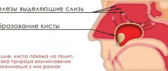
The glandular structures of the mucous membrane undergo significant changes. Among them, the most characteristic are hyperplasia of the mucous glands with the formation of microcysts and atrophy of the serous glands.
In the lamina propria of the mucous membrane there is a pronounced infiltration of lymphocytes and plasma cells involved in the implementation of the immunoregulatory function.
Thus, during a morphological study of the mucous membrane in the area of the ostiomeatal complex, at the anterior end of the middle concha, the posterior ends of the inferior concha, if the air flow from the deformed nasal septum is reflected there, the following changes are revealed:
— desquamation of the respiratory epithelium;
— sclerosis and hyalinosis of the mucous membrane;
- reduction of microcirculatory vessels;
— hyperplasia of the mucous glands with the formation of microcysts and atrophy of the serous glands;
- foci of chronic inflammation.
The mucous membrane is the first barrier in protecting the body from environmental factors. Mucociliary transport moves everything that is fixed in the nasal mucus to the stomach for further destruction of pathogens by the hydrochloric acid of gastric juice. Biochemical specific (immunoglobulins of classes A, G, M, E, D) and nonspecific (mucus glycoproteins - fucomucins, sialomucins, sulfomucins, lysozyme, lactoferrin, secretory glucosidases, interferon, complement, secretory proteases) active in nasal mucus to a certain extent moment they quite successfully protect the body from infections. The second powerful barrier is the basement membrane of the nasal mucosa. The glandular zone and the epithelium of the mucous membrane are involved in the implementation of lymphoepithelial symbiosis.
Without a sufficient amount of mucus on the surface of the mucous membrane, mucociliary transport cannot function. Mucus is produced by goblet cells - single-cell glands of the epithelium, tubular and alveolar glands of the own layer of the mucous membrane. The ratio of ciliated to goblet cells is approximately 5:1. The cilia of the ciliated epithelium move the surface layer of mucus along the more liquid, lower second layer. With vasomotor and allergic rhinitis, the ratio of cells changes, in some areas the surface layer is represented only by secreting cells, and the secretion changes its composition and becomes more watery.
The excretory function of the nasal mucosa is of great importance for protecting the body from external factors. Its violation leads to severe changes, which is observed after aggressive surgery in the nasal cavity. A so-called empty nose is formed when the dry mucous membrane is covered with crusts. In such cases, it is difficult or even impossible to help the sick person.
Air humidification plays a leading role in the nasal cavity. Passing through the nasal cavity, the air is saturated with water vapor from the surface of the mucous membrane. Active evaporation of nasal secretions is explained by the high surface temperature of the nasal mucosa, the large area of the nasal cavity and paranasal sinuses, as well as increased air movement during breathing. When evaporating, the mucous membrane transfers its temperature to the inhaled air. When exhaling, warmer air from the lower respiratory tract releases some of the moisture into the nasal cavity in the form of condensation, which is noticeable in cold weather - a watery discharge appears in the nose. This is especially noted by older people who have wide nasal passages and decreased blood flow in the mucous membrane - the “heating system” begins to work worse. When exhaling, the degree of deposition of water vapor on the surface of the mucous membrane is determined by the temperature of the inhaled air: the lower it is, the more the mucous membrane cools and the more water vapor of the exhaled air condenses.
The nasal cavity is considered as a physiological air conditioner, protecting the lower respiratory tract from exposure to cold air.
Air from –12 °C after passing through the nose is heated to +25 °C in the nasopharynx. Intense warming of the air is facilitated by the high degree of development of the capillary network reaching the epithelial layer. The presence of terminal arteries, which open with amazing speed, increasing pressure in the venous bed and accelerating venous blood flow, is an additional source of heat. All parts of the mucous membrane have a highly developed capillary network that reaches the epithelial layer. The presence of a system of cavernous venous plexuses, located between the capillary network and venules, represents physiological air heaters, heating batteries. Their rapid filling is carried out due to anastomosis with arteries, the largest of which are located in the bone marrow of the turbinates, and the presence of throttle veins, the contraction of the muscles of which prevents outflow and promotes tissue swelling. Arterial blood flows from the posterior sections forward, to the vestibule of the nasal cavity, towards the air flow. Due to the increased blood supply to the cavernous tissue of the nasal turbinates, its volume increases significantly and, consequently, the lumen of the nasal passages narrows. Because of this, the air in the nasal cavity passes through a thinner stream and comes into contact with a larger surface of the mucous membrane and, accordingly, it is warmed by blood more intensely.
The mucous membrane absorbs substances well on its surface. For example, when a tampon with atropine is inserted into the nasal cavity, pupil dilation is observed. But the absorption function is also an integral part of the informative function of the mucous membrane. Thus, various kinds of pathogens penetrate into the deep parts of the mucous membrane, which may be the beginning of the formation of adaptive (acquired) immunity.
In the area of the olfactory fissure there are peripheral neurons of the olfactory analyzer, the purpose of which is to inform the body about the presence in the environment of various chemical compounds that have an odor. Smells can be pleasant and evoke positive emotions or warn of an unfavorable situation. The smell of food stimulates the appetite. The olfactory function of the nose enriches a person's life. In many diseases of the nasal cavity, damage to the olfactory cells and olfactory filaments occurs. We do not pay enough attention to the lack of smell. It is important that the inhaled air reaches the olfactory cleft, and if the nerve receptors are not damaged, we can hope to restore the sense of smell through surgery.
And finally, about the very important function of the nasal mucosa and paranasal sinuses - informational. This concept was introduced by us and S.Z. Piskunov [1]. The essence of this function is to inform the body about the antigenic structure of the surrounding air environment. This function is aimed at the formation of adaptive immunity and the body’s immune response to the introduction of a damaging agent. It is the mucous membrane of the nose and paranasal sinuses that takes precedence in the formation of human adaptive immunity. This is quite simple to explain. The first cry of a child, signifying his arrival into this world, is associated with a breath of air. No breath, no cry, no living child. But with the first breath, the newborn’s body also receives the first portion of various types of antigens that are in the air. Having innate immunity, the child is still poorly protected. It is necessary to acquire resistance to everything aggressive that is in the surrounding air. This is a complex multi-stage path - the path to the formation of adaptive immunity.
To know what to defend against, you must obtain information about who is attacking. This information is carried by antigen-presenting cells - a group of cells endowed with the function of phagocytosis. It is phagocytosis that underlies the theory of the formation of adaptive immunity. It was believed that innate immunity is nonspecific, and specific immunity is acquired. But with the discovery of toll-like receptors, ideas about immunity and the formation of adaptive immunity received a new development. These receptors are found on the surface of many epithelial cells, including respiratory ones. Through these receptors, the body receives information through epithelial cells about the penetration of a damaging agent and forms an immune response. The epithelial cell is activated and becomes the first source of production of the corresponding cytokines [3].
We distinguish between general and mucosal immunity. Mucosal is the immunity of the mucous membranes, providing tissue homeostasis, cellular and humoral protection with the help of local lymphoid and plasma cells. The basis of mucosal immunity is a unique form of local humoral response in the form of the production of secretory antibodies.
Mucosal-associated lymphoid tissue—the MALT system—is the anatomical basis of mucosal immunity. It has its own immunoregulation, its own inductive organs and lymphocyte recycling pathways according to homing rules. The inductive organs for the upper respiratory tract are the adenoids and tonsils. Homing is the process of returning informed lymphocytes to the organ where antigenic information was received.
Lymphoepithelial relationships are an evolutionarily conservative form of immune regulation with the participation of the epithelium as a border tissue, the cells of which are the first to respond to antigenic stimulation. The relationship of informed immune cells with epithelial cells and the attachment of the secretory component forms secretory immunoglobulins, in particular class A. The concept of lymphoepithelial symbiosis has entered our lexicon relatively recently. It means the interaction of lymphoid structures (adenoids, tonsils) with epithelial cells (surface of the mucous membrane, epithelium of the glands of the mucous membrane). As a result of the interaction of these structures, secretory immunoglobulin of class A is produced - the main factor in protecting the mucous membranes from viruses and bacteria. Damage or destruction of any component of the lymphoepithelial symbiosis leads to a decrease in mucosal immunity [3].
The inflammatory process and the process of formation of adaptive immunity have much in common. The most important thing is the transfer of information from cell to cell with the subsequent response - molecular, cellular and tissue. This process ends either with the formation of immunity to a specific pathogen and full regeneration, or with the persistence of inflammation. In intercellular interaction, the transfer of information is of great importance. Depending on which cytokine carries information to which cell, this will be the tissue response and the response of the whole organism. If we master the ability to control the transmission of this information, we will learn to treat acute rhinitis, sinusitis, prevent and treat chronic inflammation.
The issue of the formation of immunity, inflammation, and the body’s response to damaging factors is very extensive and is not discussed in this work. It should be emphasized that the information function of the mucous membrane of the upper respiratory tract is fundamental and decisive. The participation of the mucous membrane of the upper respiratory tract in the formation of adaptive immunity requires fundamental research.
The paranasal sinuses are a system of reserve anatomical formations filled with air and designed to protect the body and, first of all, the contents of the orbit and cranial cavity from the effects of various unfavorable environmental factors. The first tissue and anatomical barrier of protection is the nasal cavity and its mucous membrane. In acute respiratory viral infection, inflammation of the mucous membrane usually ends with recovery. If some reasons do not allow acute inflammation of the nasal mucosa to end, it spreads to the mucous membrane of the paranasal sinuses, most often the ethmoid and maxillary sinuses. The time spent on the development of inflammation in the sinuses allows the body to build up defense forces. Anatomical structures are involved in this: from epithelial, vascular and osteochondral to humoral, forming the immune response. If this barrier becomes surmountable by the pathogen, intracranial or orbital complications occur.
Congenital or acquired violation of the anatomical structures of the nasal cavity leads to changes in aerodynamics. Constant irritation of the mucous membrane by the main (most powerful) air stream (Fig. 4) causes irritation, inflammatory changes, and the formation of a colony-stimulating factor (stimulates the activity and proliferation of granulocyte-macrophage precursors). This factor promotes the migration of lymphoid cells into the mucous membrane, and the cellular infiltration of the mucous membrane gradually increases. This leads to an increase in tissue volume. In addition, there is an increase in the amount of glands and mucus. Hypertrophy of the mucous membrane occurs - a predisposition to a chronic inflammatory process is formed, which must be taken into account when detecting unilateral difficulty in nasal breathing when the nasal septum is deviated. As a result, with the next acute inflammation, a block of the natural anastomosis of the sinus or sinuses occurs, and sinusitis is formed. It may end in recovery, but with repeated acute respiratory viral infection, inflammation in the paranasal sinuses may again occur, followed by chronicization of the pathological process, including polypous rhinosinusitis. In addition to anatomical defects, so-called biological defects—congenital disorders in the immune system and congenital syndromes with dysfunction of the mucous membrane—play an important role in the pathogenesis of polypous rhinosinusitis [4].
Rice. 4. Hypertrophy of the anterior end of the middle turbinate as a result of constant irritation by the air stream. A block of the ostiomeatal complex is formed. Fig. 4. Hypertrophy of the anterior end of the middle turbinated bone as a result of its continuous irritation by the air flow. Formation of the osteomeatal complex.
Unfortunately, we still underestimate the importance of the nasal cavity and paranasal sinuses with their anatomical features and functionally complex mucous membrane in ensuring human health.
The author declares no conflict of interest.
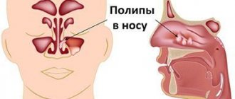
What are the causes of blocked sinuses?
There are usually several reasons:
- infections and viruses;
- polyps and tumors;
- allergies.
It has been proven that 90% of acute respiratory viral infections lead to the development of sinusitis of varying severity. And bacterial infections provoke sinusitis only in 0.5-2% of cases.
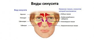
The sinuses most often hurt due to acute sinusitis, sinusitis, sinusitis or ethmoiditis. Symptoms of the disease appear as follows:
- Sinusitis. It will be “given away” by inflammation in one or more paranasal sinuses. When the inflammatory process occurs on one half of the face, we speak of hemisinusitis, when on both halves, we speak of pansinusitis. Acute sinusitis may include weakness, headache, fever, and changes in blood tests.
- Frontit. This is an inflammation of the frontal, or frontal, sinuses. With this disease, there is a characteristic increasing pain and pressure in the forehead, brow ridges and eyes. Other symptoms include general weakness, fever, and difficulty breathing.
- Ethmoiditis. It is an inflammation of the ethmoid labyrinth. The pain is localized in the center of the face. There is dark yellow-green nasal discharge, headache, high fever, swelling of the eyelids and fear of light, difficulty or complete absence of breathing.
- Sinusitis. The symptoms of sinusitis are difficult to recognize: the inflammatory process begins without fever. But with this disease there is a characteristic pain in the area of the face under the eyes. With sinusitis, there may be no nasal discharge, which also complicates diagnosis.
Contraindications:
- pregnancy (instead of tomography it is recommended to do a sinus scan);
- body weight more than 200 kg;
- thyroid diseases;
- kidney diseases;
- severe diabetes mellitus;
- multiple myeloma;
- allergy to the contrast agent administered during CT scanning.
A tomogram of the paranasal sinuses is not recommended for children, since the procedure requires perseverance and the need to lie still for several minutes. It is extremely difficult to force a child to do this.
How to treat sinus inflammation in adults and children?
Sinusitis, sinusitis and other inflammatory processes have similar symptoms. Treatment in adults will be effective if a specialist makes an accurate diagnosis and selects the optimal complex therapy, which will include:
- special medicines;
- healing procedures;
- prevention.
It is important not only to relieve the symptoms of the disease, but also to eliminate the causes of the disease. And they can be different. Some people have a weakened immune system, while others should have their body examined or give up bad habits and an unhealthy lifestyle.
In children, treatment of sinusitis, sinusitis and other inflammatory processes of the nasal sinuses also includes a whole range of measures:
- it is important to see a doctor quickly to make a diagnosis and avoid complications;
- you need to strictly adhere to the recommendations of specialists and monitor the child’s condition;
- It will be necessary to prevent inflammation in order to maintain the health of the child’s sinuses.
In the fight against serious inflammatory processes, one must not hesitate. For example, sinusitis can lead to inflammation in the skull or meninges. Acute catarrhal sinusitis develops in 70% of children who regularly suffer from ARVI. Odontogenic sinusitis develops in those who have dental problems.
Treatment
Generally, conservative treatment is recommended, which is similar to the treatment of acute inflammation in other sinuses.
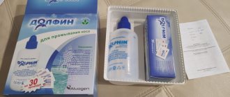
The dominant technique is still rinsing the paranasal sinuses by moving fluid because of its speed, convenience, and most importantly effect. If, after following the doctor's orders, there is no improvement within several days, hospitalization is necessary for surgical treatment - opening the cells of the ethmoid labyrinth. Endoscopic surgery is much preferable to classical ethmoidotomy, due to its higher quality and gentle (minimally invasive) approach.
How do you know if you have sinus inflammation?
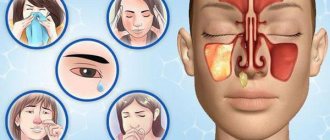
With any inflammatory process, be it sinusitis, sinusitis, ethmoiditis or frontal sinusitis, you will notice 3 characteristic symptoms:
- Problems with nasal breathing – it will be difficult to breathe due to obstruction (blocking of the lumen) of the nasal openings due to swelling.
- Characteristic nasal discharge. They can be permanent or temporary. If there is swelling of the nasal mucosa, there may be no discharge.
- Headache and soft tissue swelling. During inflammatory processes, the face swells in the area of the frontal and maxillary sinuses. Sometimes the periosteum becomes inflamed (pereosteitis).
Diagnostics
Diagnosis of acute ethmoiditis is based on identifying characteristic symptoms and data from studies.
Pain concentrated at the root of the nose and along its lateral surface, which correspond to the lower edge of the nasal bones and the frontal process of the upper jaw, may indicate damage to the anterior cells of the ethmoid labyrinth. X-rays, diaphanoscopy and computed tomography help clarify the diagnosis. Make an appointment right now!
Call us by phone or use the feedback form
Sign up
How to cure sinusitis, sinusitis and other inflammations?
What you definitely shouldn’t do is diagnose yourself and buy medications without a prescription or doctor’s recommendations. If sinusitis is mild, then the body will quickly cope with the source of the problem and defeat the infection or virus. But if sinusitis has become chronic, special therapy is needed:
- medicines that clear the air passages;
- antibacterial drugs to eliminate infection;
- rinsing the sinuses with antibiotics;
- surgical intervention in selected cases;
- regular preventive measures.
Of course, it is better to prevent sinusitis from becoming severe. Indeed, in this case, surgical intervention cannot be avoided. Why is this scary?? Just look at the image below, the hand-drawn picture of surgical techniques will give you a chill between your shoulder blades!
How the research is carried out
No special preparation is required for the procedure. You must remove all metal objects or clothing with metal elements. The patient is placed on the couch and his body is secured with belts, and his head is placed on a special headrest. You should not move during the entire procedure, as movement may affect the accuracy of the result. If the patient cannot lie still (for example, children or people with mental disabilities), the doctor may use anesthesia.
If the CT procedure requires the injection of a contrast agent, such as iodine, into the patient’s blood, you should refrain from eating for several hours before the procedure.
Then the couch on which the patient lies is pushed into the tomograph. After this, the doctor retires to another room where a computer is installed that records the tomograph signals. All this time, with the help of sound equipment, the doctor and the patient are in touch. If the patient feels any discomfort, he reports it, and the doctor hears it. The study itself lasts no more than ten minutes.
Then the specialist deciphers the tomogram and gives the patient an x-ray and a disk recording the results of the study, as well as a medical report.
CT, as a method of radiation diagnostics, involves radiation exposure to the body. Therefore, it is recommended to do CT scans no more than three times a year. The cost of this study varies from 3,000 to 5,000 rubles.
Prevention is the best remedy for sinus inflammation
Of course, no one can be 100% insured against sinusitis and more severe inflammatory processes. Prevention will help strengthen the immune system and build a protective barrier against viruses, bacteria and infections. To prevent sinusitis without symptoms in an adult or child, you should follow simple rules:
- Avoid hypothermia. Yes, low temperature in itself does not provoke colds and runny nose. But it causes a narrowing of blood vessels and makes the mucous membranes more vulnerable to viruses and infections. In cold, rainy and windy weather, it is important to properly insulate - and you will not face chronic inflammation of the sinuses.
- Rinse your nose more often. This should be done in the fall during epidemics of colds and flu, as well as in the spring during the flowering of trees and shrubs. For rinsing, ordinary saline solutions are suitable, which effectively remove bacteria and allergens.
- Eat properly. The immune system weakens due to lack of nutrients. Include more natural products in your diet - fresh meat and fish, grains and slow carbohydrates, vegetables and fruits. It’s better to avoid processed foods and fast food, or at least reduce their consumption to a minimum.
- Drink vitamins. Nowadays there are many special vitamin complexes and supplements with herbal active ingredients. They work gently but effectively: the result is noticeable within 1-2 weeks. And most importantly, they are completely safe, non-addictive and have no side effects.
What can be included in therapy during treatment?
Don’t know how to treat maxillary sinusitis besides pills? You need herbal preparations. They are good both in the treatment of existing inflammatory processes and as a preventive measure. Their effectiveness is explained by the special composition of active components. As a rule, these are herbal extracts.
Each capsule of this product contains a whole set, including up to 10 or more medicinal plants. It is this composition that helps to quickly eliminate the causes of the disease - any viruses and bacteria in the body, and also helps to cure sinusitis or sinusitis at an early stage.
Just one capsule a day - and your sinuses breathe freely, and your health remains normal even during seasonal colds!