Osteoplastic operations are becoming more and more improved every year, as their demand increases. According to statistics, bone deficiency due to one reason or another occurs in every second dental visitor. In the vast majority of cases, insufficient jaw volume is detected before dental implantation. One of the most popular and reliable operations to restore the amount of bone in the upper jaw is considered to be a sinus lift.
Indications for its implementation:
- Long-term absence of anterior premolars and molars, which leads to resorption of bone tissue;
- Resolved inflammatory processes affecting bone tissue;
- Traumatic injuries to the jaw (most often surgical tooth extraction);
- Congenital structural features of the upper jaw and maxillary sinuses (physiological deficiency of bone tissue).
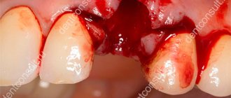
Depending on the severity of the bone tissue deficiency, open or closed volume restoration surgery is performed. The essence of the surgical intervention is to separate the mucous membrane lining the maxillary sinus, followed by filling the resulting cavity with bone grafts of various types. However, in the process of preparing for surgery, and sometimes during its implementation, obstacles to the procedure may be discovered. Most often, such unfavorable factors are cysts in the maxillary sinus.
What is a cyst in the maxillary sinus called?
A maxillary sinus cyst is a benign formation that has a hollow structure containing fluid inside. Cysts of the maxillary sinuses are quite common, but are almost always found at random. Their location and origin may differ, which will determine the characteristics of the clinic and even treatment.
Odontogenic cyst
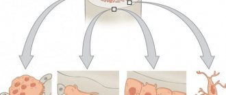
Among the many benign tumors of the jaw tissue, the cyst is found most often and represents a pathology quite dangerous for the bone structure. Odontogenic formations in the vast majority of cases remain undiagnosed for a long time, and therefore reach enormous sizes. The growth of the cyst provokes atrophic processes in the bone tissue, which thins, becomes fragile, and then disappears completely.
Such formations are characterized by a long latent course until the cavity grows to gigantic proportions. Then a characteristic crunch begins to be noted in the jaw, especially during chewing loads. Ignoring the problem leads to spontaneous fractures of the jaw, compression of the maxillary sinus with disruption of its functions, exacerbation of the inflammatory process with damage to the structures of the maxillary sinus and the formation of sinusitis.
Tooth cyst growing into the maxillary sinus
Damage to the dental canal involving the root and surrounding tissues is a consequence of an infectious process of bacterial origin. In the absence of adequate and timely treatment, a gradual delimitation of the inflammatory reaction from healthy tissues by a wave of cellular structures occurs.
This creates a cavity called a dental cyst. It gradually increases and destroys the bone tissue of the jaw. When localized in the anterior part of the upper jaw, gradual penetration into the maxillary sinus occurs.
Retention
The most common form of neoplasm in the maxillary sinus, which is also called a true cyst. It is located in the lower wall of the sinus and has an epithelial lining - a distinctive feature of true cystic cavities. The formation is clearly determined by X-ray examination randomly, as it does not manifest itself clinically.
When the cyst reaches a significant size, problems begin with the functioning of the maxillary sinus. The structure and physiology of the microvasculature is disrupted, resulting in the formation of edema. This symptom is manifested by nasal congestion, often without pronounced discharge from it. Due to the lack of specificity of the clinical picture, even the presence of subjective signs of the disease is not a reason to consult a doctor.
Mycetoma
non-invasive fungal sinusitis of the left maxillary sinus
The hypointense MR signal of a mycetoma may be mistaken for air in the paranasal sinus; Non-invasive fungal sinusitis does not look the same in different sequences.
If they are large, they cause headaches due to the pressure of the cyst shell on the walls of the sinus.
Often combined with allergic rhinitis, hypertrophy of the nasal turbinates and deviated nasal septum
Large cysts located in the lower parts of the maxillary sinus may be asymptomatic, while a small cyst located on the upper wall, in the area of the 2nd branch of the trigeminal nerve, can cause headaches.
Mucocele of the ethmoid labyrinth and frontal sinus on the right
This is a large formation of the paranasal sinus, lined with epithelium and filled with mucus, which is formed as a result of obstruction of the main sinus canal.
The most typical symptom: expansion of the paranasal sinus with smooth, clear contours with thinning and remodeling of the adjacent bone plate.
How to identify a cyst in the maxillary sinus?
Since cystic cavities are benign neoplasms, they do not cause any systemic manifestations. The local clinical picture manifests itself several months after the onset of the pathological process. But even its occurrence does not always become a reason to go to the doctor. Most often, maxillary sinus cysts are discovered by a dentist during a preliminary examination to perform a sinus lift or implantation.
The main clinical manifestations of cysts in the maxillary sinus:
- Painful sensations in the infraorbital region of a pressing nature (pain intensifies when changing body position);
- Discharge from the nasal passages is purulent or mucopurulent in nature, the amount of which depends on the presence and severity of the inflammatory process;
- Gradually increasing facial asymmetry (when the cyst reaches a large volume);
- Constant exacerbations of infectious lesions of the maxillary sinus;
- Swelling of the nasal passages (severe nasal congestion);
- Pain in the upper jaw (as well as: a characteristic crunch when pressed or loaded);
- Headaches (most often in the forehead or temples).
The clinical picture of cystic formations is practically indistinguishable from acute sinusitis, so doctors always differentiate these two pathologies using instrumental studies. For dental specialists, it is especially important to make the correct diagnosis, since the presence of a cyst is not always a contraindication for sinus lifting, but an acute infection does not allow surgical intervention.
Identification of cystic formations occurs by performing x-rays of the sinuses or in the process of studying the bones of the skull on a computed tomography scan. Additionally, an endoscopic examination may be prescribed, which is both diagnostic and therapeutic. Histological examination using targeted biopsy is mandatory, which allows us to have an idea of the origin of the tumor and exclude malignant oncology.
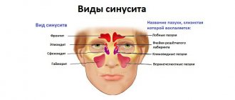
Types and types of maxillary sinusitis
Depending on the etiology (cause of occurrence) of the disease, rhinogenic, odontogenic, traumatic and allergic maxillary sinusitis are distinguished.
Rhinogenic sinusitis occurs against the background of rhinitis (when the nasal mucosa becomes inflamed). The mucous membrane is the main obstacle to infections. When bacteria gets on it, a runny nose or rhinitis develops. The causes of rhinitis are different: viruses, hyperthermia, allergies.
, decreased protective properties of the body, penetration of synthetic agents, influence of dry air, too long use of drugs with a vasodilator or vasoconstrictor effect.
Rhinitis is characterized by congestion, nasal discharge, impaired circulation in the nasal cavity, and the development of congestive blood phenomena. There are 4 types of rhinitis: allergic
, chronic, acute, vasomotor.
Odontogenic sinusitis occurs due to inflammation of the mucous membrane of the maxillary sinus due to infection from an unhealthy tooth, the tissues around it, and the communication formed between the sinus and the oral cavity after tooth extraction. Symptoms of this disease can be apathy, loss of appetite, headache, aching temples, discharge from the nose or ear, cough, runny nose and others. Depending on the course of the disease, different treatment methods are chosen: antibacterial therapy and washing of the maxillary sinuses, pumping out pus or surgery.
Injury to the maxillary sinus or jaw can also lead to sinusitis.
The cause of allergic sinusitis is the body's hypersensitivity to one of the irritants. The disease begins in the nasal cavity and then spreads to the maxillary sinuses. What usually serves as an allergen? This is pollen during the flowering period, fur and excrement of pets, dust mites, medicines, household chemicals, perfumes, cosmetics, chemicals, dirty city air.
According to the duration of the disease, sinusitis is divided into:
- acute (less than 3 months);
- recurrent acute (can repeat up to four times a year);
- chronic (more than 3 months);
- exacerbation of chronic sinusitis (adding new symptoms to existing ones).
According to the severity of symptoms, sinusitis is:
- lungs
- moderate severity
- heavy.
Treatment of maxillary cysts
There are no conservative methods for treating cystic formations in the maxillary sinus. The doctor chooses one of the options for surgical intervention, which depends on the type of cyst, its location, the professional qualities of the specialist and the individual characteristics of the patient.
In modern medicine, the following operations are widely used to treat cysts:
- Open surgery. A manipulation that involves general anesthesia and has many complications and risks. The anterior wall of the maxillary sinus is opened, and the cyst is removed through the resulting hole. The main problem is the overgrowing of the access with scar tissue. This completely contradicts the physiology of the body and does not allow sinus lifting and dental implantation in this area in the future.
- Endoscopic treatment. One of the most advanced and successful in modern medicine. Using a special instrument, the doctor enters the maxillary sinus through its natural opening without damaging the bone tissue. The cyst is then sucked out or desquamated.
- Classic removal. A typical open sinus lift approach is performed, through which the cyst is removed. The operation is performed in cases where the cause of the development of the formation is a diseased tooth.
Since cystic formations are most often discovered during the preliminary examination for sinus lift, the doctor must choose a treatment method that will not interfere with further bone grafting. The final decision always belongs to the patient, so he needs to be told the advantages and disadvantages of each therapeutic technique.
Paranasal sinus cyst - symptoms and treatment
Treatment depends on the type of cyst, its location and size. If it is less than 1 cm and there is no discharge, then the doctor suggests conservative methods: regular examination, tablets, sprays and drops. If the cyst is large or there is purulent discharge, then surgery will be required.
Drug treatment of cysts
The method allows you to eliminate unpleasant symptoms and causes of cyst formation: swelling, inflammation and blockage of the sinus anastomosis. Drug treatment is effective and can alleviate the patient's condition in the early stages of the disease. However, it will not help when the fibrous membrane of the cyst has become too dense.
Drugs used:
- vasoconstrictor drops - quickly reduce swelling in the anastomosis area and normalize ventilation of the sinuses of the nasal cavity; medications in this group are addictive and can be safely used for no more than 5 days in a row;
- antihistamines - will help if the cyst is caused by allergies;
- mucolytics - normalize the outflow of mucus;
- antiseptics - help fight inflammation and cleanse the surface of the nasal mucosa;
- topical glucocorticosteroids are most effective in the treatment of allergic rhinosinusopathy, prolonged sinusitis, hyperplasia of the mucous membrane in the area of the anastomosis and ostiomeatal complex;
- painkillers - reduce pain caused by the pressure of the cyst on surrounding tissues [6];
- probiotics - normalize the microflora of the nasal cavity and nasopharynx, which is important in the treatment of chronic inflammation of the upper respiratory tract.
Surgery
Indications:
- cyst more than 8 mm;
- ineffectiveness of conservative treatment;
- signs of suppuration inside the cyst [7].
The most popular method of treating cysts is puncture or puncture. The surgeon pierces the wall of the cyst with a thin needle and pumps out the purulent discharge [1]. However, this method only has a temporary effect. Piercing the cyst, as well as spontaneous leakage of its contents, relieves pain for a while. But gradually the cyst grows together again and begins to accumulate purulent fluid. To remove it completely, more serious surgery will be required [8].
Maxillary sinusotomy according to Caldwell-Luke
It is performed through an incision under the upper lip in the mouth. This is a classic method for removing sinus cysts, but now it is practically not used due to its high morbidity: after the operation, scars, adhesions can form and the functioning of the sinuses can be disrupted.
Microsinusrotomy
A minimally invasive surgical technique that is performed through the anterior wall of the maxillary sinus. A 4–8 mm hole is formed in the bone, and the cyst is removed with special instruments. Performed under general anesthesia. The disadvantage is the risk of damage to the branches of the trigeminal nerve and the formation of persistent facial neuralgia.
Functional Endoscopic Sinus Surgery (FESS surgery)
This method is considered the most gentle and effective: during the procedure, unnecessary incisions and punctures are not made on the patient’s face. It is performed through the nasal cavity under endoscopic control. Allows you to reach all paranasal sinuses, including the sphenoid, frontal sinuses and cells of the ethmoid labyrinth. The operation is performed under local and general anesthesia [17].
Additional benefits:
- can be used to treat children;
- reduce the likelihood of cyst recurrence;
- you need to stay in the hospital for no more than two days;
- the incision heals quickly;
- the operation leaves no scars;
- there is no risk that adhesions will appear [18].
Relative disadvantages: trained specialists and special equipment are required, which makes the operation expensive.
Cyst in the maxillary sinus and sinus lift
Many patients are interested in the question of whether it is possible to simultaneously extract a cystic formation and perform bone grafting of the upper jaw. The answer to this question depends on several factors, which include:
- The degree of severity of bone tissue atrophy at the site of the cyst (in some cases, the deficiency is so pronounced that sinus lifting becomes impossible, and patients can only hope for the installation of removable dentures);
- The presence of an active inflammatory process (cysts are often complicated by infectious pathologies, especially sinusitis, which are a direct contraindication for surgery);
- The size of the cystic formation (small cavities can be removed immediately before sinus lifting, but if the cyst occupies the entire space of the sinus, then bone grafting is out of the question);
- The location of the cyst (if it is located on the lower wall, then problems with removal during sinus lifting usually do not arise, but if the formation is located higher, the dentist simply will not be able to reach it without injuring the walls of the sinus).
Performing a sinus lift with preliminary removal of a cystic formation is performed quite rarely and only in cases where the process has just begun to develop and was accidentally discovered by instrumental studies. The doctor must correctly weigh the benefits and risks of future surgery. Most often, patients are transferred for treatment to ENT specialists, and the sinus lift is postponed to a later time.
Treatment
Etiotropic therapy
Prescribing antibacterial agents (the main causative agents of acute sinusitis) helps to achieve rapid results in therapy:
- β-lactams: amoxicillin, clavulanate, cefaclor, cefuroxime axetil, sulbactam;
- fluoroquinolones: levofloxacin, gatifloxacin, moxifloxacin;
- macrolides: azithromycin, clarithromycin.
Puncture
This method is used if the disease cannot be cured with medication. It is the most famous method for removing pus from the maxillary sinuses. Quite painful, unlike other procedures.
"YaMiK"-catheterization
A fairly effective remedy in the fight against maxillary sinusitis, it does not cause complications. The procedure is quite painful, like puncture; patients cannot always tolerate it well.
Important: the operation is not advisable for isolated lesions of one paranasal sinus, since infection can be introduced into healthy paranasal sinuses.
Possible risks
A cyst in the maxillary sinus and sinus lifting can be complicated in different ways and seriously interfere with any dental interventions by the doctor. When a cystic formation is removed, the risk of perforation of the mucous membrane of the maxillary sinus only increases, as do subsequent associated problems. It is also possible for the cyst to accidentally open and spill its contents into the sinus. This threatens further infection and the development of acute or chronic sinusitis.
When performing a sinus lift for a maxillary sinus cyst, the doctor must remember that the formation promotes the resorption of bone tissue. The thinner the alveolar bone, the less load it can bear. Therefore, any careless movement can lead to a fracture of the bone crest.
During the sinus lifting process, a barrier membrane is necessarily installed that will protect the operated area from unfavorable external factors and the additional impact of osteoclasts (cells that destroy bone).
In addition, only the safest and highest quality grafts are used in the bone grafting process with minimal risk of rejection. This precaution is due to the fact that a significant deficiency of bone tissue will not suffer additional problems in the form of poor engraftment. The doctor will not be able to perform a second operation, and he will have to look for alternative solutions.
Diagnosis of sinus cysts
In addition to the standard questioning and examination of the patient, all doctors at the Lor-Plus clinic are proficient in endoscopic examination techniques. Using an endoscope - a long thin tube with a miniature video camera, the doctor can conduct the most complete examination of the ENT organs, determine the presence of additional structural features of the nasal cavity, changes in the nasal mucosa in the deep sections that may interfere with normal breathing.
Currently, the gold standard for diagnosing sinus cysts is computed tomography . This method allows you to determine the size of the cyst and its location in the sinus with millimeter accuracy, which is very important for choosing a method for removing the cyst.