Pain in the buttocks indicates the presence of pathologies, such as:
- lumbosacral radiculitis,
- formation of phlegmon and abscesses,
- osteomyelitis,
- furuncle
- or is considered a consequence of muscle overexertion.
Pain in the buttocks can also be caused by:
- osteochondrosis,
- coccyx cyst,
- intervertebral hernia.
If there is a disease in the lower parts of the spine, due to the location of the nerves, the pain may radiate to the legs or buttocks.
To diagnose the cause of pain, the patient should determine the nature, intensity and painful location. We recommend that you contact experienced proctologists.
The buttocks are considered symmetrical parts of the body and represent, figuratively speaking, a “layer cake”. The first, top layer is the skin. The second is the tissue of the left and right gluteal muscles, respectively. The third layer is represented by subcutaneous fat, which is located directly under the muscles and, in comparison with other parts of the body, is considered the most developed.
Pain in the butt occurs in any of the balls. A painful process in the buttocks indicates the consequences of an injury, the presence of an infectious or inflammatory process in the body, or muscle pathologies.
Symptoms that accompany pain in the butt
Pain in the butt is accompanied by a number of symptoms that worry the patient.
- discomfort and pain during bowel movements;
The appearance of pain in the butt is caused by anal fissures and hemorrhoids. The causes of anal fissures are poor hygiene, constipation, and mechanical damage to the mucous membranes.
Symptoms of cracks are:
- constant pain in the butt,
- bleeding,
- difficulty in defecation and discomfort.
Anal fissures often appear in women after natural childbirth.
- pain in neighboring areas;
In addition to the gluteal region, the patient has pain in the lower back, sacrum, coccyx, and thigh. The pain syndrome is accompanied by increased tone in the gluteal muscles, muscles of the lower back and lower extremities. As a result of changes in the spine and increased muscle tension, movement in the spinal column and hip joint is limited.
- general disturbance of health: malaise, weakness;
The patient feels severe weakness of the thigh and lower leg muscles, decreased sensitivity, and discomfort. Sometimes pain in the body is accompanied by discomfort in the buttock area. The patient complains of lack of appetite and worsening condition after eating. In this case, advice from a nutritionist and urologist is necessary.
- elevated temperature;
The patient's condition is accompanied by an increase in temperature. This process indicates inflammation or infection.
- headaches and dizziness;
There are complaints of headaches, migraines, dizziness. Sometimes cases of loss of consciousness are possible.
- nausea and vomiting;
Inflammatory processes in the buttocks are closely related to the gastrointestinal tract. The patient complains of nausea and vomiting, and belching.
- bowel dysfunction;
The patient develops fecal disturbances, upset and constipation, flatulence, bloating in the abdomen, discomfort in the buttocks and other organs.
- loss of consciousness and loss of coordination;
In severe cases, sharp pain in the buttock and increased body temperature are accompanied by impaired consciousness - the patient falls into a coma;
Treatment
Pre-hospital assistance
Patients with pain in the buttock are given rest. For severe pain, an anesthetic is given. For diagnosed diseases of the spine and neurological diseases, the use of warming and anti-inflammatory ointments is effective. Intense pain, poor general condition, and hyperthermia are reasons to immediately contact a specialist.
Conservative therapy
Conservative treatment includes drug and non-drug therapy. Patients may be prescribed:
- Protective mode
. Bed rest or exercise limitation is recommended. Sometimes the use of additional devices is indicated: walkers, crutches, canes. - NSAIDs
. Used for rheumatic and degenerative processes. Used in the form of tablets, injections, topical agents: gels, ointments, creams. - Antibiotics
. Necessary for infectious diseases. Treatment of specific infections is carried out according to special protocols. For nonspecific infections, broad-spectrum drugs are prescribed; after determining the sensitivity of the pathogen, antibiotic therapy is adjusted. - Physiotherapy
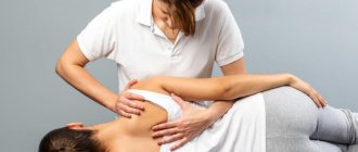
. To reduce pain, stimulate resorption, activate blood circulation, accelerate healing or improve nerve conduction, referrals are given for electrophoresis, laser therapy, UHF, and electrical stimulation. According to indications, exercise therapy, massage, kinesiotaping, acupuncture, and manual therapy are used.
Causes of pain in the buttocks
Medical experts identify the following causes of butt pain:
- herniated intervertebral discs in the lumbar region;
Acute severe pain radiating to the buttock is observed with herniated intervertebral discs in the lumbar region. Pain first occurs in the lower back - directly in the place where the affected disc is located, then goes down to the buttock and lower along the back of the thigh. The pain occurs only on the right or left, depending on which side the nerve is affected. Weakness occurs in the leg on the affected side, skin sensitivity is impaired, and discomfort is noted in the anal area.
- lumbosacral radiculitis;
The patient feels discomfort in the buttocks. The condition is accompanied by nagging pain.
- excessive muscle tension;
Severe pain in the butt indicates excessive muscle tension. Such symptoms are observed in athletes and people involved in physical labor.
- sciatic nerve neuralgia;
With neuralgia of the sciatic nerve, the patient experiences severe pain in the thigh and anus. The pain intensifies with movement and turning.
- infections of the female genital tract;
Severe cutting pain in the anus can be observed in women who have problems with the female genital organs. To establish an accurate diagnosis, you should visit a gynecologist.
- arthritis and arthrosis;
With arthritis and arthrosis, inflammation of the joints of other organs is recorded in the patient. The main symptom of this disease is pain between the buttocks while walking. To prevent the development of arthritis and arthrosis, the patient should visit a neurologist and orthopedist. Health care providers will prescribe medications, healing ointments, exercise therapy, and physical therapy.
- infection;
Pain in the anus after diarrhea indicates the presence of an infectious disease in the human body. The patient undergoes tests and visits a urologist.
Pinched pudendal nerve
The sacrotuberous ligament arises from the ischial tuberosity and attaches to the sacrum and coccyx, while the sacrospinous ligament lies at a 90-degree angle to it, deeper to the sacrotuberous ligament and attaches to the ischium. The thickness of these ligaments can lead to pinching of the pudendal nerve, called Alcock's canal syndrome or cyclist's syndrome.
In addition to pain in the gluteal region, symptoms of pudendal neuralgia include sexual dysfunction, rectal pain, fecal incontinence, and urinary incontinence. Thus, a pinched pudendal nerve can significantly impact quality of life.
This may be caused by prolonged sitting, especially when riding a bicycle, or a recent change of bicycle saddle. Symptoms are typically worsened by sitting, but sitting on the toilet has been reported to relieve pain by relieving pressure on the nerve.
Diagnosis and treatment of pain in the butt
The treatment process for pain in the buttocks depends entirely on the nature of the pathology. The patient needs to see a proctologist and attend an initial consultation. The doctor will examine the affected organ and perform palpation. If purulent discharge, bleeding and infectious processes are detected, then we suggest performing an operation to remove anal fissures.
For pain in the butt resulting from an injury, the patient is prescribed painkillers and warming ointments that can relieve swelling. Non-steroidal medications relieve pain and promote rapid healing of soft tissues.
For boils, patients are prescribed Vishnevsky ointment and ichthyol ointment. In untreated cases, medical workers use massages, warm compresses, and physical therapy.
After consulting a doctor, the patient begins to engage in physical therapy. Improves muscle tone and strengthens. Doctors at the private clinic “KDS Clinic” have developed a special gymnastic system that will relax injured muscles and reduce pain.
Diagnosis of piriformis syndrome
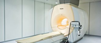
In addition to the examination, checking tendon reflexes, posture and gait, the doctor prescribes instrumental research methods to the patient. Among them, the most informative is considered to be radiography of the spine in the lumbar region, as well as the joints of the sacrum with the pelvic bones. In addition, comprehensive results can be obtained by magnetic resonance scanning of the lumbar and sacral area. Radioisotope scanning is used in cases of suspected cancer or infection in the area of the piriformis muscle and nearby organs.
A clear confirmation of piriformis syndrome is the diagnostic injection of an anesthetic solution into the muscle, which can be done under the guidance of an x-ray or computed tomography. If the pain disappears after the injection, then the diagnosis is made without doubt.
Exercises
Principles for selecting exercises:
- Try limiting exercise to 15 or 20 minutes per day to improve patient compliance and adherence.
- Avoid stretching at the beginning of treatment for tendinopathy to reduce the strain on the tendon and possibly return to it later when the pain has subsided.
- When treating tendinopathy, aim to keep pain below 5 on a scale of 10 and not worsen for 24 hours afterwards, especially when performing functional exercises such as step-ups, single-leg squats, dynamic lunges and split squats. .
- When performing the piriformis stretch with the hip flexed at 90 degrees, the hip should be externally rotated. The piriformis muscle is an abductor and external rotator of the hip below 45-60 degrees of hip flexion, but functions as an internal rotator above 60 degrees of hip flexion.
- Combine exercises with neurodynamic techniques such as sciatic nerve glides.
- Recommend glute strengthening in the form of bird-dog exercises, split squats, and functional loading exercises.
- Progressive loading with emphasis on hip extensors, abductors and external rotators.
Causes and features of the development of arthrosis of the hip joint
Osteoarthritis of the hip joint (HJ) is actually a group of diseases that have different origins, but are similar in the mechanism of development of the pathological process, changes in joint tissues and main symptoms. The disease can be primary (the causes of this disease are not fully established) and secondary.
Reasons for the development of secondary coxarthrosis:
- Exogenous – factors of external influence. This is hard physical labor, sports, accompanied by increased stress on the legs and microtraumas. This also includes the consequences of macrotraumas - fractures, dislocations, ligament ruptures.
- Internal causes are various common diseases, one of the manifestations of which is coxarthrosis. Such diseases include chronic infectious-inflammatory and autoimmune (rheumatoid, reactive, psoriatic arthritis), as well as metabolic (gout) pathological joint processes. Over time, along with inflammation in the joints of the legs, degenerative-dystrophic processes develop - arthrosis-arthritis (osteoarthrosis).
- Congenital diseases - dysplasia (impaired joint formation) and osteochondropathy (malnutrition of the joint with subsequent bone necrosis) can also result in coxarthrosis, for example, aseptic necrosis of the femoral head (Perthes disease) - the causes of these diseases have not been precisely established.
- Genetic predisposition – hereditary structural features of the hip joint and genetic pathology of connective tissue.
- Age-related physiological processes accompanied by hormonal changes, including a decrease in the content of female sex hormones (women get sick more often than men), excess body weight, and less physical activity.
Under the influence of the listed factors (often several at once), changes gradually occur in the articular cavity at the cellular level: the metabolism in the cells of cartilage tissue changes, the processes of destruction in them begin to prevail over the processes of synthesis. The volume of joint fluid that nourishes the cartilage tissue decreases. The joint space narrows.
Damage to the hip joint due to coxarthrosis
This leads to a gradual thinning and then cracking of the articular hyaline cartilage and the growth of connective tissue in the subchondral zone of the bone. The bones along the edges of the articular surfaces begin to grow (protective reaction), forming growths (osteophytes) and deforming the leg. Degenerative-dystrophic processes occur in the articular cavity, periodically aggravated by aseptic (without infection) inflammation. Over time, the articular surfaces partially grow together due to the proliferation of connective tissue, which prevents the leg from bending, straightening and rotating inward. The surrounding muscles are constantly under tension, protecting the joint from additional injury and at the same time increasing pain, which leads over time to their atrophy (decrease in volume).
Restriction and pain of movements contribute to the fact that the patient takes a forced position when walking with a displacement of the pelvis, the head of the femur and the axis of movement in the leg. This leads to changes in the knee and ankle, and the development of flat feet.
Individuals at risk:
- whose work involves lifting weights - loaders, professional heavyweight athletes;
- those suffering from chronic infectious and inflammatory diseases of the joints or having close relatives suffering from such pathology;
- those suffering from spinal diseases – osteochondrosis, scoliosis, etc.;
- those who are overweight and lead a sedentary lifestyle;
- aged 50 years and older.
Hamstring tendinopathy
Hamstrings start at the ischial tuberosity (very close to the sciatic nerve). Proximal hamstring tendinopathy is common among long-distance runners and athletes performing sagittal plane exercises (eg, sprinting, hurdles) or change-of-direction exercises such as soccer and hockey movements.
Symptoms:
- History of repeated flexion loads. During flexion movements such as deadlifts and other flexion activities, the proximal hamstring tendon is subjected to tensile loading at its insertion on the ischial tuberosity.
- Deep localized pain in the area of the ischial tuberosity.
- The pain is worse when sitting, driving, lifting heavy objects, and running uphill. This occurs due to shear forces between the hamstring attachment and the ischial tuberosity as hip flexion increases. During running, force peaks in the late swing phase and has a second peak in the early stance phase.
- Positive straight leg raise test.
- A positive stoop test, which indicates sciatic nerve compression but does not rule out hamstring tendinopathy.
- Thickening on palpation around the ischial tuberosity.
Pain assessment should be performed as stress assessment tests are performed:
- Transition from a bridge with one leg bent at the knee to a bridge with a long lever.
- Deadlift on one leg.
- Three passive stretch tests (flexed-knee stretch, modified flexed-knee stretch, and Puranen-Orava test) have moderate to high validity and high sensitivity and specificity for diagnosing proximal hamstring tendinopathy.
Differential diagnosis
The following table presents conditions that may have symptoms similar to OHSS.
- Reactive hamstring tendon, bursitis. Running uphill, deadlifting, lifting boxes/other bending load, feeling like one is sitting on a boggy mass.
- Non-discogenic sciatic nerve entrapment. Radicular pain in the leg when flexing the hip.
- Pudendal neuralgia. Increased cycling time or changing saddles is accompanied by pain. Sitting on the toilet relieves pain.
- Ischiofemoral impingement syndrome, pain in the lumbar region, pain in the sacroiliac joint, pain when extending the hip or increasing step length. History of hip injury or surgery.
- Tendinopathy of the gluteus maximus, obturator or gemellus muscles. Limping after sitting for a long time.
What is coxarthrosis
Arthrosis of the hip joint or coxarthrosis is a chronic, slowly progressive degenerative disease, accompanied by the destruction of cartilage and bone tissue of the joint, the growth of bone osteophytes that deform the joint, as well as the involvement of periarticular tissues in the pathological process. All this gradually leads to complete loss of leg function and disability. Women suffer from this type of arthrosis more often.
For a long time, the disease was considered age-related, but it has now been proven that in many people this pathology of the legs begins to develop after 20 years, and sometimes in childhood, remaining asymptomatic for a long time, appearing after 40 - 50 years. In terms of incidence, coxarthrosis is second only to arthrosis of the knee joint. But in terms of the duration of periods of incapacity, it is much ahead of it, since it is more severe. Coxarthrosis of both joints and one joint develop equally often.
In some cases, the disease is detected in late stages, but this is not a death sentence: conservative and surgical treatment methods have been developed for any stage. The code for coxarthrosis according to ICD 10 (International Classification of Diseases, 10th revision) is M16.
Approach to treating the disease in our clinic
Moscow Medical Specialists have extensive experience in treating degenerative-dystrophic diseases of the leg joints. If coxarthrosis is suspected, a full examination of the patient is first performed using the most modern diagnostic methods, including MRI. And only after the final diagnosis and stage of the disease is established, comprehensive treatment is prescribed.
A special feature of the treatment in our clinic is that doctors use not only the most modern methods of treating leg diseases, but also the use of traditional oriental techniques that have been successfully used for centuries by doctors of Ancient China and Tibet. To treat coxarthrosis we use:
- PRP therapy is an advanced modern technique for activating restoration processes in the hip joint by introducing the patient’s platelets into the pathological focus; This method can restore cartilage tissue;
- methods of reflexology (RT) – influence in various ways on points on the human body that are reflexively connected with the tissues of the hip joint; techniques such as acupuncture, moxibustion with wormwood cigarettes, acupressure, auriculotherapy (impact on points in the ear area), etc. are used;
- manual therapy – a chiropractor uses his hands to correct the position of the hip bones;
- hirudotherapy - treatment with leeches that improve blood circulation in the leg, which leads to the restoration of cartilage tissue of the joint;
- kinesiotherapy – restoration of motor function of the hip joint with the help of specially selected therapeutic movements.
And these are not all the methods used. An integrated approach to treatment allows not only to quickly eliminate symptoms, but also to prevent exacerbations and improve overall well-being. If you have problems with your leg joints, contact the Paramita clinic, we will definitely help you!
We combine proven techniques of the East and innovative methods of Western medicine.
Read more about our unique method of treating arthrosis
Reviews
07/15/2019 I would like to express my deep gratitude to the doctors of the clinic! Usenko Nikita Sergeevich is a unique doctor, he explained everything and gave advice on further restoration of the joint. There should be more doctors like this!!!! And also many thanks to Bondar T. and Abramov S. for making the correct diagnoses, which they could not do at the hospital on the Vavilovs. I will recommend your center and will also use your services myself. THANK YOU!!!!!
Lidia Anatolyevna
04/01/2019 I would like to say a huge thank you to the RIORIT center for their good attitude, responsiveness, and help. Thanks to all the staff and especially orthopedist Usenko.
Svetlana Anatolyevna
10/05/2017 I chose the RIORIT center near the Ozerki metro station solely because of the discounts. Well, I don’t have the amount that central clinics ask for to do an MRI. And it had to be done urgently. I have two children in my arms, and at the age of 30 I began to feel like an old wreck. My hands are shaking, my vision periodically gets dark, and I started talking. In this state, I am neither a mother nor an employee. So thanks to the guys for helping out. And they made an appointment for me at a convenient time, almost out of turn, and treated me with understanding and conducted a high-quality examination. Now I am undergoing plasma therapy treatment, there is progress. Thank you very much for your help.
Karapityan
01/15/2018 I am very grateful to all the employees for their attentive attitude towards people and the qualifications of a neurologist.
Povozhilova
Read all reviews
