Testicle
The testicle is formed by parenchyma enclosed in a dense connective tissue tunica albuginea; From the tunica albuginea into the thickness of the gland there are testicular septa, septula testis, which divide the gland into testicular lobules, lobuli testis. The septa are located radially, running from the anterior edge and lateral surfaces of the testicle to the posterior edge of the testicle, in the upper part of which they connect in the mediastinum. The mediastinum of the testis, mediastinum testis, is a thickening of the tunica albuginea in the form of a wedge-shaped body of a spongy structure. The number of lobules in the testicle ranges from 100 to 250. The shape of the lobules is similar to a cone; their apex faces the mediastinum. The lobules contain convoluted seminiferous tubules, tubuli seminiferi contorti. Each lobule has 3–4 seminiferous tubules, the length of each of them reaches 70–100 cm, and the diameter is 140 microns. The seminiferous tubules contain the seminiferous elements from which sperm develop. At the apex of the lobule, 3–4 seminiferous tubules merge into straight seminiferous tubules, tubuli seminiferi recti. Having entered the mediastinum of the testicle, the straight seminiferous tubules anastomose with each other, forming the rete testis, rete testis. From this network in the mediastinum, up to 18 efferent testicular canaliculi, ductuli efferentes testis, are formed, which pierce the tunica albuginea and enter the head of the epididymis. The testicle with its epididymis is enclosed in the tunica vaginalis testis. forming a closed serous cavity around them. Like all intraperitoneally located organs, the testicle is directly covered by a visceral plate, lamina visceralis, which passes along the posterior edge of the testicle into the parietal plate, lamina parietalis. The visceral plate is firmly fused with the tunica albuginea along its entire length; only along the posterior edge, passing onto the appendage, does it leave an uncovered area through which nerves and vessels enter the testicle.
The epididymis, epididymis, is a long, narrow, paired structure lying along the posterior margin of each testicle. The epididymis forms the main mass of the vas deferens. It distinguishes the upper part - the head of the epididymis, caput epididymidis. wide and slightly blunt, protruding beyond the upper end of the testicle; the middle thinnest part is the body of the epididymis, corpus epididymidis, which has a triangular shape, and the lower part is the tail of the epididymis, cauda epididymidis, which directly continues into the vas deferens, ductus de ferens.
The head of the epididymis consists of lobules (cones) of the epididymis, lobuli epididymidis (coni epididymidis). On the head of the epididymis there is sometimes a connective tissue formation - appendix testis, appendix testis (appendix epididymidis), a rudimentary organ. Above the head there is a rudimentary remnant of the primary kidney in the form of a small formation - the epididymis appendix testis, paradidymis, consisting of convoluted tubules. In the appendage one can find blind protrusions, the so-called deviating ducts, ductuli aberrantes (upper deviating duct, ductulus abberans superior), which have lost connection with the excretory duct. The epididymis is covered with a visceral plate - the tunica vaginalis of the testicle, and since the serous layer extends between the body of the epididymis and the testicle, a slit-like sinus of the epididymis, sinus epididymidis, is formed here. The upper and lower boundaries of the fissure are the serous folds, the upper and lower ligaments of the epididymis, ligg. epididymidis superius et inferius. Innervation: plexus celiacus, renalis, aorticus, hypogastricus. Blood supply: a. testicularis.
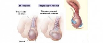
Testicular diseases: varicocele
Varicose veins of the spermatic cord, caused by defects in the venous valves, are a common cause of infertility. The scrotum has many vessels that drain blood away from the testicle. If their valves do not function, blood stagnation and varicose veins occur.
Varicocele
Slow blood flow causes an increase in scrotal temperature. Overheating of the testicle provokes changes in the composition of sperm and hormonal disorders.
Varicose veins of the testicle are not very painful. There is a feeling of heaviness in the testicle and a nagging pain in the groin. A doctor can easily recognize the disease by examining the testicles and performing an ultrasound.
Varicose veins come in different sizes, from small ones that you can barely feel to large ones that look like a bunch of grapes. The only effective method of getting rid of the pathology is surgery (classical or laparoscopic).
Reasons for downsizing
Small size of the tissues is a signal that pathological changes are occurring in the male body. The normal size is 4-6 cm in length and 2.5-3.5 cm in width. Size reduction in men occurs for the following reasons:
- influence of harmful factors during intrauterine development;
- pathologies of pregnancy, in particular from the fact that a woman uses different medications;
- infectious pathologies or autoimmune processes negatively affect the tissue (sometimes the human immune system is able to “eat” the tissues of its own body, including the testicles);
- injuries;
- harmful effects of ionizing radiation;
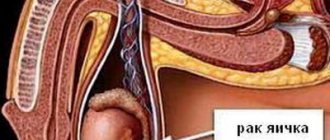
- cancer processes;
- Klinefelter or Shereshevsky-Turner syndrome (and other hereditary diseases);
- hypotrophy or atrophy of the testicles due to cryptorchidism (this often results in small genitals);
- inflammation of the testicles, varicocele and other diseases of the male genital area;
- uncontrolled use of anabolic steroids by athletes.
A true decrease in size indicates that the tissue is gradually atrophying. Accordingly, the production of sperm and testosterone decreases in them. These processes negatively affect fertility and male hormonal balance. The disease is diagnosed by palpation (palpation). The doctor makes conclusions about testicular disease only after a comprehensive examination.
The danger of severe atrophy is that it stops producing enough hormones, which negatively affects health. In some cases, this is a prerequisite for cancer.
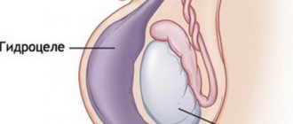
Diagnosis of hydrocele
To diagnose this disease, a physical examination (examination of the patient by a doctor using the senses, possibly using auxiliary medical instruments) of the scrotum is performed in a lying and standing position. Physical examination is necessary to establish a preliminary diagnosis. However, hidden pathologies cannot be detected using this method. For this purpose, instrumental diagnostics are used. This includes ultrasound of the scrotum and testicle (this study helps to visualize the tumor, understand where it is located, determine the size of the tumor, study its internal structure), sometimes CT and MRI (this method uses magnetic fields and radio waves and allows you to create a three-dimensional image of the area under study, MRI of men genital organs is absolutely safe for the patient, at the same time, the method allows the specialist to detect and identify the smallest structural elements of the organs, identify possible pathologies and neoplasms at an early stage). The patient is also recommended to undergo tests and undergo PCR diagnostics. The essence of this method is to collect different types of biomaterial. To do this, take a scraping or smear, as well as urine. Another diagnostic method is diaphanoscopy (a technique for examining tissues by shining them with a narrow beam of light). Diaphanoscopy makes it possible to see the transparent nature of the liquid filling the membrane, as well as the contours of the dark testicle.
Main functions
The main functions of the testicles are the production of testosterone and the formation of sperm. In medicine, there are two types of functions - endocrine and spermatogenesis. Testicles simultaneously perform the function of internal secretion organs - they form hormones and carry them into the blood.
Endocrine
The endocrine function of the testicles is intrasecretory; it consists in the formation of sex hormones: testosterone, androsterone, dihydrotestosterone, dehydroapiandrosterone, which enter directly into the blood. Androgens are synthesized by Leidig cells lying in the interstitium between the convoluted seminiferous tubules.
The endocrine function of the testicles is as follows:
- development of a male body type, for example, broad shoulders, a rough voice, physical endurance, strong muscles;
- formation of courageous character, activity, endurance, desire for leadership, strong muscles;
- regulates cholesterol levels;
- responsible for the normal level of sexual activity;
- development of internal and external genital organs;
- hair development.
Reference! The production of testosterone is influenced not only by the gonads, but also by a man’s lifestyle - his diet, weight, physical activity, health and environmental factors.
The endocrine function of the testicles precedes the function of spermatogenesis, since under the influence of sex hormones the formation of the functioning of the genital organs occurs.
Spermatogenesis
Spermatogenesis is an important process that is responsible for the function of childbirth and occurs in men throughout their lives. During puberty in boys, the process of development of germ cells occurs, which lasts until old age.
There are cases where hundred-year-old men became fathers.
Spermatogenesis consists of four periods:
- Reproduction (carried out in the prenatal period, represents the division of germ cells after birth and until puberty. Upon reaching sexual development, some of the cells continue the process of forming new cells, the second part moves closer to the center of the tubule).
- Maturation (biochemical processes that are involved in preparing cells for the formation of sperm).
- Formation (sperm produce special enzymes that dissolve the shell of the egg, which allows them to penetrate inside and carry out the fertilization process).
Spermatogenesis lasts 75 days. This process is very delicate at the biochemical level, overly sensitive to the effects of adverse factors, such as:
- increased body or air temperature;
- chronic stress;
- taking hormones, antibiotics and other medications;
- excessive drinking;
- smoking;
- sedentary lifestyle;
- prolonged abstinence or sexual abuse.
Treatment
Testicular diseases in men are eliminated using modern methods:
- drug therapy. Having the patient’s research data and symptoms in hand, the doctor can prescribe anti-inflammatory drugs or new generation antibiotics with local action, hormonal drugs. When choosing medications, individual characteristics, the presence of chronic diseases and the patient’s age are taken into account;
- folk remedies. Creams, decoctions and ointments based on medicinal herbs are most often used as additional measures. As a primary means, they may be ineffective, and they are not always able to restore reproductive functions in full;
- surgical intervention. Modern surgery allows the operation to be performed as quickly, efficiently and as minimally traumatic as possible. Depending on the specifics of the surgical procedure, it can be performed under local or general anesthesia.
The specificity of scrotal diseases is such that they can be corrected quite well. Therefore, it will not be difficult for doctors to restore full male health and reproductive functions.
Testicular diseases: inflammation of the testicles - orchitis
Inflammation of the testicle is rare, but it is very painful. The pathology is accompanied by swelling of the testicles and high temperature, reaching up to 40 ° C.
Infection occurs in two ways:
- when bacteria or viruses enter the nucleus with blood from another source of inflammation in the body;
- pathogens enter through the vas deferens, usually caused by urinary tract infections.
Almost always, the epididymis is affected first, then the testicle itself. The swelling continues until a hydrocele forms (the content of the scrotum increases due to the accumulation of serous fluid).
Orchitis
In young men, a common cause of orchitis is mumps, a viral disease. The symptoms are the same as for a bacterial infection, but treatment occurs without antibiotics. After a few days, the inflammation goes away on its own.
In contrast, acute bacterial orchitis is treated with antibiotics. This is necessary to avoid an abscess that requires surgery.
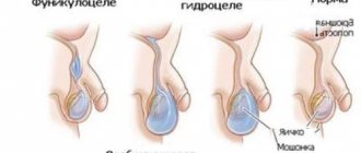
Classification of hydrocele
Physiological . Exists from birth. Over time, the opening of the peritoneal layer, which remains unclosed since birth, becomes obliterated, that is, overgrown due to the spread of connective tissue. The lymphatic apparatus undergoes changes and improves, and the fluid in the serous membrane is absorbed.
Congenital . It may communicate with the peritoneum or its vaginal process, which is subsequently directly transformed into the tunica vaginalis, or it may not communicate. Then there will be no common canal with the abdominal cavity, and the fluid is produced (formed) by the cells of the processus vaginalis themselves.
Purchased . There are two types of congenital dropsy (hydrocele). Primary, or idiopathic, if the cause cannot be determined, and secondary, or symptomatic, the symptoms of which appear during a person’s life, and which is related to a pathogenetic factor.
Based on localization, hydrocele of the ovary is distinguished between unilateral and bilateral. If the cavity with the resulting fluid contains more than 200 ml of exudate (serous fluid), then the ovarian hydrocele is considered large. The maximum possible amount of liquid during dropsy can reach 3000 ml, in which case the dropsy of the ovary will be gigantic.