Introduction
Tuberculosis is an infection caused by mycobacteria, most commonly Mycobacterium tuberculosis.
This infection most often affects the lungs. When the skin is affected, it is called cutaneous tuberculosis. This is a rare disease. The history of cutaneous tuberculosis dates back to 1826, when it was first reported by Laennec. He reported lesions on his arm caused by penetration of the pathogen into the skin. However, the causative agent of the disease was not known until 1882, when M. tuberculosis was first discovered by Robert Koch. It was later isolated from a cutaneous lesion of an affected individual (1, 2, 3).
Classification
Although various classification systems exist, the most universally used classification of cutaneous tuberculosis variants is based on the following:
· Exogenous tuberculosis of the skin: Tuberculous chancre and warty tuberculosis of the skin
· Endogenous cutaneous tuberculosis: Contact or autoinoculation (scrofuloderma, official tuberculosis and some cases of lupus vulgaris); hematogenous dissemination (lupus vulgaris, tuberculous gumma and acute miliary tuberculosis)
· Tuberculides: Papulonecrotic tuberculosis; lichen scrofulous
· Cutaneous tuberculosis: Secondary vaccination with Bacillus Calmette–Guerin (BCG)
Etiology
Infection with M. tuberculosis causes cutaneous tuberculosis. This is a contagious disease that is transmitted from person to person. It can also be caused by Mycobacterium bovis and, less commonly, by the BCG vaccine (4, 5, 6).
Epidemiology
Unlike pulmonary tuberculosis, cutaneous tuberculosis is rare. Of all patients with extrapulmonary manifestations of tuberculosis, 1% to 2% suffer from cutaneous tuberculosis. It is more common in parts of the world where human immunodeficiency virus (HIV) infection is common or where people are immunocompromised for other reasons.
Pathophysiology
Skin invasion by M. tuberculosis leads to the development of the disease. This invasion can be exogenous or endogenous. Endogenous invasion is usually caused by the spread of pulmonary tuberculosis through hematogenous or lymphogenous dissemination. Exogenous invasion is caused by direct inoculation of bacteria. This leads to an immune cascade in skin cells that leads to the formation of granulomas characteristic of tuberculosis. They are present in various forms of clinical lesions.
Histopathology
Early lesions may present with superficial ulcerations, epidermal hyperplasia, and a dense dermal infiltrate consisting of neutrophils, lymphocytes, and plasma cells. Over time, cells involved in chronic inflammation and foci of granulomatous inflammation are replaced by neutrophils with variable caseous necrosis. Acid-fast bacilli are usually readily detected in early lesions but are rarely seen after granulomas have developed.
Clinical and physical examination
The patient's medical history may reveal various symptoms. The patient may have pulmonary tuberculosis along with cutaneous tuberculosis and may have a history of cough, sputum, night sweats, fever, weight loss, hemoptysis, chest pain and fatigue, among others, along with skin symptoms. In disseminated cutaneous tuberculosis, abdominal pain and diarrhea may indicate abdominal tuberculosis. Headache may indicate tuberculous meningitis. Direct inoculation of bacteria results in skin symptoms only.
A history may also reveal immunodeficiency due to underlying diseases such as AIDS, uncontrolled diabetes, malignancies, and end-stage renal disease (ESRD). Other causes of immunodeficiency include intravenous drug use and immunosuppressive therapy.
On examination, objective findings may include various types of skin lesions. There may be inflammatory papules, ulcers, nodules, pustules, verrucous plaques or any other type of lesion.
Survey
Assessing the symptoms of cutaneous tuberculosis requires a history and laboratory examination along with a complete examination. Testing may include tuberculosis skin test (TST) or Mantoux reaction, serum QuantiFERON-TB Gold (QFT-G), PCR, and skin biopsy (7, 8, 9).
The tuberculosis skin test (TST) involves inoculating 0.1 ml of a liquid containing a purified protein derivative (PPD) into the skin. The injection site is the skin of the outside of the wrist or the volar surface of the forearm. The test is interpreted after 48 to 72 hours. The diameter of the skin infiltrate is measured and recorded.
The QFT-G test is an alternative to the tuberculosis skin test (TST). This is a blood test that detects both active and latent tuberculosis using an interferon gamma release assay.
Skin biopsy is a reliable test for diagnosing cutaneous tuberculosis. The skin biopsy is assessed in two ways. First, sections are prepared and examined under a microscope to detect acid-fast bacilli. Next, the tissue is cultured at low temperature to detect the growth of M. tuberculosis.
Other tests that can help make a diagnosis include a chest x-ray and sputum examination. Sputum examination includes microscopy of acid-fast bacilli AfB and culture.
Treatment/Management
Treatment of cutaneous tuberculosis is similar to the treatment of systemic tuberculosis. It also includes treatment with multiple drugs. The most commonly used are isoniazid, rifampicin, pyrazinamide, ethambutol, or streptomycin. Treatment consists of 2 stages.
· Intensive phase, which includes a rapid decrease in the number of M. tuberculosis
Continuation phase, also called sterilization phase
The intensive therapy phase lasts about 8 weeks. After this phase, the patient is no longer contagious, but still requires additional treatment to achieve resolution of the infection. The continuation phase is designed to kill remaining bacteria and lasts 9 to 12 months. Treatment of tuberculosis requires the patient to strictly adhere to the treatment regimen.
Treatment results depend on the patient's immunity, general health, stage of the disease, type of skin lesions, patient compliance, duration of treatment and the presence of any side effects.
Differential diagnosis
Differential diagnosis of skin tuberculosis includes:
- Chronic ulcerative-vegetative pyoderma
- Blastomycosis (endemic deep mycosis - ed.)
- Chromomycosis (subcutaneous mycosis - ed.)
- Reactions to drugs
- Granulomatosis with polyangiitis (Wegener's granulomatosis)
- Granulomatous rosacea
- Syphilitic gumma
- Keratosis spinosa
- Papular eczema
- Coccidioidomycosis (endemic deep mycosis - ed.)
- Glossitis
- Atypical mycobacterial infection
- Chronic granulomatous disease
- Lupoid rosacea
- Nodular vasculitis (Weber-Christchen disease)
- Nodular chills
Many different diseases can be included in the list of differential diagnoses depending on the manifestations of cutaneous tuberculosis. It is difficult to diagnose this condition due to its similarity to many other skin lesions.
Forecast
The prognosis for cutaneous tuberculosis is favorable in patients who do not have a weakened immune system. However, even active treatment may not be effective in immunocompromised and multidrug-resistant patients.
Other aspects
Prevention
Prevention of cutaneous tuberculosis includes BCG vaccination, examination, isolation and treatment of the person who is the source of infection, as well as the use of sterilized instruments and other items. Since immunodeficiency is the main cause of skin tuberculosis, one should try to avoid it by managing diabetes and other diseases. In cases where drug therapy is the cause of immunodeficiency, preliminary testing and treatment of latent tuberculosis with antibiotics is indicated.
Compliance with recommendations
Following treatment recommendations is extremely important when it comes to treating tuberculosis. A patient who does not follow recommendations may develop drug resistance. Multiple treatment modalities may be required, and directly monitored treatment (DOT) is recommended in these patients.
Improving the performance of medical personnel
The diagnosis of cutaneous tuberculosis can be made by a specialist in the field. Since there are particularities in the diagnosis and treatment of skin diseases, the patient should be referred to a consultation with an infectious disease specialist, dermatologist or therapist. Treatment of cutaneous tuberculosis is similar to that of systemic tuberculosis. It also includes treatment with multiple drugs. The most commonly used are isoniazid, rifampicin, pyrazinamide, ethambutol, or streptomycin. Treatment consists of 2 stages.
· Intensive phase, which includes a rapid decrease in the number of M. tuberculosis
Continuation phase, also called sterilization phase
Treatment results depend on the patient's immunity, general health, stage of the disease, type of skin lesions, patient compliance, duration of treatment and the presence of any side effects.
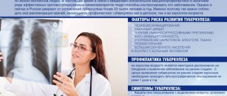
Symptoms and first signs of tuberculosis in children
First of all, tuberculosis in children is manifested by such clinical signs as:
- weakness;
- stopping weight gain;
- irritability;
- fatigue during school hours;
- absent-mindedness;
- lagging behind peers in studies;
- body temperature – up to 37.5°C;
- enlarged lymph nodes;
- sweating;
- chills.Source: N.M. Koretskaya Tuberculosis in children and adolescents in modern conditions // Siberian Medical Review, 2010
Forms of tuberculosis and early signs
| Form | Symptoms |
| Tuberculosis of the bronchial glands | Cough, elevated body temperature for a long time, lethargy, fatigue, pallor, thinness. |
| Pulmonary form | Cough, shortness of breath, weakness after sleep, apathy, decreased performance, absent-mindedness, unhealthy blush, thinness, pale skin, sweating, chills, dry (and then “wet”) cough for more than 3 weeks, sputum with blood. |
| Lymph node involvement | An increase in the size of the lymph nodes, their softening, suppuration (the contents leak out), the formation of fistulas in places where pus comes out, scrofuloderma (skin lesions in the form of tumors with subsequent release of the contents). |
| Involvement of bones and joints in the process | Slow development, pain when moving, changes in gait, lameness. |
| Damage to the meninges (tuberculous meningitis) | Anxiety, lethargy, loss of appetite, bad mood, headaches, fever, vomiting, convulsions. |
| With damage to the gastrointestinal tract | Constipation or diarrhea, increased body temperature, blood in the stool, pain. Source: A.V. Mordyk, E.A. Tsygankova, L.V. Puzyreva, A.A. Turitsa Tuberculosis in children of the Russian Federation at the present stage // Pediatric pharmacology, 2014, v. 11, no. 3, pp. 27-30 |
Clinical manifestations by age of children
| Age | Symptoms |
| Infancy (up to one year) | Low mobility, apathy, weakness, attacks of suffocation or coughing, retraction of part of the chest, weight loss (including muscle mass), loss of appetite, cessation of crying, insomnia. Early detection and diagnosis are extremely important, because at this age this pathology is most dangerous. |
| 5-8 years | Decreased activity, weakness, lack of sleep and appetite, loss of body weight, cough, condensation of the chest. |
| 8-15 years | Rapid appearance of pain in the lungs against a background of apathy and weakness, active urge to cough, shortness of breath even at rest, thinning or discoloration of the skin, the appearance of cracks, wounds, hemoptysis, changes in lymph nodes in size and structure, intoxication of the lungs (at the last stage) . |
Manifestations of the chronic form:
- sleep disorders;
- liver enlargement;
- lagging behind peers in physiological development;
- dry and pale skin;
- mild euphoria.
Ulcerative skin tuberculosis (Tuberculosis cutis ulcerosa)
The disease most often affects men who already had tuberculosis of internal organs. Tuberculosis bacilli enter the skin from the urine, feces or sputum of the patient himself, affecting areas of natural external openings: the skin around the anus, glans penis, nose, area around the mouth, mucous membrane of the tongue.
Symptoms of the disease
Merging, tuberculous tubercles form small yellowish nodules. Over time, the nodules suppurate and open with the formation of painful ulcers that make natural acts difficult.
The ulcers have soft, undermined edges. The color of the ulcers is pale red. The size of the ulcers is up to 1.5 cm, the bottom is granular. Javas often bleed. Sharply painful. As the disease progresses, tuberculous tubercles reappear at the bottom of the ulcers and around them. The tissues are destroyed, forming yellowish microabscesses, the so-called “Trel grains”, containing a large amount of MBT. The disease is difficult. Ulcers heal slowly. In their place, thin scars form, located below the skin level (atrophic scars).
Rice. 21. Ulcerative tuberculosis.
Papulonecrotic tuberculosis of the skin (tuberculosis cutis papulo-nectrotica)
Papulonecrotic tuberculosis of the skin is more often recorded in young people. The disease is manifested by the appearance of papules, the size of which does not exceed 3 cm in diameter. The skin of the buttocks, abdomen, extensor surfaces of the limbs and chest is affected. The skin over the papules becomes pinkish-bluish in color. Over time, the papules become ulcerated. At the site of the ulcers, a grayish-white crust appears, which is replaced by a whitish scar.
Rice. 25. Tuberculosis cutis papulo-nectroticа. Multiple bluish-purple infiltrates and nodes on the legs.
Rice. 26. Papulonecrotic tuberculosis of the skin of the legs. Multiple bluish-purple infiltrates and nodes on the legs.
Indurative tuberculosis of the skin (indurated erythema)
This form of the disease manifests itself in 2 varieties - Bazin's erythema nodosum and Hutchinson's ulcerative erythema. The disease often affects patients with tuberculosis of internal organs.
Erythema nodosum of Bazin
The disease is more often registered in women aged 16 to 40 years. Dense nodes or flat, extensive infiltrates that appear on the skin of the legs, thighs, buttocks and upper extremities are bluish-red in color. The size of the seals is 3 – 8 cm. Seals and infiltrates are often located symmetrically, in the deep layers of the skin and subcutaneous fatty tissue. With regression of tuberculous elements, ring-shaped atrophic areas and pigmentation remain.
Hutchinson's ulcerative erythema
Sometimes the lesions merge, forming an extensive lesion with ulcerations in the center and dirty gray granulations. This form is named after the English dermatologist who first described it - Hutchinson's ulcerative erythema. Without treatment it lasts for a long time, sometimes even years. After healing, sunken pigmented scars remain.
Rice. 29. The photo shows indurative tuberculosis of the skin (indurated erythema of Bazin).
Rice. 30. Ulcerative erythema of Hutchinson.
Types and forms of pathology
According to the period of occurrence in medicine, stages are distinguished:
- infiltration;
- decay;
- contamination;
- resorption;
- seals;
- the appearance of scars;
- calcification.
| Form | Peculiarities |
| Chronic and early | It occurs against a background of body temperature up to 40℃, cough, pain in the lungs, uneven breathing, wheezing, decreased appetite, and loss of strength. |
| Respiratory damage | Includes pathologies:
|
| Other localizations | There may be lesions:
|
How does the disease progress?
The mechanism of development of such an infection includes several stages:
- entry of the pathogen into the upper respiratory tract (on the mucous membranes), from where, with a weak immune response, they enter the lungs;
- penetration into the alveoli of the lungs and further through their walls (without specific changes);
- entry into the lymph nodes and lymphatic pathways with further reproduction (here the infection can take a latent form and persist without external phenomena);
- circulation of microbacteria through the bloodstream without reproduction (about 2 weeks);
- spread of infection through tissues and organs, manifestation of a primary disease or latent infection;
- formation of immunity - formation of granulomas around microbacteria;
- infection (if the process does not progress, the granuloma becomes covered with connective tissue or resolves, the microbacterium inside it turns into the L-form);
- disease – transition of L-forms into rods.
Diagnosis of skin tuberculosis
Rice. 31. View of Mycobacterium tuberculosis in the light of a fluorescent microscope.
The diagnostic process consists of the following components:
- Bacteriological diagnosis (detection of MBT in discharge from ulcers and lymph node punctures).
- Diagnostic biopsy followed by histological examination, which allows to identify the morphological components of the tuberculous tubercle.
- Detection of tuberculosis of internal organs.
- Detection of scars on the skin as a result of a previous tuberculosis lesion.
- Tuberculin diagnostics.
- Trial treatment.
Diagnostic biopsy together with bacteriological examination is the most significant method in diagnosing tuberculosis.
Dermatologists are prescribed to detect skin tuberculosis. Knowledge of risk groups, which include patients in this category, early symptoms of the disease and diagnostic methods helps doctors to detect the disease in a timely manner.
Identification of the pathogen in the early stages of the disease will allow the patient to be successfully cured.
Treatment
Chemotherapy, antibiotics
Bactericidal and bacteriostatic drugs are used to achieve complete recovery. In this case, the correct combination of drugs is important, taking into account the resistance of some bacteria to this type of therapy. First, treatment is aimed at suppressing the growth of bacteria and eliminating their resistance to medications. Residual infection in the cells is then eliminated. Duration of therapy is 6-12 months.
Surgery
Pulmonary resection is practiced as a radical technique. The operation is performed for bronchial stenosis, fibrous-cavernous lesions, pleural empyema, abscesses in the lungs, tuberculomas prone to progression, etc. In addition, decortication is used, that is, removal of fibrous layers, and cavernotomy - cleansing of the opened cavity.
DOTS
A treatment system consisting of several levels: bacterioscopic examination, chemotherapy, anti-tuberculosis treatment.
Sources:
- N.M. Koretskaya. Tuberculosis in children and adolescents in modern conditions // Siberian Medical Review, 2010.
- A.V. Mordyk, E.A. Tsygankova, L.V. Puzyreva, A.A. Turitsa. Tuberculosis in children of the Russian Federation at the present stage // Pediatric pharmacology, 2014, v. 11, no. 3, pp. 27-30.
- https://www.ncbi.nlm.nih.gov/pubmed/24548085 Marais BJ. Tuberculosis in children // J Paediatr Child Health. 2014 Oct;50(10):759-67. doi: 10.1111/jpc.12503. Epub 2014 Feb 19.
- V.N. Krivohizh. Modern methods of early detection of tuberculosis among children and adolescents // Health is the basis of human potential: problems and solutions, 2013, pp. 570-585.
The information in this article is provided for reference purposes and does not replace advice from a qualified professional. Don't self-medicate! At the first signs of illness, you should consult a doctor.
Lichenoid tuberculosis of the skin (Tuberculosis cutis lichenoides seu lichen scrofulosorum)
Lichenoid tuberculosis of the skin often develops in weakened children, less often in adults with tuberculosis. The disease is manifested by the appearance of tubercles, which are covered with light gray scales. The tubercles are located symmetrically, most often on the skin of the abdomen and limbs. After healing, pigmentation or small scars remain at the site of the rash.
Rice. 28. The photo shows lichenoid tuberculosis of the skin.