Home » Department of Orthopedics » Diseases » Chest deformity
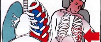
The chest is a part of the human body. It is formed in front by the sternum, behind by the spine, on the sides and partially in front and behind by the ribs. It is also formed by muscles that connect these bones to each other and to other bones. The rib cage contains the chest cavity, which contains the most important organs of the human body.
The chest contains the lungs and heart, as well as the aorta , the largest artery in the body. If these organs stop functioning, the person dies within a few minutes. That is why, during evolution, they settled in such a protected place. Due to the rigidity of the chest, the organs inside it are in a constant position, which ensures the stability of their functioning.
However, in some cases the chest changes its shape. Depending on the degree of change in shape, the position of the organs inside changes, their transformation occurs as they adapt to changing conditions. It should be noted that the chest is deformed, in most cases, in children, or is completely congenital. And with intensive growth, changes occur very quickly. This can lead to serious consequences for the entire body.
Types of chest deformation
Chest deformities can be divided into two groups.
- Congenital - usually caused by a violation of skeletal development in the prenatal period (changes in the shape of bones, absence of elements of the chest walls): - funnel-shaped deformity of the chest; – keeled chest deformity; – flat chest deformity.
- acquired (more common) - are the consequences of injuries, lung diseases, bone tuberculosis, rickets, scoliosis: - emphysematous; – paralytic; – scaphoid; – kyphoscoliotic.
Poland syndrome
Poland syndrome is named after Albert Poland, who first described this type of chest deformity as a result of observations in school and refers to a spectrum of diseases that are associated with underdevelopment of the chest wall. This syndrome includes abnormalities of the pectoralis major, pectoralis minor, serratus anterior, ribs, and soft tissues. In addition, deformation of the arm and hand may occur.
The incidence of Poland syndrome is approximately 1 case per 32,000 children born. This syndrome is 3 times more common in boys than girls, and the right side is affected in 75% of patients. There are several theories regarding the etiology of this syndrome, which include abnormal migration of fetal tissue, subclavian artery hypoplasia, or intrauterine trauma. However, none of these theories have proven their validity. Poland syndrome is rarely associated with other diseases. Some patients with Poland syndrome have leukemia. There is a definite association of this syndrome with Moebius syndrome (unilateral or bilateral palsy of the facial nerve, abducens ophthalmic nerve).
Symptoms of Poland syndrome depend on the degree of the defect and in most cases they are cosmetic complaints. In patients with significant bone defects, the lung may bulge, especially when coughing or crying. Functional and respiratory problems may occur in some patients. The lungs themselves are not affected by this syndrome. In patients with significant muscle and soft tissue defects, decreased exercise capacity may become apparent.
Congenital chest deformities
The reliable causes of congenital chest deformity are currently unknown. However, any teratogenic factors negatively affect the development of the fetus in the womb.
The causes of congenital deformity are currently divided into three groups.
As a rule, with the development of deformation, a combination of these is observed.
- Uneven growth of costal cartilage and sternum.
- Pathology on the part of the diaphragm - too short parts of this muscle are able to “pull” the developing sternum inward.
- Developmental pathology of connective tissue and cartilage.
Funnel chest deformity
Deformation of the chest, manifested in retraction of the sternum, anterior ribs and costal cartilages. It occurs more often than other congenital malformations of the chest. You can also find its name as “ shoemaker's breast ”. Previously, shoemakers, when working, rested the heel of their shoes on the lower third of the sternum. This led to professional deformation of the chest.
Similar pathologies are often observed in the patient’s relatives, which indicates the genetic nature of this pathology.
A deformed chest is the cause of other diseases. As the child actively grows, the shape of other bones, in particular the spine, changes. There is also a change in position and disruption of the functioning of internal organs.
There are three degrees of pectus excavatum:
- I – depth of depression up to 2 cm, organs are not displaced;
- II – funnel 2-4 cm. deep, the heart is displaced up to 3 mm;
- III – depression more than 4 cm, heart displaced more than 3 mm.
Symptoms
At first, the violation is hardly noticeable. As the child grows, a “funnel” forms. Children lag behind their peers in development and suffer more often from colds. The deformation reaches its maximum by 3-4 years. Afterwards it gradually stops progressing. By this time, the child additionally develops kyphosis and scoliosis, disorders of the chest organs, asymmetry of the chest develop, and decreased excursion of the chest is noted.
Pileated chest deformity
The pathology is a protrusion of the sternum forward, which gives the breast cell a keel shape. Protrusion is caused by excessive growth of costal cartilages, which “push” the sternum forward, increasing the anteroposterior size of the chest.
There are three degrees of severity of deformation:
- I – protrusion of the keel up to 2 cm above the normal contours of the chest;
- II – protrusion from 2 to 4 cm;
- III – protrusion from 4 to 6 cm.
Symptoms
The main symptom is protrusion of the sternum forward. The degree of deformation increases with age. Unlike the previous pathology, keeled breasts do not cause serious problems. This is explained by the fact that the volume of the chest does not decrease and compression of the internal organs does not occur. The position of the heart changes. It occupies a vertical position and takes the shape of a “hanging drop”. The child complains of shortness of breath and fatigue.
3. Diagnosis and classification of keeled breasts
Chicken breast is detected by the surgeon during a physical examination. Additionally, radiography is prescribed, the data of which allows us to judge the nature and degree of deformation. The following abnormalities are usually visible on an x-ray:
- increase in the chest space;
- teardrop-shaped heart, its rotation along its own axis and displacement;
- delimitation of the sternum segments from each other in the lateral projection./li>
If the patient complains of difficulty breathing and cardiac activity, additional studies are carried out aimed at assessing the effect of the bone defect on the functioning of the cardiovascular system and respiratory function:
- electrocardiography;
- sonography;
- diagnostics of external respiration function.
When classifying the “chicken breast” defect, they proceed from the degree of influence of this pathology on the position of the heart:
- 1st degree - the heart is not displaced;
- 2nd degree - there is a displacement of the heart by no more than 3 cm;
- 3rd degree - the position of the heart is deviated from the norm by 3 cm or more.
In addition, the specific type of abnormal breast structure is determined:
- corpocostal (the lower part of the chest is convex);
- manubriocostal (the upper part of the chest protrudes);
- costal (the defect is localized in the area of the costal cartilages).
About our clinic Chistye Prudy metro station Medintercom page!
Acquired chest deformities
Emphysematous chest
Emphysema is an increase in the airiness of the lung tissue. The disease is accompanied by an increase in lung size. This entails a gradual change in the shape of the chest. The anteroposterior size of the chest increases. The patient's chest takes on a rounded shape, the so-called “barrel” chest. Chest excursion decreases.
Paralytic chest
It is observed in chronic diseases of the lungs and pleura, characterized by organ shrinkage. The anteroposterior and lateral dimensions of the chest decrease. There is a retraction of the intercostal spaces, which indicates difficulty in breathing in patients. The collarbones and shoulder blades are also clearly visible - this is due to a change in their location relative to the ribs and sternum. The symmetry of movements of the right and left halves of the chest is disrupted.
Scaphoid chest
It is observed in patients with a rare disease - syringomyelia. This disease causes cavities to form in the spinal cord. One of its manifestations is a change in the composition of bones due to the leaching of calcium salts from them. Bones become less rigid and are able to deform. Patients have a scaphoid depression localized in the upper and middle thirds of the sternum.
Kyphoscoliotic chest
The reason for this is disorders of the spine. This can be a consequence of diseases such as spinal tuberculosis, rickets, scoliosis, or as a result of injuries. When the shape of the spine changes, pathology in the chest organs may be observed.
Also worth mentioning is deformation due to trauma and surgery.
Cystic fibrosis
An emphysematous chest may be a sign of cystic fibrosis. This is a severe hereditary disease associated with a gene mutation. With cystic fibrosis, mucus accumulates in all organs, including the bronchi. Patients experience a severe cough with viscous sputum and difficulty breathing.
Typically, this disease is diagnosed in children in the first months of life. The pathology is often complicated by chronic pulmonary failure.
Treatment of chest deformity in Israel
Most acquired deformities of the chest are a consequence of chronic diseases of the chest organs and do not pose a threat to the patient’s life.
Congenital deformities are treated
Conservative treatment of chest deformity is ineffective. Surgical treatment is indicated for disorders of the chest organs or for severe cosmetic defects. It is carried out when the child reaches the age of 6-7 years. By this time, the formation of the defect usually stops.
Chest reconstruction
Reconstruction of the chest is being carried out. The sunken parts return to their place and are fixed mechanically. With keeled deformity, the costal cartilages are truncated (this pathology is less often an indication for surgical intervention).
Magnet implantation
New treatments involve implanting a magnet in the area that needs to be corrected. The second magnet is positioned so that their interaction generates a force aimed at eliminating the defect.
Some deformities can be masked by implanting silicone over the area of deformation.
Currently, operations to eliminate chest defects with minimal consequences for the body are being successfully carried out in Israel.
Attention! All form fields are required. Otherwise we will not receive your information. Alternatively use
Sternum defects
Sternal defects can be divided into 4 types and all are rare: thoracic ectopia cordis, cervical ectopia cordi, thoracoabdominal ectopia cordi, and cleft sternum. Thoracic ectopia cordis is an anomaly in the location of the heart outside the chest, and the heart is completely unprotected by dense bone tissue. The survival rate of patients with thoracic ectopia cordis is very low. Only three successful cases of surgical treatment out of 29 operations with this anomaly have been described.
Cervical ectopia of the heart differs from thoracic ectopia only in the localization of the abnormal location of the heart. As a rule, such patients have no chance of survival. In patients with thoracoabdominal ectopia, the heart is located inferior to the sternum. The heart is covered by a membrane or thin skin. Downward displacement of the heart is the result of a semilunar pericardial defect and a diaphragmatic defect. Abdominal wall defects are also common.
A cleft sternum is the least serious of the 4 abnormalities because the heart is almost closed and in a normal position. There is a partial or complete split of the sternum over the heart, with partial split being more common than complete splitting. Associations with heart defects with this anomaly are quite rare. In most children, cleft sternum usually does not cause particularly noticeable symptoms. In some cases, respiratory symptoms are possible as a result of paradoxical movement of the sternal defect. The main indication for surgical treatment is the need to protect the heart.
Treatment methods
Barrel-shaped breast deformation is just one of the symptoms of various diseases. You can get rid of such a defect only after treating the underlying pathology.
For chronic obstructive respiratory diseases and bronchial asthma, patients are shown the following bronchodilators:
- "Foradil."
- "Serevent."
- "Atrovent N".
- "Salbutamol."
These medications come in the form of inhalers. They relieve bronchospasm and make breathing easier.
For severe obstructive diseases and asthma, drugs with corticosteroid hormones are prescribed:
- "Prednisolone."
- "Dexamethasone."
Hormonal drugs are used both in oral and inhalation forms.
If it is difficult to discharge sputum, mucolytic medications are indicated:
- "Ambroxol".
- "ACC."
- "Carbocisteine".
These agents thin the mucus and facilitate easier removal of mucus from the bronchi.
If the use of inhalers does not have the desired effect, then drug treatment is supplemented with oxygen therapy sessions. This helps to significantly improve the condition of patients.
Treatment for cystic fibrosis can only be symptomatic. Modern medicine cannot cure a gene mutation. However, the patient's condition can be significantly alleviated. Patients are prescribed bronchodilator and mucolytic drugs. These medications must be taken throughout your life. In case of severe blockage of the respiratory tract with mucus, the bronchi are washed with a solution of sodium chloride.
For osteoarthritis, chondroprotectors and intra-articular injections of drugs with hyaluronic acid are prescribed. In case of severe pain, taking non-steroidal anti-inflammatory drugs (Diclofenac, Naise, Ibuprofen) is indicated.
Patients are often interested in the possibility of plastic surgery for curvature of the chest wall. If the deformity is caused by serious lung diseases, then it cannot be eliminated through cosmetic surgery. After all, the volume of the chest in this case increases due to air retention in the respiratory organs. Usually, after remission is achieved, the shape of the chest returns to normal.
Prevention
How to prevent chest wall deformation? To do this, it is necessary to protect the respiratory system from harmful effects. Pulmonologists advise adhering to the following recommendations:
- completely stop smoking;
- avoid exposure to allergens, dust and toxic gases;
- when working in hazardous industries, undergo regular medical examinations;
- promptly cure inflammatory bronchopulmonary diseases.
If you have a systematic cough, wheezing in the chest and difficulty breathing, you should immediately consult a doctor. This will help avoid serious complications such as heart and pulmonary failure.