Human sex cells, chromosomes, fertilization
Sex cells - gametes (from the Greek gametes - “spouse”) can be found already in a two-week human embryo. They are called primordial germ cells . At this time, they are not at all similar to sperm or eggs and look exactly the same. It is not possible to detect any differences inherent in mature gametes at this stage of embryo development in primary germ cells. This is not their only feature. Firstly, primary germ cells appear in the embryo much earlier than the sex gland itself (gonad), and secondly, they arise at a considerable distance from the place where these glands will form later. At a certain moment, an absolutely amazing process occurs - the primary germ cells rush together to the gonad and populate, “colonize” it.
After the future gametes enter the gonads, they begin to divide intensively, and their number increases. At this stage, germ cells still contain the same number of chromosomes as “bodily” ( somatic ) cells - 46. However, to successfully carry out their mission, germ cells must have 2 times fewer chromosomes. Otherwise, after fertilization, that is, the fusion of gametes, the cells of the embryo will contain not 46, as established by nature, but 92 chromosomes. It is not difficult to guess that in subsequent generations their number would progressively increase. To avoid this situation, the developing germ cells undergo a special division, which in embryology is called meiosis (Greek meiosis - “reduction”). As a result of this amazing process the diploid (from the Greek diploos - “double”) set of chromosomes is, as it were, “pulled apart” into its constituent single, haploid sets (from the Greek haploos - single). As a result, from a diploid cell with 46 chromosomes, 2 haploid cells with 23 chromosomes are obtained. Following this, the final stage of the formation of mature germ cells begins. Now, in a haploid cell, the existing 23 chromosomes are copied and these copies are used to form a new cell. Thus, as a result of the two divisions described, 4 new ones are formed from one primary germ cell.
Moreover, in spermatogenesis (Greek genesis - origin, development), as a result of meiosis, 4 mature sperm with a haploid set of chromosomes appear, and in the process of egg formation - in oogenesis (from Greek oon - “egg”) only one. This happens because the egg cell does not use the second haploid set of chromosomes formed as a result of meiosis to form a new mature germ cell - an oocyte, but “throws” them out as “extra” in a kind of “garbage container”, which is called a polar body. The first division of the chromosome set is completed in oogenesis with the release of the first polar body just before ovulation. The second replication division occurs only after the sperm penetrates the egg and is accompanied by the release of the second polar body. For embryologists, polar bodies are very important diagnostic indicators. The first polar body is present, which means the egg is mature, the second polar body appears, and fertilization has occurred.
Primary germ cells found in the male gonad do not divide for the time being. Their division begins only during puberty and leads to the formation of a cohort of so-called diploid stem cells, from which sperm are formed. The supply of stem cells in the testicles is constantly replenished. Here it is appropriate to recall the feature of spermatogenesis described above - 4 mature sperm are formed from one cell. Thus, after puberty, a man produces hundreds of billions of new sperm throughout his life.
The formation of eggs proceeds differently. Having barely populated the gonad, the primary germ cells begin to divide intensively. By the 5th month of intrauterine development, their number reaches 6-7 million, but then mass death of these cells occurs. In the ovaries of a newborn girl there are no more than 1-2 million of them, by the age of 7 - only about 300 thousand, and during puberty 30-50 thousand. The total number of eggs that will reach a mature state during puberty will be even less. It is well known that during one menstrual cycle, only one follicle usually matures in the ovary. It is easy to calculate that during the reproductive period, which lasts for women 30-35 years old, about 400 mature eggs are formed.
If meiosis in spermatogenesis begins during puberty and is repeated billions of times during a man’s life, in oogenesis the forming female gametes enter meiosis during the period of intrauterine development. Moreover, this process begins almost simultaneously in all future eggs. It begins, but does not end! Future eggs reach only the middle of the first phase of meiosis, and then the division process is blocked for 12 - 50 years! Only with the advent of puberty will meiosis continue in oogenesis, and not for all cells at once, but only for 1-2 eggs monthly. The process of meiotic division of the egg will be completed, as mentioned above, only after its fertilization! Thus, the sperm penetrates into an egg that has not yet completed division and has a diploid set of chromosomes!
Spermatogenesis and oogenesis are very complex and largely mysterious processes. At the same time, their subordination to the laws of interrelation and conditionality of natural phenomena is obvious. To fertilize one egg in vivo (lat. in a living organism), tens of millions of sperm are needed. The male body produces them in gigantic quantities almost throughout his life.
Carrying and giving birth to a child is an extremely difficult burden on the body. Doctors say that pregnancy is a health test. How a child will be born directly depends on the state of health of the mother. Health, as you know, does not last forever. Old age and illness, unfortunately, are inevitable. Nature gives a woman a strictly limited, irreplaceable number of germ cells. A decrease in fertility develops slowly, but gradually along an incline. We receive clear evidence that this is indeed the case by daily assessing the results of ovarian stimulation in ART programs. Most of the eggs are usually used up by age 40, and by age 50 the entire supply is completely depleted. Often, so-called ovarian depletion occurs much earlier. It should also be said that the egg is subject to “aging”; over the years, its ability to fertilize decreases, and the process of chromosome division is increasingly disrupted. Having children at a late reproductive age is risky due to the increasing risk of having a child with a chromosomal abnormality. A typical example is Down syndrome, which occurs due to an extra 21 chromosome remaining during division. Thus, by limiting the reproductive period, nature protects the woman and takes care of healthy offspring.
According to what laws does chromosome division occur? How is hereditary information transmitted? In order to understand this issue, we can give a simple analogy with cards. Let's imagine a young married couple. Let's call them conventionally - He and She. Each of its somatic cells contains chromosomes of the black suit - clubs and spades. He received a set of clubs from six to ace from his mother. A set of spades - from my dad. In each of its somatic cells, red chromosomes are diamonds and hearts. She received a set of diamonds from six to ace from her mother. A set of worms - from my dad.
In order to obtain a sex cell from a diploid somatic cell, the number of chromosomes must be halved. In this case, the sex cell must contain a complete single (haploid) set of chromosomes. Not a single one should get lost! In the case of cards, such a set can be obtained as follows. Take one at random from each pair of black cards and thus form two single sets. Each set will include all cards of the black suit from six to ace, however, what kind of cards these will be (clubs or spades) is determined by chance. For example, in one such set the six may be a spades, and in another it may be a club. It’s not hard to imagine that in the example with cards, with such a choice of a single set from a double set, we can get 2 combinations to the ninth power - more than 500 options!
In the same way, we will make a single set of her red cards. We will get more than 500 different options. From his single and her single set of cards we will make a double set. It will turn out to be, to put it mildly, “variegated”: in each pair of cards, one will be of a red suit, and the other will be black. The total number of such possible sets is 500×500, that is, 250 thousand options.
Nature does approximately the same thing, according to the law of random sampling, with chromosomes during the process of meiosis. As a result, from cells with a double, diploid set of chromosomes, cells are obtained, each of which contains a single, haploid complete set of chromosomes. Let's say that as a result of meiosis, a sex cell is formed in your body. Sperm or egg - in this case it does not matter. It will definitely contain a haploid set of chromosomes - exactly 23 pieces. What exactly are these chromosomes? Let's take chromosome 7 as an example. This could be the chromosome you received from your father. It could just as likely be a chromosome you got from your mother. The same is true for chromosome No. 8, and for any other.
Since in humans the number of haploid chromosomes is 23, then the number of possible variants of sexual haploid cells formed from diploid somatic cells is equal to 2 to the power of 23. This results in more than 8 million variants! During the process of fertilization, two germ cells unite with each other. Therefore, the total number of such combinations will be 8 million x 8 million = 64,000 billion options! At the level of a pair of homologous chromosomes, the basis of this diversity looks like this. Let's take any pair of homologous chromosomes of your diploid set. You received one of these chromosomes from your mother, but it could be from either your grandmother or your maternal grandfather. You received the second homologous chromosome from your father. However, it can again be, regardless of the first chromosome, either your grandmother’s or your paternal grandfather’s. And you have 23 pairs of such homologous chromosomes! This results in an incredible number of possible combinations. It is not surprising that one pair of parents gives birth to children who differ from each other in both appearance and character.
By the way, a simple but important conclusion follows from the above calculations. Every person currently living, or who has ever lived in the past on Earth, is absolutely unique. The chances of a second one appearing are almost zero. Therefore, there is no need to compare yourself with anyone. Each of you is unique, and that makes you interesting!
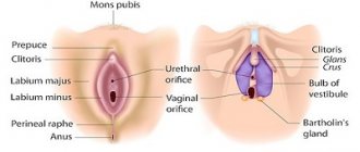
However, let's return to our reproductive cells. Each diploid human cell contains 23 pairs of chromosomes. Chromosomes from 1 to 22 pairs are called somatic and they are the same in shape. The chromosomes of the 23rd pair (sex chromosomes) are the same only in women. They are designated by the Latin letters XX. In men, the chromosomes of this pair are different and are designated XY. In the haploid set of an egg, the sex chromosome is always only X, while the sperm can carry either an X or a Y chromosome. If the egg is fertilized by X sperm, a girl will be born, if Y sperm, a boy will be born. It's simple!
Why does meiosis in an egg take so long? How is a monthly selection of a cohort of follicles that begin their development and how is the leading, dominant, ovulatory follicle selected from them, in which the egg will mature? Biologists do not yet have clear answers to all these difficult questions. The process of formation of mature eggs in humans awaits new researchers!
The formation and maturation of sperm, as already mentioned, occurs in the seminiferous tubules of the male reproductive gland - the testicles . The formed sperm has a length of about 50-60 microns. The sperm nucleus is located in its head. It contains the paternal hereditary material. Behind the head there is a neck, in which there is a large convoluted mitochondrion - an organelle that ensures the movements of the tail. In other words, this is a kind of “energy station”. There is a “cap” on the head of the sperm. Thanks to it, the shape of the head is oval. But, it’s not about the form, but about what is contained under the “cap”. This “cap” is actually a container and is called an acrosome , and it contains enzymes that are capable of dissolving the shell of the egg, which is necessary for the sperm to penetrate inside - into the cytoplasm of the egg. If the sperm does not have an acrosome, its head is not oval, but round. This sperm pathology is called globulospermia (round-headed sperm). But, again, the trouble is not in shape, but in the fact that such a sperm cannot fertilize an egg, and a man with such a disorder of spermatogenesis was doomed to childlessness until the early 90s of the last century. Today, thanks to ART, infertility in these men can be overcome, but we will talk about this later in the chapter on micromanipulation, in particular ICSI.
The movement of the sperm is carried out due to the movement of its tail. The speed of sperm movement does not exceed 2-3 mm per minute. It would seem not much, however, in 2-3 hours in the female reproductive tract, sperm travel a distance 80,000 times greater than their own size! If a person were in the place of the sperm in this situation, he would have to move forward at a speed of 60-70 km/h - that is, at the speed of a car!
The sperm in the testicle are immobile. They acquire the ability to move only by passing through the vas deferens under the influence of the fluids of the vas deferens and seminal vesicles, and the secretion of the prostate gland. In the female genital tract, sperm remain motile for 3-4 days, but they must fertilize the egg within 24 hours. The entire development process from a stem cell to a mature sperm lasts approximately 72 days. However, since spermatogenesis occurs continuously and a huge number of cells enter it at once, the testicles always contain a large number of sperm at different stages of spermatogenesis, and the supply of mature sperm is constantly replenished. The activity of spermatogenesis varies from person to person, but decreases with age.
As we have already said, eggs are located in the follicles of the ovary. As a result of ovulation, the egg enters the abdominal cavity, from where it is “caught” by the fimbriae of the fallopian tube and transferred to the lumen of its ampullary section. This is where the egg meets the sperm.
What structure does a mature egg have? It is quite large and reaches 0.11-0.14 mm in diameter. Immediately after ovulation, the egg is surrounded by a cluster of small cells and a gelatinous mass (the so-called corona radiata ). Apparently, in this form it is more convenient for the fimbriae of the fallopian tube to capture the egg. In the lumen of the fallopian tube, with the help of enzymes and mechanical action (beating of the cilia of the epithelium), the egg is “cleaned” from the corona radiata. The final release of the egg from the corona radiata occurs after it meets the sperm, which literally stick around the egg. Each sperm secretes an enzyme from the acrosome that dissolves not only the corona radiata, but also acts on the membrane of the egg itself. This shell is called the pellucida, which is what it looks like under a microscope. By secreting the enzyme, all sperm strive to fertilize the egg, but the zona pellucida will allow only one of them to pass through. It turns out that by rushing towards the egg and acting on it collectively, the sperm “clear the way” for only one lucky person. The role of the zona pellucida is not limited to the selection of sperm; in the early stages of embryo development, it maintains the ordered arrangement of its cells (blastomeres). At some point, the zona pellucida becomes tight, it ruptures and hatching (from the English hatching - “hatching”) - the hatching of the embryo.
The embryo is ready for implantation into the endometrium.
Diagnosis of various types of aneuploidy.
The only reliable way for early intrauterine diagnosis of chromosomal abnormalities in the fetus is genetic research. Confirmation of a diagnosis suspected on the basis of risks identified during screening is carried out with invasive sampling of material, which requires clearly substantiated indications for the procedure.
Using the non-invasive prenatal test Prenetix, you can examine fetal DNA in the venous blood of the expectant mother starting from the 10th week of pregnancy. The reliability and specificity of the method allows us to classify Prenetix as an expert-level screening, which significantly increases the information content of traditional first trimester screening.
How is X chromosome analysis performed?
X chromosome analysis allows us to determine the following connections:
- father - daughter;
- mother Daughter;
- sister - sister (based on the genetic material of a common father);
- aunt – niece (if the aunt is the father’s sister);
- grandmother – granddaughter (if grandmother is father’s mother).
Genetic examination involves the study of individual sections of the X chromosome (STR loci). These are short chains consisting of several nucleotide repeats. These chains are unique for each person and coincide only with close relatives. Based on the coincidences, conclusions are drawn about the relationship. Since the procedure is complex, it is not completed in one day.
Upon completion, the result is reported in a report, which describes the research methods and answers the important question: whether there are family ties between two people or not. To increase the accuracy of the result, the study is performed by two independent expert groups. After completing the work, they compare the results.
What is needed for analysis?
DNA analysis requires genetic material from each party: from the person whose relationship is to be established and from his alleged relative. The main source of information here is the buccal epithelium, which is quickly obtained by scraping from the inner surface of the cheek.
This is a simple and painless procedure, it can be easily performed at home, but it is optimal to come to the clinic so that a specialist takes the material and immediately sends it for examination.
There are other, less traditional materials on the basis of which DNA text is made. They are contacted if the alleged relative refuses to assist in the examination, because these sources can be collected without his knowledge:
- hair with follicles;
- saliva (for example, on a toothbrush);
- blood (a blood stain on the fabric is enough);
- clipped nails.
With a little creativity, genetic materials will be extracted and delivered to the clinic for research. DNA testing is ordered for both personal and forensic purposes. Depending on this, placing an order and issuing the result differ.