Difficulty breathing, or shortness of breath, is a fairly common symptom that can occur in healthy people, for example, after exercise. However, if the problem appears at rest, in a lying position, it can be caused by a number of diseases, including serious ones. Why is it hard to breathe while lying down? There can be many reasons for this.
For example, shortness of breath can occur with a rare hereditary pathology - Pompe disease. Pompe disease belongs to the so-called storage diseases. In this disease, due to a gene mutation, there is a deficiency of the enzyme acid glucosidase, which breaks down glycogen. It accumulates and progressive muscle weakness develops.
Usually, weakness of the leg muscles first occurs, and gradually the arm muscles and muscles involved in the breathing process are affected, which leads to shortness of breath1,2. Pompe disease is rare and is classified as an orphan disease. Much more often, shortness of breath when lying down is caused by other pathologies. What diseases can cause it and what to do in such cases?
Mechanism of shortness of breath in the supine position
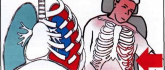
Shortness of breath while lying on your back, or orthopnea, occurs due to increased pressure in the blood vessels of the lungs. When lying down, blood flows from the lower extremities to the heart and then to the lungs. In healthy people, this distribution of blood does not cause any difficulties. But with a number of diseases, the heart's ability to pump excess blood out of the heart may decrease. As a result, blood accumulates in the pulmonary circulation passing through the lungs3.
Increased blood pressure in the pulmonary artery can contribute to the release of fluid into the alveoli and the development of pulmonary edema. This makes breathing while lying down even more difficult3.
Orthopnea is not a disease, but a symptom that occurs only in a horizontal position. An attack can develop at night, when a person wakes up due to lack of air. This phenomenon is called nocturnal paroxysmal (sudden) shortness of breath. In a sitting or reclining position, breathing, as a rule, becomes easier or completely normalized3.
Symptom of chest compression with VSD
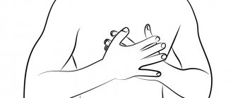
Most people with vegetative-vascular dystonia experience side effects that can be confused with a number of concomitant diseases. One of these is a symptom of VSD - a feeling of squeezing in the chest area.
When to expect an attack?
A feeling of pressure in the chest can occur at absolutely any time of the day and does not depend at all on what the person was doing before the attack, whether he was physically active, engaged in mental work, or simply resting. Also, such attacks are not uncommon during sleep, but not in its intense phase, but during awakening.
Prerequisites for the manifestation of a symptom
The main condition for the onset of pain is that the person must be hungry. Compression during VSD differs from the manifestations of heart diseases of various etymologies in that heart attacks begin just after a person has eaten abundantly and all the blood has rushed to the stomach, depriving the heart and its area of the necessary nutrition and the required volume of oxygen.
How does an attack of chest constriction manifest during VSD?
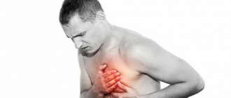
An attack can begin quite suddenly, and often this occurs against the background of states of complete rest and absence of stress and depression. The pain begins to manifest itself in the chest area, less often in the stomach, and as it increases in the chest, it is localized in the left half, where the heart is located. A person may suspect gastritis or a heart disease, but when pressing on the stomach area, the pain does not become more acute, and if an electrocardiogram is taken at the time of the attack, it will not show any abnormalities.
In this case, the pain is dull, aching, and increasing in nature, although it can be tolerated. The attack lasts about 15-30 minutes, while weakness increases, health worsens and the person feels the desire to lie down to restore his strength and wait for the end of the painful sensations in a state of complete rest. Then the pain stops as suddenly as it began, while drug intervention does not in any way affect the speed at which the attack ends.
Naturally, squeezing the chest can cause a feeling of lack of oxygen, which, in turn, can cause a panic attack, also characteristic of VSD. And even if everything went without suffocation and fear, then panic can begin after it’s all over. Questions begin to spin chaotically in my head, what is happening to the body. I would like to find out the causes of the pain as quickly as possible and cure them as quickly as possible. It is from this moment that useless visits to doctors, finding out the reasons, and useless tests begin. But all you need to do is admit that the patient suffers from VSD and such a manifestation as chest compression is one of the symptoms of this disease, which now affects many people, although most do not even suspect it.
How to treat chest compression due to VSD
The occurrence of such attacks during VSD indicates that the prescribed treatment is insufficient or ineffective. Or, as an option, psychotropic drugs were selected incorrectly. The thing is that when medications do not work or act incorrectly, and the patient suffers from VSD, his nervous system is in constant tension and it increases, but the drugs do not relieve it. So a wide variety of attacks occur, as accompanying the underlying disease, that is, VSD.
The main reason for triggering an attack is the contraction of the muscles of the esophagus, and this only happens if the patient’s stomach is empty. As soon as the attack begins, drink a couple of glasses of water and it will pass within 30-60 seconds.
If you take medications during an attack, such as Corvalol or phenazepam, they will have no effect and the attack will go away on its own within 15-30 minutes.
And you will mistakenly think that the medicine worked. Therefore, as soon as you feel the beginning of an attack, run and drink water. And if this does not work, then this becomes a reason to think and act further. Author: K.M.N., Academician of the Russian Academy of Medical Sciences M.A. Bobyr
Why does orthopnea develop?
Shortness of breath while lying down can occur with various disorders, primarily with lung diseases. Predisposing factors are disturbances in ventilation and gas exchange in the lungs and changes in their blood circulation4. Let's look at some of the most common reasons.
Chronic obstructive pulmonary disease (COPD) is a common disease in which inflammation of the airways develops, often accompanied by an infectious process. Due to narrowing or complete blockage of the airways and loss of elasticity in the lungs, airflow is limited. Constant shortness of breath appears, including in a horizontal position, which becomes stronger over time. Along with it, a chronic wet cough occurs5.
Pulmonary edema is a condition that occurs when plasma rapidly moves from the capillaries of the lungs to the alveoli. It can develop against the background of heart disease - rhythm disturbances, arterial hypertension, heart failure and other pathologies. With pulmonary edema, there is a lack of air, orthopnea, anxiety, often a cough with bloody sputum, and pallor. The patient needs immediate medical attention6.
Pain in the esophagus
General information
The esophagus is part of the alimentary canal.
It is a hollow muscular tube flattened in the anteroposterior direction, through which food from the pharynx enters the stomach. The esophagus of an adult has a length of 25-30 cm. It is a continuation of the pharynx, begins in the neck at the level of the VI-VII cervical vertebra, then passes through the chest cavity in the mediastinum and ends in the abdominal cavity at the level of the X-XI thoracic vertebrae, emptying into the stomach. Accordingly, the areas of occurrence in the esophagus are distinguished: cervical, thoracic and abdominal parts.
The lumen of the esophagus is not uniform; along its length it is customary to distinguish three narrowings. The first of them is located at the junction of the pharynx with the esophagus, the second at the intersection of the esophagus with the left main bronchus, and the third at the passage through the diaphragm. In the upper part of the esophagus there is the upper esophageal sphincter, in the lower part, respectively, the lower esophageal sphincter, which play the role of valves that ensure the passage of food through the digestive tract in only one direction and prevent the entry of aggressive stomach contents into the esophagus, pharynx, and oral cavity. The wall of the esophagus is made of:
- mucous membrane;
- submucosa;
- muscular and adventitial membranes.
The muscular lining of the esophagus consists of two layers: the outer longitudinal and the inner circular. In the upper part of the esophagus, the muscular layer is formed by striated muscle fibers. At approximately the level of one third of the esophagus (counting from the top), striated muscle fibers are gradually replaced by smooth muscle fibers. In the lower part, the muscular layer consists only of smooth muscle tissue.
The mucous membrane is covered with stratified squamous epithelium; in its thickness there are mucous glands that open into the lumen of the organ. Pain in the esophagus is perceived as a feeling of discomfort in the chest and may be accompanied by dysphagia and regurgitation.
Characteristics of pain in the esophagus
In diseases of the esophagus, pain is usually localized behind the sternum, radiates to the back, to the right and left of the sternum, is associated with eating and is accompanied by difficulty swallowing. More often the pain is caused by damage to the esophagus:
- perforation;
- burns with concentrated solutions of caustic alkalis or strong acids;
- neuromuscular diseases (cardia achalasia, esophagospasm, diverticulum);
- tumors;
- inflammatory and peptic changes (esophagitis, peptic ulcer of the esophagus, hiatal hernia, peptic strictures).
Causes of pain in the esophagus
Pain in the esophagus may be a consequence of reflux esophagitis. This is an inflammatory process in the distal part of the esophagus, caused by the action of gastric juice, bile and intestinal contents on the mucous membrane of the organ during gastroesophageal reflux and accompanied by the appearance of characteristic symptoms (heartburn, retrosternal pain, dysphagia).
Pain in the esophagus is observed with its injuries, which are divided into internal (closed) from the mucous membrane and external (open), with penetrating wounds of the neck and chest.
Injuries to the esophagus from the inside are also possible when foreign bodies enter it. Bedsores of the esophageal wall occur when probes are in it for a long time, from the pressure of the cuff of an endotracheal or tracheostomy tube.
Perforation of the esophageal wall can occur due to various diseases:
- tumors;
- peptic ulcer;
- chemical burns.
When malignant tumors , damage to the esophagus occurs due to tumor disintegration.
The first symptom of esophageal perforation is a sharp pain behind the sternum , which tends to increase, intensifying with the following reflex actions of the body:
- cough;
- swallowing;
- deep breath.
With spontaneous rupture of the esophagus, unbearable pain occurs most often during vomiting , is localized at the xiphoid process, and radiates to the epigastric region, back, and left shoulder. Subcutaneous emphysema (above the collarbone and on the neck) and sometimes bloody vomiting .
Esophageal rupture
Blunt injuries to the neck, chest and abdomen can lead to malnutrition and necrosis of the esophageal wall as a result of compression between the sternum and vertebral bodies. In this case, damage to neighboring organs is often noted. Direct trauma to the esophagus can occur during operations on the mediastinal and pulmonary organs.
Experts also observe cases of spontaneous rupture of the esophagus. Factors predisposing to spontaneous rupture of the esophagus are:
- alcohol intoxication;
- binge eating;
- vomiting during gagging movements;
- abdominal muscle tension;
- contraction of the diaphragm and muscular lining of the stomach.
As a result of a significant increase in pressure in the esophagus, a longitudinal or transverse rupture of its wall occurs, most often directly above the diaphragm (posterior left in 95%, right in 5% of cases). A rupture of the esophagus occurs when trying to restrain vomiting or when there is a lack of coordination in the work of the above sphincters as a result of severe alcohol intoxication or diseases of the central nervous system.
The rupture most often takes the form of a linear wound and can spread to the stomach. With spontaneous ruptures of the esophagus, pain occurs suddenly (usually during vomiting) in the area of the xiphoid process, and can radiate to the epigastric region, back, or left shoulder. Bloody vomiting, mediastinal and subcutaneous emphysema are noted, shock develops, and body temperature rises.
Foreign bodies in the esophagus
Most often, meat, fish and bird bones, pins, coins, buttons, needles, paper clips, and less often pieces of wood, glass, dentures, nails, badges and other objects are retained in the esophagus. The reasons for foreign bodies entering the esophagus are different:
- negligence in the cooking process, when foreign objects may get into food products;
- hasty eating, inattention while eating;
- insufficient chewing of food;
- the habit of workers of some professions (shoemakers, tailors, carpenters) to hold needles and nails in their mouths while working;
- deliberate ingestion of foreign objects by mentally ill people.
When a foreign body is swallowed into the esophagus, the pain is localized behind the sternum, intensifies with swallowing, and increased salivation occurs. Subsequently dysphagia purulent mediastinitis may subsequently develop Retention of a foreign body in the esophagus threatens complications even after a long period - from several months to several years.
Burns of the esophagus
Burns of the esophagus occur either from accidental ingestion of caustic substances or from a suicide attempt. Most often, burns of the esophagus are observed with concentrated solutions of alkalis and acids, less often with phenol, iodine, sublimate and other chemicals. Patients experience severe pain in the esophagus, epigastric region, oral cavity and pharynx. With peptic esophagitis, pain in the esophagus can occur both during swallowing saliva and during the passage of food through the esophagus. Pain is localized in the following parts of the body:
- behind the sternum or under the xiphoid process;
- radiates to the back of the interscapular space, up the esophagus, into the neck, jaw, left half of the chest;
It often resembles coronary pain, differing from it in the lack of connection with physical activity, more often in dependence on the intake and nature of food, the position of the patient’s body, as well as the lack of effect from nitroglycerin.
Hiatal hernia
With a hiatal hernia, when gastroesophageal reflux , pain in the esophagus can mimic angina . In patients, often without physical or nervous stress, often after a heavy meal, in a horizontal position, pain appears behind the sternum, sometimes intense, often with typical radiation to the left arm. With large hernias, pain can radiate to the spine. The pain is not relieved by nitroglycerin, but becomes less intense when the patient is in an upright position. Attacks of pain may be accompanied by shortness of breath.
Neuromuscular diseases of the esophagus
Sometimes spontaneous pain occurs in the esophagus in the form of a “pain crisis.” With achalasia cardia, a pain crisis occurs in the early period of the disease, often at night, without an immediate cause. The pain is intense, radiating to the back, up the esophagus, neck, jaw, and lasts from several minutes to several hours.
The crisis occurs 1-3 times a month. Some patients with achalasia cardia experience pain when trying to swallow food. In these cases, the passage of food through the cardia leads to short-term, sometimes quite severe, usually cutting pain in the area of the xiphoid process spreading upward beyond the sternum, a feeling of fullness of the esophagus , then the pain disappears, and the patient continues eating.
Esophageal carcinoma
With esophageal cancer, a pain crisis occurs in advanced stages when the tumor into the surrounding tissue. The pain is usually not associated with swallowing, is more pronounced at night, is localized behind the sternum or at the xiphoid process, and can radiate to the
- back;
- neck;
- left half of the chest.
Attacks of severe pain in the lower retrosternal region can be caused by retrograde prolapse of the gastric mucosa into the esophagus. If this pain is not associated with eating and is not accompanied by dysphagia, it is difficult to distinguish from angina at rest. Frequently recurring chest pain, reminiscent in localization and radiation of the pain of angina pectoris, can be caused by esophagospasm (esophageal dyskinesia).
In many patients, it is possible to determine the connection between pain and dysphagia . In some cases, esophagospasm occurs outside of meals, against the background of emotional or physical stress. It is difficult to distinguish esophageal dyskinesia from an attack of angina, especially since taking nitroglycerin, due to its relaxing effect on smooth muscles, has a positive effect in both cases.
If you have pain in the esophagus, you should immediately seek help from a gastroenterologist. In case of injuries, you may need to consult a traumatologist and surgeon. Pain in the esophagus can be accompanied by serious complications throughout the body, so treatment of the disease should be taken responsibly.
Other causes of shortness of breath while lying down
Orthopnea may indicate a number of other diseases, primarily damage to the heart and blood vessels. “Cardiac” dyspnea develops against the background of impaired blood outflow from the pulmonary circulation and left ventricular failure due to damage to the heart muscle (myocardium), heart valves and/or coronary vessels4.
A common cause of orthopnea is heart failure. This is a syndrome that develops as a result of various diseases of the cardiovascular system associated with a decrease in the pumping function of the heart. In this case, the heart is not able to pump as much blood as the body needs for the proper functioning of all organs and systems. Blood can accumulate (stagnate) in the veins, lungs, and other tissues, putting even more strain on the heart7.
Heart failure can be asymptomatic for many years. As the disease progresses, signs of illness appear. The most common are shortness of breath, including when lying down, palpitations, fatigue, and chest discomfort7.
In addition, orthopnea can develop when3,8:
- Large accumulation of fluid in the abdominal cavity - ascites. Its causes may be severe liver damage, malignant neoplasms, and heart failure.
- Bilateral paralysis of the diaphragm due to trauma to the chest, upper spine and a number of diseases.
- Pompe diseases. According to the American Association of Neuromuscular and Electrodiagnostic Medicine, it may be indicated by a complex of symptoms: weakness of the lumbar girdle, especially the pelvis, winged shoulder blades, orthopnea and weakness of the back muscles, weakness of the respiratory muscles.
- Severe obesity.
- Severe pneumonia (pneumonia).
But osteochondrosis can cause shortness of breath during exertion and in an upright position, which is associated with damage to the thoracic spine9.
Causes of chest heaviness
Physiological factors
As a rule, a person experiences an unpleasant heaviness in the chest when there is severe fear or anxiety. There is a feeling that everything is shrinking inside, nagging or aching pain appears. Symptoms are accompanied by interruptions in work or a feeling of cardiac arrest. Discomfort in the chest is sometimes perceived as the onset of a heart attack, but the condition returns to normal immediately after the person calms down and the traumatic factor disappears.
Depression
Heaviness with aching pain in the chest, which is most pronounced in the first hours after waking up, is pathognomonic for masked depression. Patients are concerned about discomfort in the precordial area, sometimes the condition is aggravated by pressing or squeezing pain. Characterized by the absence of mood changes typical for depression, patients are confident that they have a cardiac disease.
Diseases of the cardiovascular system
Heaviness and discomfort in the chest projection are often associated with cardiac problems. In such cases, symptoms are more typical for middle-aged and elderly people. Unpleasant sensations are often combined with tingling or pain in the heart area, shortness of breath during exercise, and swelling of the lower extremities. Heaviness in the chest is a manifestation of heart lesions such as:
- IHD.
Stable angina is characterized by periodic episodes of chest tightness and squeezing pain. Severity develops at the beginning of paroxysm, provoked by physical activity or stress. Sometimes the symptom continues to bother the patient even in the periods between attacks of angina. - Myocarditis.
The pathology manifests itself suddenly and is more common in young patients. A person complains of heaviness with moderate aching chest pain, accompanied by shortness of breath and fever. Sometimes there are interruptions in the heart rhythm. - Pericarditis.
With exudative inflammation of the pericardium, patients feel increasing heaviness, tightness in the chest, and progressive shortness of breath. Changing body position does not affect the intensity of symptoms. Swelling of the neck veins and puffiness of the face are usually noticeable. - Cardiomyopathy.
Non-inflammatory heart disease is characterized by constant or periodic heaviness in the chest, which bothers a person for several months or even years. The manifestations are moderate and do not interfere with normal life, so patients consult a doctor only if the problem worsens and heart failure develops. - Cardiac tamponade.
Patients experience sudden heaviness in the chest, inability to breathe deeply, severe weakness and cold sweat. The condition is life-threatening and, without medical attention, ends in collapse or acute heart failure. - Post-infarction syndrome.
The disease is characterized by the appearance of pressing pain and heaviness in the chest 2-4 weeks after myocardial infarction. Patients notice a deterioration in their health, an increase in shortness of breath and weakness, and discomfort in the chest cavity. Often the clinical picture is supplemented by fever.
Heaviness in the chest
Respiratory diseases
Damage to the bronchopulmonary system is another common cause of heaviness, pressing and squeezing sensations in different parts of the chest. The symptom most often occurs as part of typical inflammatory processes: acute and chronic bronchitis, pneumonia. Discomfort increases during coughing attacks or attempts to take a deep breath. Heaviness in the chest is accompanied by fever, weakness, and the release of mucous or purulent sputum.
In chronic processes - tuberculosis, pneumoconiosis - patients complain of periodic tightness in the chest area, incomprehensible discomfort or aching sensations. Manifestations occur without any visible provoking factor and persist for many months. When the pathology worsens, the heaviness in the chest is replaced by a dull pain.
Attacks of heaviness and a feeling of chest compression are observed during exacerbation of bronchial asthma. Unpleasant symptoms develop as harbingers of a paroxysm of suffocation and are accompanied by nasal congestion, itching, and coughing. During the interictal period, patients with moderate and severe asthma experience heaviness in the chest.
Mediastinal lesion
Heaviness and periodic pain in the chest occur with chronic mediastinitis. Symptoms are mild, periodically they intensify and are supplemented by fever. Patients complain of pressing or dull pain without clear localization, discomfort when taking a deep breath and coughing. Similar manifestations occur with mediastinal neoplasms: thymoma, lymphoma, cysts.
Rare causes
- Poisoning
: citrate intoxication, hydrogen sulfide poisoning. - Digestive diseases
: GERD, achalasia cardia, dilatation of the esophagus. - Emergency conditions
: anaphylactic shock, heat stroke, toxic pulmonary edema.
When to see a doctor?
If it becomes difficult to breathe while lying down, this is an alarming symptom, and you should not delay consulting a doctor. In order to diagnose the disease in time and begin treatment, it is important to contact a pulmonologist, cardiologist or therapist as early as possible. The doctor conducts an examination and, if necessary, refers to other specialists. Based on diagnostic data, a treatment regimen is drawn up.
References
- Klyushnikov S.A. et al. Clinical case of Pompe disease with late onset //Nervous diseases, 2015. No. 2.
- Sukhorukov V.S. et al. Diagnosis of Pompe disease // Russian Bulletin of Perinatology and Pediatrics, 2010. T. 55. No. 6.
- McGee S. Evidence-based physical diagnosis e-book. – Elsevier Health Sciences, 2012; p.145-155.
- Radiation methods for diagnosing heart disease / Manfred Thelen, Raimund Erbel, Karl-Friedrich Kreitner, Jörg Barkhausen; lane with him. ; under general ed. prof. V.E.Sinitsyna. – M.: MEDpress-inform, 2011. – 408 p. : ill.
- Belovol A. N., Knyazkova I. I., Gridasova L. N. Diagnosis of chronic heart failure in patients with chronic obstructive pulmonary disease // Scientific bulletins of Belgorod State University. Series: Medicine. Pharmacy, 2014. T. 28. No. 24 (195).
- Chuchalin A.G. Pulmonary edema: treatment programs //Practical Pulmonology, 2005. No. 4.
- Frolova E. B., Yaushev M. F. Modern understanding of chronic heart failure // Bulletin of modern clinical medicine, 2013. T. 6. No. 2.
- Pompe Disease More Common Than Previously Believed, Experts Say. Pompe Disease News. URL: https://pompediseasenews.com/2019/05/08/pompe-disease-more-common-than-previously-believed-experts-say/ (accessed 09/13/2019).
- Shchukina S.V. et al. Frequency and degree of shortness of breath in patients with ankylosing spondylitis // Siberian Medical Journal (Irkutsk), 2007. T. 74. No. 7.
GZEA.PD.18.09.0435q