Most often, sudden cardiac death occurs outside a medical institution, with 80% of cases occurring in public and work places where there is no possibility of providing emergency resuscitation care [1, 2].
The probability of survival in patients with ventricular fibrillation, according to modern literature, decreases by 7–10% in each subsequent minute. Primary resuscitation measures (ABC) cannot convert fibrillation into hemodynamically effective heart contractions in such patients without electrical pulse therapy (EPT). Therefore, early defibrillation in such situations is a chance to restore an effective heart rhythm, i.e. save the patient's life.
In recent years, there has been increasing support among experts for the concept of early defibrillation using an automatic external defibrillator (AED). This concept implies the presence of AEDs in public places (airports, supermarkets, educational institutions, etc.), which will allow even an unqualified eyewitness to help a patient with sudden cardiac arrest before the arrival of an emergency medical team (EMS).
I would like to remind the reader that electropulse therapy is a procedure for stopping cardiac arrhythmias (arrhythmias) by exposing the myocardium to an electrical discharge. There are two types of EIT: defibrillation and synchronized electrical cardioversion (ECV). These two types of EIT differ significantly in the mechanism for applying an electric discharge.
Defibrillation is the application of a pulse of direct electrical current with sufficient energy to depolarize the myocardium, which allows the first order pacemaker to resume control of the heart rhythm. Defibrillation is one of the integral components of resuscitation measures and should be carried out, if necessary, by nursing staff.
Electrocardioversion is the effect of a direct electric current pulse on the myocardium, synchronized with the least vulnerable phase of the electrical systole of the ventricles. Electrocardioversion can be planned - restoring the rhythm with stable hemodynamics, when preparing the patient for the procedure, and emergency - performed for various tachyarrhythmias with unstable hemodynamics.
Defibrillators-cardioverters
A defibrillator is a device that generates a single charge of direct electrical current that can be passed through the myocardium to eliminate ventricular fibrillation.
An automatic external defibrillator is a device that generates a single charge of direct electrical current and is used to conduct an electrical impulse through the chest wall to the heart. The AED is a computer that evaluates the patient's heart rhythm and decides whether EIT is necessary. Specificity in recognizing the rhythm subject to EIT approaches 100%. Currently, AEDs guide unskilled rescuers through the entire resuscitation process, from assessing the patient's viability to performing CPR. The AED operation protocol includes a sequence of visual and voice prompts that are aimed at helping the rescuer during resuscitation, and also has a function for recording the course of events, which allows you to subsequently retrospectively analyze the use of the device. The effectiveness of AEDs has been proven and recommended for use by the ERC (2010) [4].
An implantable defibrillator cardioverter ( IDC) is The IDK constantly monitors the patient’s heart rhythm and, when life-threatening arrhythmias are detected, stops them using differentiated electrical impulses. The choice of electrical impulse by the device depends on the type of rhythm disturbance. Heart rate analysis and therapy takes seconds for the IDC. The effectiveness of the device has been confirmed by SMASHVT studies [5] in the USA and Europe. IDC can significantly prolong the life of patients with episodes of hemodynamically ineffective rhythms. The need to replace the device every 5–8 years (Gem III VR, Maximo VR).
Cardioverter is a complex of a defibrillator and an electronic synchronizer device, which makes it possible to deliver a discharge at a certain phase of the electrical systole of the ventricles (usually 20–30 ms after the apex of the R wave). When eliminating tachyarrhythmia, there is a danger of applying an electrical pulse in the most vulnerable phase of cardiac activity (the period of repolarization of the ventricles of the heart on the ECG corresponds to the apex of the P wave), which can cause ventricular fibrillation (VF). To prevent VF in the treatment of tachyarrhythmia, a cardioverter is used.
Recommendations on the form and energy of the electrical shock for defibrillation are set out in the ERC (2005).
During the procedure
The nurse or technician will place several electrodes on the surface of your chest. The electrodes are connected by wires to a medical device called a defibrillator. The defibrillator records the rhythm of your heartbeat and sends electrical shocks to the heart muscle to restore a normal rhythm.
Before administering the shocks, the nurse will insert an intravenous catheter into you that will give you medications that will make you sleepy. You will be asleep during the procedure and will not feel any pain. The doctor may also give medication through an intravenous catheter to normalize your heart rhythm.
Once you are sedated, the electrical cardioversion procedure itself will only take a few minutes.
Indications for electropulse therapy
At the prehospital stage, indications for EIT include cardiopulmonary resuscitation (CPR), unstable hemodynamics, increasing symptoms of acute left ventricular failure, syncope, severe anginal attack with various types of supraventricular and ventricular tachyarrhythmias (atrial flutter or fibrillation, paroxysmal supraventricular tachycardia, ventricular tachycardia) . In emergency situations, the main indication for EIT is the picture of unstable hemodynamics or CPR.
Success of EIT
The effectiveness of EIT largely depends on the transthoracic impedance (transthoracic resistance). Typically, 10–20% of the discharge energy passes through the heart; an increase in transthoracic impedance leads to a decrease in the current reaching the myocardium, which impairs the effectiveness of EIT. Transthoracic resistance is influenced by chest size and hair cover; size and location of the electrodes, the force of their pressing to the chest; conductive material between the electrodes and the patient’s skin; number of shocks applied; previous surgical interventions on the chest by the patient and a number of other factors. Modern cardioverters have the option of automatic compensation of transthoracic impedance, which allows a discharge close to the optimal one to be passed through the heart.
You should lubricate the defibrillator electrodes with a special conductive gel or use special disposable electrodes, this will reduce the resistance between the electrodes and the patient's skin.
The electrodes must be pressed tightly against the patient’s body with a force of 10 kg, since even a small layer of air between them and the skin is a good insulator, and this will lead to burns.
The discharge must be applied during the exhalation phase; the lungs filled with air increase the resistance of the chest tissues.
EIT in “wet” patients is ineffective, since water on the chest dissipates the charge over the surface and the current discharge will spread in the superficial tissues and, therefore, will not reach the myocardium.
Rational implementation of electropulse therapy
— Before performing EIT, all patients with preserved consciousness must be provided with complete pain relief. Inhalations are carried out with 100% humidified oxygen. As a premedication, 0.1 mg of fentanyl is prescribed, and for elderly or weakened patients, 10–20 mg of promedol.
— Medicinal sleep is carried out with the help of sibazon (Seduxen, Relanium), 5 mg of which is administered intravenously in a slow stream, and then 2 mg is added every 1–2 minutes to medicinal sleep. It is advisable to use intravenous anesthesia with propofol 2–2.5 mg/kg. In exceptional cases, in the absence of these drugs, the use of ketamine (0.5–1 mg/kg) is acceptable.
— Before and after EIT, an ECG is recorded in leads that are informative for rhythm analysis (II, V1, according to Lian).
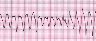
— The required discharge energy to stop the rhythm disturbance is selected depending on the type of arrhythmia: for supraventricular tachycardia and atrial flutter - 50 J, for atrial fibrillation - 75 J, for ventricular tachycardia - 100 J, for the diagnosis of polymorphic ventricular tachycardia or ventricular fibrillation - 200 J .
— If EIT is ineffective, repeat after administration of an antiarrhythmic drug indicated for this arrhythmia.
— After EIT, the heart rhythm is assessed. If the arrhythmia continues, a second shock is given at a higher energy level; if ventricular fibrillation is detected, then defibrillation is performed; if sinus rhythm is recorded, then an ECG is recorded and the patient is hospitalized in a hospital.
results
In most patients, cardioversion can normalize the heart rate. In some patients, the arrhythmia recurs within minutes or days after the procedure. In such cases, if possible, the procedure is repeated to restore normal rhythm.
To keep your heart activity normal, you will need to make changes to your usual lifestyle. This is necessary to improve general health and to prevent or treat pathological conditions that cause arrhythmias. These include, for example, high blood pressure.
Your doctor will advise you to:
- Eliminate caffeine from your diet
- Eat less table salt (sodium). This will help lower your blood pressure levels
- Spend more time on physical activity
- If you smoke, then quit
- If you drink alcohol, don't drink more than one drink a day if you're a woman and no more than two drinks a day if you're a man.
- Eat healthy, heart-healthy foods and maintain a healthy weight.
Indications for hospitalization
All patients who experienced clinical death in the prehospital stage should be urgently hospitalized after hemodynamic stabilization. Hospitalization should be carried out with reliable venous access, with the obligatory possibility of performing EIT during transportation. Patients who have experienced clinical death are transferred “hand to hand” to the resuscitator on duty.
After the procedure
Electrical cardioversion does not require further hospitalization. You can go home the same day. You will spend about an hour in a special ward, where staff will monitor your condition to prevent complications.
Because sedatives are used during the procedure, your consciousness will remain clouded. You will need a chaperone who can take you home. Your reaction will remain slow for several hours after the procedure.
Even if no blood clots were found in your heart before cardioversion, you will need to take blood thinners after the procedure. This is necessary to prevent the formation of new blood clots.
Safety precautions
Persons who have studied the safety rules when working with electronic medical devices are allowed to work with a defibrillator-cardioverter. Remember, a defibrillator is a device of increased, and sometimes fatal, danger! During EIT, it is important not only to help the patient, but also to protect yourself and others. The discharge of the released electrical energy can cause fatal rhythm disturbances in the rescuer or in the person who inadvertently touched the patient.
Necessary:
- remove all strangers (neighbors, relatives, etc.) from the premises;
- wipe the patient’s chest dry;
— remove oxygen from the defibrillation zone.
Prohibited:
— Hold both electrodes of the device in one hand!
— Charge the defibrillator if the electrodes are not placed on the patient’s chest!
— Direct or indirect contact with the patient during EIT!
How to use a defibrillator
1. Turn on the defibrillator.
2. Apply a sufficient amount of conductive gel to the electrodes. If there is not enough gel under the electrodes, burns will occur.
3. Select the required energy level.
4. Install electrodes: one - under the right clavicle marked Sternum, the second - above the area of absolute cardiac dullness marked Apex; with force (10 kg) press the electrodes to the patient’s chest.
5. Give the command “Get energy!”
6. Avoid touching the patient while the shock is being applied.
7. Give a loud command “Discharge!” Perform a discharge by simultaneously pressing both trigger buttons on the electrodes.
8. Check the result of the EIT. Register an ECG.
9. If necessary, resolve the issue of repeated discharge.
Complications of cardioversion include: ventricular fibrillation; aspiration of gastric contents; laryngospasm; hypoventilation; skin burns; electric shock to medical personnel.
Electropulse therapy in the treatment of arrhythmia
Defibrillation and cardioversion are types of electrical impulse therapy. Despite all their similarities, they have some differences. Defibrillation is the process of stopping ventricular fibrillation by applying an electrical discharge; it is the most important resuscitation measure. Cardioversion is a method of treating tachyarrhythmias, which is based on stopping the circulation of excitation in the myocardium by applying an electrical discharge at a certain phase of the cardiac cycle. Cardioversion requires synchronization - applying an impulse at the moment of registration of the R wave, since otherwise, applying a discharge in another phase of the cardiac cycle can lead to ineffectiveness of the procedure and even to the development of ventricular fibrillation. Cardioversion can be planned, when the rhythm is restored with stable hemodynamic parameters if other treatment methods are ineffective, and emergency - with paroxysms with unstable hemodynamics, with ventricular tachycardia without a pulse (in the latter case, it is carried out without synchronization and is equivalent to defibrillation).
Electrical methods for treating arrhythmias have been known since the beginning of the second half of the 18th century. The first officially documented case of using electrical impulses to help with sudden death dates back to 1774, when Mr. Squires, a resident of London, tried to help a three-year-old girl who had fallen from the first floor, using discharges of electricity from Leyden jars. Over the next few days, the girl experienced stupor, but after about a week she was completely healthy.
Defibrillation was subsequently studied by Luigi Galvani, Charles Kight, John Snow, Jean-Louis Prevost and Frederic Batelli and other scientists. In 1947, American surgeon Claude Beck performed successful defibrillation during heart surgery on a fourteen-year-old boy. Developed by Claude Beck, the defibrillator operated on alternating current and allowed only open defibrillation.
Laying the scientific foundations for understanding EIT, as well as the first serious experiments in this area, were carried out by Paul Zoll. While studying cardiac pacing, he hypothesized that the application of a strong external electrical shock could interrupt ventricular fibrillation, and already in 1956 Zoll and his colleagues made the first clinical demonstration of successful transthoracic defibrillation. In his research, he used a self-designed defibrillator that generated alternating current. In 1960, Bernard Lown developed his first DC defibrillator. This defibrillator was the first in a line of modern devices of this type. Lown also proposed the method of cardioversion - the use of electrical discharges synchronized with the cardiac cycle to treat tachyarrhythmias.
Preparation for planned EIT
- If AF lasts more than 48 hours and there is no adequate anticoagulant therapy during the last 3 weeks, before restoring sinus rhythm with ECV, preliminary transesophageal echocardiography is necessary to exclude intra-atrial thrombosis.
- All patients should refrain from eating for 6-8 hours.
- Cancel cardiac glycosides 3–4 days before the procedure
- Normalization of electrolyte balance (EIT for hypokalemia is less effective and is more often complicated by ventricular fibrillation)
EIT methods
External EIT is the main method. Both electrodes are placed on the chest so that the heart is covered by the electric field of the capacitor discharge. The ERC and AHA guidelines establish recommended energy values for the first shock during defibrillation. They are (for adults): when using a monopolar impulse - 360 J, when using a bipolar impulse - 120-150 J., in children, shocks are used at the rate of 2 J / kg of body weight. When carrying out defibrillation, the anterior or standard arrangement of electrodes is now predominantly used; the electrodes must be lubricated with a special conductive gel, and care must be taken that it does not spread over the surface of the chest between the electrodes. It is permissible to use wipes moistened with saline solution. During the procedure, one electrode marked “Apex,” or red (positive charge), is placed exactly above the apex of the heart or below the left nipple; another electrode labeled “Sternum,” or black (negative charge), is placed just below the right collarbone. An anteroposterior arrangement of electrodes is also used - one electrode plate is located in the right subscapular region, the other in front above the left atrium. There is also a postero-right subscapular location of the electrodes. The choice of electrode location depends on the specific situation; the benefit or harm of any of the described locations has not been proven.
Before administering a shock, make sure that no one touches the patient or the bed on which he is lying. Modern control and diagnostic equipment is protected from defibrillator impulses. At the moment the discharge is applied, the monitor readings change and the patient’s reaction is noted - muscle contraction, shuddering, and sometimes a scream. It is strictly forbidden to touch the patient or objects in contact with him at the time of delivering the shock, as this is dangerous for the personnel. After the discharge is completed, the monitor readings are assessed and, if necessary, the issue of repeated discharge is decided.
If the patient is conscious, then general anesthesia is mandatory. The objectives of general anesthesia during cardioversion are to ensure the loss of consciousness for a short period of time and provide amnesia for the period of the manipulation. As a rule, they are limited to the use of short-acting hypnotics in small doses administered intravenously (thiopental 100-250 mg or propofol 50-100 mg).
Internal EIT – electrodes are applied directly to the heart. In this case, a significantly smaller discharge value is required (for an adult patient, about 500 V or 12.5–25 J).
Transesophageal EIT - one of the electrodes is inserted into the esophagus to the level of the atria, the other is placed in the precordial region. Discharge energy 12–25 J. Transesophageal EIT is indicated for severe supraventricular tachyarrhythmias that are resistant to transthoracic discharges, as well as for suppressing severe ventricular tachyarrhythmias with low-energy discharges.
Transvenous intracardiac EIT using a multipolar electrode, which is installed in the right ventricle, is used in intensive care units for recurrent ventricular tachycardia. The discharge energy during endocardial EIT varies from 2.5 to 40 J. To relieve atrial fibrillation, intracardiac EIT can also be used, which can be of two types: high and low energy. When using high energy (200–400 J), one electrode is placed in the right atrium, the other on the surface of the body. Efficiency up to 100%. When using low energy 2–4.5 J, one electrode is placed in the right atrium, the other in the coronary sinus.
Complications of cardioversion
ECV may be complicated by thromboembolism and arrhythmias, and complications of general anesthesia may also occur. The incidence of thromboembolism after defibrillation is 1-2%. It can be reduced by adequate anticoagulation before elective cardioversion or by ruling out left atrial thrombosis. A common complication is skin burns. Patients with sinus node dysfunction, especially older adults with organic heart disease, may develop prolonged sinus arrest. Dangerous arrhythmias such as ventricular tachycardia and ventricular fibrillation may occur in the presence of hypokalemia, cardiac glycoside toxicity, or inadequate timing. The use of anesthesia may be accompanied by hypoxia or hypoventilation, but arterial hypotension and pulmonary edema are rare.
Electrical cardioversion in patients with implanted cardiac pacemakers and defibrillator
It is clear that the presence of such a device in a patient somewhat changes the technique of the procedure, but is by no means a contraindication to external defibrillation. If the patient has an implanted pacemaker-cardioverter, then the position of the electrodes should be slightly changed. The electrode for external cardioversion must be located at a distance of more than 6-8 cm from the implantation site of the pacemaker or cardioverter-defibrillator. Anterior-posterior placement of electrodes is recommended. It is preferable to use a biphasic defibrillator, since in this case a lower energy discharge is required to stop AF. In pacemaker-dependent patients, it is necessary to take into account a possible increase in the stimulation threshold. Such patients should be closely monitored. After cardioversion, the implanted device should be tested using an external programmer.
Recurrence of arrhythmia after electrical cardioversion
Factors predisposing to recurrent AF include age, duration of AF before cardioversion, number of previous relapses, enlarged left atrium or decreased left atrium function, presence of coronary artery disease, pulmonary disease, or mitral valvular disease. Atrial extrasystoles with changing coupling intervals and the so-called early extrasystoles “P” to “T”, sinus tachycardia, intra-atrial and inter-atrial conduction disorders also increase the risk of relapse of AF. Antiarrhythmics given before cardioversion increase the likelihood of restoration of sinus rhythm and reduce the risk of immediate and early relapses. To prevent late relapses, constant long-term use of antiarrhythmic drugs is necessary. The most effective means of such prevention is amiodarone, which is superior in its effectiveness to all other means of antiarrhythmic therapy. 69% of patients maintain sinus rhythm within a year of amiodarone use. For sotalol and propafenone this figure is 39%. Some patients whose episodes of AF occur with severe clinical symptoms, but do not recur frequently (1-2 times a year), prefer repeated cardioversion to long-term anti-relapse antiarrhythmic therapy or treatment aimed at reducing heart rate in conditions of persistent arrhythmia.
Automatic external defibrillators and the concept of early defibrillation
In this regard, the concept of early defibrillation using a “publicly available defibrillator-monitor” has recently become increasingly popular among specialists. According to this concept, automatic defibrillators should become publicly available, allowing even an unqualified user to provide first aid to a patient with cardiac arrest before the arrival of a medical team. Several case reports of successful defibrillation at airports have already been published. At two Chicago airports, automatic defibrillators are located throughout the terminal and in the baggage area. All airport personnel, including security, are trained to use defibrillators and have the appropriate certificates. As a result of this organization of assistance, 69% of passengers who suffered cardiac arrest as a result of ventricular fibrillation at the airport survived. Thus, only early defibrillation is the only chance in these situations to restore hemodynamically effective cardiac contractions and save the patient.
Selecting a device for EIT
A wide range and price differences make choosing a device for EIT a difficult task. First of all, you need to decide who will use this device: a paramedic, an emergency rescue worker or a doctor. An AED is a necessary tool for performing high-quality CPR, which does not require extensive personnel training. The price is determined by the quality of electronic components and batteries. Professional cardioverter defibrillators are multifunctional devices that are equipped with an ECG monitor, a built-in thermal printer, an external pacemaker module, and a pulse oximeter for determining SpO2 saturation.
It is necessary to clarify the availability of children's electrodes. Remember that modern devices for EIT generate a biphasic pulse shape, and monophasic defibrillators have not been produced since 2005.
There are no contraindications to the use of emergency cardioversion in a critically ill patient.