Local therapist
Burnatskaya
Svetlana Nikolaevna
33 years of experience
Local therapist, occupational pathologist. Member of the Russian Scientific Medical Society of Therapists
Make an appointment
Agranulocytosis is a severe pathological condition. With it, there is a critical decrease in granulocytes in the blood. Granulocytes are an important fraction of the leukocyte series (neutrophils, eosinophils and basophils). At the same time, the concentration of basophils and eosinophils in the blood in the normal state is a small percentage, so changing their number does not have a significant effect. Thus, it can be argued that it is the decrease in the concentration of neutrophils that leads to the development of blood agranulocytosis. Hence the second name of the pathology – neutropenia.
Experts raise the question of agranulocytosis in a patient when the leukocytes in the blood decrease below 1×10⁹/l and granulocytes below 0.75×10⁹/l. Since leukocytes and granucolites perform a protective function against pathogens, a decrease in their concentration reduces the body’s ability to resist infections and viruses. The development of agranulocytosis is almost always accompanied by the development of various infectious processes, these can be ulcerative stomatitis or tonsillitis, pneumonia, hemorrhagic manifestations, etc.
Depending on the concentration of granulocytes, the severity of peripheral blood pathology is determined. The lower their concentration, the more severe the degree; there are three degrees: mild, moderate and severe. In severe cases, the granulocyte concentration is below 0.4 × 10⁹/l.
In women, agranulocytosis is more common. In men, this disease is diagnosed on average 2-3 times less often. In most cases, the manifestation of pathology occurs after the age of 40 years.
Etiology and pathogenesis
According to the mechanism of occurrence, AGRANULOCYTOSIS can be myelotoxic and immune.
Myelotoxic
AGRANULOCYTOSIS occurs as a result of suppression of the growth of prestage granulocytes in the bone marrow, including stem cells. In this regard, there is a decrease in the blood not only of granulocytes, but also of platelets, reticulocytes and lymphocytes. Myelotoxic AGRANULOCYTOSIS can develop as a result of exposure of the body to ionizing radiation, chemical compounds with cytostatic properties (antitumor drugs, benzene and others), waste products of a Fusarium type fungus that multiplies in overwintered grain (see Aleukia alimentary-toxic).
Immune
AGRANULOCYTOSIS develops as a result of accelerated death of granulocytes under the influence of anti-leukocyte antibodies; stem cells are not affected. Anti-leukocyte antibodies are formed under the influence of medications that can play the role of haptens (see Haptens). With repeated administration of such a medication, agglutination of leukocytes appears.
The development of immune agranulocytosis depends little on the dose of the drug; the most important role in its occurrence is played by the unusual sensitivity of the body. On the contrary, in myelotoxic AGRANULOCYTOSIS the decisive role is played by the magnitude of the damaging effect. Among the drugs that cause immune agranulocytosis, the main place belongs to amidopyrine. Immune agranulocytosis can also be caused by butadione, phenacetin, atophan, analgin, diacarb, barbamyl, sulfonamides, PAS, tubazide, ethoxide, streptomycin, pipolfen and some other drugs. Long-term use of the hapten drug can cause the destruction of not only mature granulocytes, but also myelocytes and promyelocytes. The development of autoimmune AGRANULOCYTOSIS, more often leukopenia, is observed with collagenosis (especially disseminated lupus erythematosus, rheumatoid polyarthritis), as well as with some infections.
A special place is occupied by AGRANULOCYTOSIS with systemic lesions of the hematopoietic apparatus - leukemia (see), hypoplastic anemia (see), as well as with metastases to the bone marrow of a cancerous tumor and sarcoma.
What is agranulocytosis
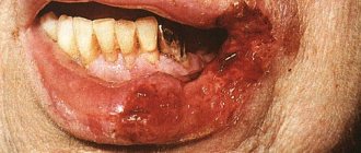
The agranulocytic type of sore throat is one of the manifestations of agranulocytosis, which is quite rare.
Make an appointment right now!
Call us by phone or use the feedback form
Sign up
With agranulocytosis, there is a sharp decrease in the number of granular leukocytes in the blood (up to their complete disappearance). Leukocytes, as we know, are the main defenders of our body from viruses and bacteria that have penetrated it. As soon as the infection enters the body, the immune system receives a signal about the invasion, and increased production of leukocytes begins, which capture the “enemy” and destroy it. Of course, if there are very few white blood cells in the blood, a person becomes much more susceptible to the effects of pathogens.
The manifestation of agranulocytic tonsillitis cannot be considered an independent disease. This is a syndrome that occurs against the background of certain diseases. It is impossible to distinguish this type of tonsillitis from others “by eye”; laboratory tests must be carried out.
Most often, the disease manifests itself in people under the age of forty. Children and young people are diagnosed with this diagnosis quite rarely. It is noteworthy that women suffer from the disease twice as often as men.
The main reason for the development of the disease is the incorrect prescription of antibacterial agents and cytostatics (drugs whose action is aimed at inhibiting and suppressing the process of cell division; most often these drugs are used in the treatment of malignant neoplasms).
Pathological anatomy
Pathological signs of agranulocytosis are necrotic-ulcerative changes, most often found in the oral cavity and pharynx. The tonsils are enlarged, loose, gray-dirty in appearance, with fibrinous deposits and ulcerations. In the area of the soft and hard palate, foci of necrosis are found, sometimes with perforation of the soft palate. Necrotic changes are detected in the skin, at injection sites, in the perineal area, and around the anus. Foci of necrosis are described in the conjunctival sac, in the mucous membrane of the larynx, esophagus and stomach. With the development of necrosis in the mucous membrane of the small or large intestine, including the appendix, intestinal bleeding and perforation are observed. Necrotic ulcers can be in the wall of the bladder, in the genitals, especially in the vaginal wall, as well as in the tissue of the liver and other organs. Microscopic examination shows that there are no neutrophilic leukocytes in areas of necrosis. The demarcation strip around the necrosis is not detected; lymphohistiocytic and plasma cell accumulations can be seen near the areas of necrosis. Pneumonia is fibrinous-hemorrhagic in nature. In this case, fibrinous deposits are also located on the pleura. In the area of pneumonia, areas of tissue decay (gangrene) may be detected. Microscopically, desquamated epithelial cells, bacteria, yeast cells and their mycelium are visible in the lumens of the alveoli. Lymph nodes are usually not enlarged. With necrotic changes in the oral cavity, a slight enlargement of the cervical and submandibular lymph nodes may be observed. Their microscopic structure is relatively preserved. In the cortex and especially in the area of the medullary cords, a large number of plasma cells are detected. Proliferation and swelling of reticuloendothelial cells in the sinuses is sharply expressed. The spleen is often unchanged. The spleen tissue is of a soft consistency; on the section there is a pink-gray pulp with a large scraping. Microscopic examination reveals a uniform decrease in the number of red pulp cells of the spleen. The bone marrow of flat bones is macroscopically more common than usual, somewhat dry, but there may be various foci of hemorrhage - from small to extensive; in the lower and middle third of the tubular bones, the bone marrow is fatty.
Microscopically, small foci of resorption of bone beams with the formation of small lacunae are revealed. Osteoblast proliferation may be observed in areas of bone resorption. The ratio of adipose and hematopoietic tissue is different. More often there is a decrease in the number of hematopoietic cells and an increase in the number of fat cells in the bone marrow. In the cellular composition, attention is drawn to a sharp decrease in the number of young, band and segmented granulocytes. There may be some predominance of young forms of granulocytes. Megacarpocytes and red row cells are usually preserved. In the most severe course of AGRANULOCYTOSIS, the picture of the bone marrow is the same as in hypoplastic anemia (see).
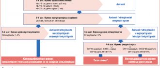
Tests and diagnostics
Doctors may suspect neutropenia in people who have frequent, severe infections or unusual infections. This condition is also highly likely to occur in patients undergoing radiation therapy and receiving cytotoxic drugs.
- Anamnesis must be taken into account: the doctor must know what medications or toxic substances the patient took.
- Granulocytopenia is determined by conducting a laboratory blood test to determine the leukocyte formula. A blood test can detect agranulocytosis.
- Culture may also be done to determine infection.
- However, in the diagnostic process, it is important not only to determine the presence of this phenomenon, but also to find out the mechanism and reason for its development. For this purpose, the doctor conducts a physical examination and questioning of the patient.
- A urine and stool test is performed.
- An informative method of investigation is chest radiography.
- People who are immunocompromised have a chest CT scan.
- Other tests are also ordered depending on signs of infection.
Bone marrow testing is performed to determine whether the development of neutropenia is due to decreased production of neutrophils by the bone marrow.
Clinical picture
Myelotoxic AGRANULOCYTOSIS begins gradually: without any subjective signs. Immune Agranulocytosis can clinically have different variants depending on the factor that caused it. AGRANULOCYTOSIS against the background of collagenosis develops gradually and is characterized by persistent progression. The onset of drug-induced immune AGRANULOCYTOSIS is acute in most cases.
The first manifestations of any AGRANULOCYTOSIS are fever, stomatitis, and sore throat. With myelotoxic syndrome, there is also usually a moderately severe hemorrhagic syndrome (bruising and bleeding gums, nosebleeds): severe hemorrhagic syndrome is an infrequent complication of AGRANULOCYTOSIS. Damage to the mucous membranes (necrosis and thrush) of the oral cavity and gastrointestinal tract is the most constant sign of AGRANULOCYTOSIS. In myelotoxic agranulocytic, it is due to the fact that, on the one hand, the disappearance of granulocytes makes microbial invasion possible, on the other hand, the suppression of mitoses of epithelial cells of the mucous membrane by cytostatic factors violates its integrity.
In the peripheral blood, the number of all forms of leukocytes decreases (often up to hundreds of cells in 1 μl), as well as platelets and reticulocytes. The number of plasma cells is usually increased. There may be anemia. Sometimes granulocytes disappear completely. Leukopenia in immune AGRANULOCYTOSIS is moderate - 1000-2000 cells per 1 μl, but the number of granulocytes, as a rule, decreases to zero; thrombocytopenia is absent. Anti-leukocyte antibodies are detected in the serum.
At the height of myelotoxic AGRANULOCYTOSIS, both granulocytic elements, erythronormoblasts, and megakaryocytes almost completely disappear in the bone marrow; lymphoid, reticular and plasma cells are preserved. 2-3 days before exiting the state of AGRANULOCYTOSIS, promyelocytes and single normoblasts appear in the bone marrow in huge numbers. In peripheral blood, the first sign of activation of hematopoiesis is the detection of young elements - myelocytes and metamyelocytes, sometimes plasma cells. Often 2-3 days before the appearance of granulocytes, the number of platelets and reticulocytes increases.
In the bone marrow with immune AGRANULOCYTOSIS, a decrease in cellular elements is observed exclusively due to the granulocytic germ. Exit from immune AGRANULOCYTOSIS is characterized by the appearance in the peripheral blood of young cells - myelocytes, metamyelocytes, and sometimes promyelocytes.
The duration of AGRANULOCYTOSIS varies and depends both on the degree of bone marrow damage and on the individual characteristics of the patient’s body.
Diagnosis of agranulocytosis in patients
If the patient is at risk or corresponding symptoms appear without other visible causes, agranulocytosis is diagnosed using the following methods.
- A general blood test is the very first and most important test that allows you to determine the concentration of granulocytes and leukocytes in the bloodstream.
- Antibody test - prescribed to identify autoimmune agranulocytosis, allows you to determine antineutrophil antibodies.
- Myelogram - allows you to determine the decrease in myelokaryocytes and other indicators of the condition of peripheral blood.
If necessary, when diagnosing agranulocytosis, other research methods are prescribed to determine the degree of damage to organs and systems caused by this pathology. In particular, the following may be prescribed:
- X-rays of the lungs;
- visiting a dentist;
- blood chemistry;
- blood sterility tests;
- consultation with an otolaryngologist.
Complications
The most common are sepsis (often staphylococcal), intestinal perforation (usually the ileum, since it is more sensitive to cytostatic effects), mediastinitis, pneumonia, noma; less often - severe swelling of the intestinal mucosa with the formation of obstruction and peritonitis. A serious complication is acute epithelial hepatitis, which often develops after the elimination of AGRANULOCYTOSIS. The absence of granulocytes gives a unique character to the course of infectious complications - the absence of ulcers, the predominance of necrosis. Pneumonia occurs against the background of scanty physical data: dullness is barely noticeable, there may be no wheezing or pronounced bronchial breathing, only crepitus is heard over the affected area. X-ray changes are very scarce.
Neutropenia in children
Neutropenia in children under one year of age and older can occur due to various reasons. Pediatrician Komarovsky and other well-known experts note that this condition can develop as a result of severe viral, bacterial and fungal lesions, as well as exposure to radiation, radiation therapy, and toxic damage. Neutropenia in infants can also be associated with genetic disorders, as well as with treatment with drugs that interfere with hematopoiesis. A rare condition is congenital agranulocytosis , where a child develops severe immunodeficiency .
As a rule, severe symptoms of this condition do not appear in children, so most often the problems are determined only after a blood test. But if agranulocytosis is not detected for a long time, then a number of symptoms associated with intoxication of the body develop. Therefore, as pediatrician Komarovsky and other doctors note, it is important to monitor the condition of children who have an increased risk of developing neutropenia, and take timely measures to normalize the body’s condition in order to avoid complications.
Diagnosis
The diagnosis is made on the basis of anamnestic data, a characteristic clinical picture, data from a study of peripheral blood and bone marrow puncture. The diagnosis of immune AGRANULOCYTOSIS can be confirmed by serological studies: detection of anti-leukocyte antibodies (see Blood groups, leukocyte groups). AGRANULOCYTOSIS must be differentiated from acute leukemia in the aleukemic stage and hypoplastic anemia. In the first case, the decisive factor in diagnosis is the study of bone marrow puncture, the second is an indication of the suddenness of the development of AGRANULOCYTOSIS due to its immune nature or information about long-term use of drugs with myelotoxic action (sometimes interrupted several weeks before the development of AGRANULOCYTOSIS). Observation of the patient allows us to come to a final conclusion about the nature of bone marrow devastation.
Risk factors
The risk group primarily includes patients who are frequently exposed to severe infections, as well as unusual, rare infections. Another risk group is patients who, as part of the treatment of other diseases, receive drugs or therapy (for example, radiation therapy), which can cause the development of agranulocytosis. Patients at risk should be especially attentive to their well-being and pay attention to alarm bells. Accurate laboratory diagnostics are required to confirm the diagnosis.
Treatment
Treatment of A. of any origin requires immediate elimination of the cause that caused it: a cytostatic drug, ionizing radiation, a hapten drug that provoked an immune conflict, and so on. Therapy for AGRANULOCYTOSIS, which complicates the course of rheumatoid arthritis or disseminated lupus erythematosus, should first of all be directed against the underlying disease.
In myelotoxic agranulocytosis, steroid hormones are not indicated. When the number of leukocytes decreases to 50-200 cells per 1 μl, replacement therapy is necessary - transfusion of leukocyte concentrate. More than 15-20 billion cells are transfused at the same time. In cancer patients, a good effect was observed when using leukocyte mass obtained from patients with chronic myeloid leukemia in the advanced stage of the disease. Bone marrow transplantation is used only in acute total irradiation at doses exceeding 600 rads per bone marrow. Along with the use of pathogenetic agents, pisymptomatic therapy is used: in the case of persistent hyperthermia up to 39-40° with myelotoxic AGRANULOCYTOSIS (but not immune!), patients are given analgin up to 2-3 g per day or acetylsalicylic acid - 2 g per day.
In the treatment of immune AGRANULOCYTOSIS, the decisive role belongs to prednisolone at a dose of 1-1.5 mg per 1 kg of weight or its analogues in adequate doses, and in the absence of effect - 2-3 times more. In case of severe damage to the mucous membranes, the drug is administered parenterally. Course - 7-10 days or until agranulocytosis is eliminated. Replacement therapy with leukocyte transfusions is indicated only for infectious complications.
To prevent infection, patients must be placed in boxes or isolation wards where aseptic conditions are created (see Medical ward). Prevention of infectious complications with antibiotics is necessary when the number of granulocytes drops to 750 in 1 μl. The non-absorbable antibiotic neomycin is prescribed orally up to 2-3 g per day (against E. coli), polymyxin B 150-200 mg per day (against Pseudomonas aeruginosa). In addition, a broad-spectrum antibiotic is prescribed - garamycin intramuscularly 40-80 mg 2-3 times a day, or oxacillin 4-6 g per day orally, or oletethrin up to 2 g per day orally. Be sure to take nystatin up to 100,000 units per day. For systematic rinsing of the mouth, use a solution of levorin 1: 500. Ulcers on the mucous membrane of the mouth and lips are lubricated with sea buckthorn oil. With the development of staphylococcal sepsis, the administration of antistaphylococcal gamma globulin and antistaphylococcal plasma is indicated. If diarrhea, bloating, or persistent severe pain in the abdomen occurs, the patient must be transferred to parenteral nutrition, and surgical supervision becomes necessary. Perforation of intestinal ulcers, characterized by mild signs of irritation of the peritoneum, increasing bloating, loss of intestinal motility, and the appearance of effusion in the abdominal cavity, makes urgent surgical intervention necessary.
Prevention of myelotoxic AGRANULOCYTOSIS consists of careful hematological monitoring during therapy with cytostatic drugs. If the number of leukocytes decreases to 1000-1500 per 1 μl or their rapid decrease, it is necessary to stop cytostatic therapy or take a break in treatment. However, with leukopenic variants of acute leukemia, therapy must be carried out with small or medium doses of cytostatic drugs, despite the low numbers of leukocytes, focusing on their dynamics and platelet levels. A persistent decrease in the latter indicates the need for a break in cytostatic treatment. Prevention of immune AGRANULOCYTOSIS is aimed at avoiding repeated use of drugs that previously turned out to be the cause of AGRANULOCYTOSIS in a given patient.
Symptoms and signs
Since the pathology has several forms, the symptoms of agranulocytosis of different types differ. The immune type is characterized by acute agranulocytosis with the following symptoms:
- severe weakness and sweating;
- body temperature up to 39-40°C;
- pallor;
- the appearance of infectious stomatitis;
- inflammation of the pharynx and tonsils;
- inflammation of the gums;
- sore throat and spasm of the chewing muscles.
Myelotoxic and autoimmune types progress gradually and also gradually develop symptoms:
- hemorrhagic symptoms in the form of nosebleeds, bleeding gums, etc.;
- presence of blood in the urine;
- the occurrence of bruises, hematomas;
- abdominal pain and bloating, diarrhea if the intestines are affected;
- possible occurrence of chest pain when breathing.