Diseases that start with the letter “M”: Lesser chorea, Slow infections of the central nervous system, Medulloblastoma, Herniated disc, Thoracic disc herniation, Lumbar disc herniation, Cervical disc herniation, Intercostal neuralgia, Meningeal syndrome, Meningioma, Meningitis, Meningococcal meningitis, Meningomyelitis , Meningoencephalitis, Metabolic myopathy, Metastatic brain tumors, Metachromatic leukodystrophy, Weather sensitivity, Myasthenia gravis, Migraine stroke.
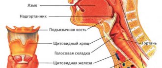
Meningoencephalitis is a disease of a neuroinfectious nature, occurring with simultaneous damage to the substance of the brain and its membranes. It is expressed by variable focal, membrane, infectious symptoms. The diagnosis is made on the basis of a neurological examination, assessment of cerebrospinal fluid, MRI and CT results of the brain, and examinations designed to identify the pathogen. Complex treatment consists of prescribing antibacterial, antiviral, antifungal drugs in combination with symptomatic and pathogenetic drugs.
Etiology
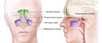
The main cause of pathology is infection. Neurotropic pathogens enter the brain areas, thus causing primary infection. Secondary infection is the result of the expansion of the process from infected foci (sinusitis, otitis), with general processes (rubella, influenza). The cause of the disease is bacteria and viruses, sometimes pathogenic fungi. Infection occurs when:
- Entry of the pathogen into the nasopharynx through alimentary or airborne droplets. Through the bloodstream, it is possible to bring the pathogen into the cranial cavity, where it causes inflammatory changes and the further formation of meningoencephalitis.
- Insect bite (tick, mosquito). Viral meningoencephalitis and encephalitis have a transmissible transmission route (tick-borne encephalitis, Japanese mosquito encephalitis, St. Louis encephalitis). The pathogen is released from an insect bite into the blood and enters the brain, which causes the development of meningoencephalitis.
- Presence of infection in the body. Meningoencephalitis can become a complication of tuberculosis, purulent maxillofacial diseases, and syphilis. In some cases, it develops as a consequence of certain acute respiratory viral infections.
- Traumatic brain injury. Contact infection occurs with open injuries with skull fractures. Meningoencephalitis, as a post-traumatic complication, occurs in 1.3-3.5% of patients.
- Vaccination. The infection develops when the immune system is weakened, after the administration of a live vaccine. The pathogen passes the blood-brain barrier and causes meningoencephalitis.
- The pathogen does not always lead to the development of the disease. This becomes possible in the presence of a weakened, immature immune system, a weakened body, or the presence of initial or secondary immunodeficiency.
Meningoencephalitis: ICD code 10
ICD-10 is a normative document with a generally accepted statistical classification of medical diagnoses that is used in healthcare.
According to the international classification, meningoencephalitis is included in the following codes:
- G04.2 - Bacterial meningoencephalitis and meningomyelitis, not elsewhere classified;
- G05 - Encephalitis, myelitis and encephalomyelitis in diseases classified elsewhere. Includes: meningoencephalitis and meningomyelitis.
Development of the disease
Inflammation is a response to the introduction of a pathogen into brain tissue. Its purulent or serous nature is associated with the type of infectious factor. An inflammatory focus forms around blood vessels, which causes cerebrovascular accident. As a result, secondary tissue ischemia and a compensatory increase in the production of cerebrospinal fluid occur, which affects the development of intracranial hypertension.
The membranes of the brain damaged by infection become irritated and cause meningeal syndrome. Inflammatory foci can have varying degrees of distribution in the cerebral substance. Focal manifestations are formed due to neurological insufficiency of brain functions. Persistent neurodeficiency is the result of massive death of brain neurons.
Classification
Neurology divides meningoencephalitis according to the type of etiology, the nature of the changes caused, and the type of development. Confirmation of the type of disease occurs at the diagnostic stage, which is a central factor in choosing treatment tactics.
Etiological types of disease:
- Viral—the source includes herpes simplex, influenza, measles, rabies, cytomegalovirus, and enterovirus viruses. It has a serous type of inflammatory consequences.
- Bacterial - has a purulent inflammatory nature caused by pneumo-, strepto-, meningococci, Haemophilus influenzae, Klebsiella.
- Protozoan—a rare species caused by protozoa (amoeba, toxoplasma).
- Fungal—diagnosed mainly in individuals with secondary immune deficiency. Often accompanies the development of neuroAIDS.
According to the type of inflammatory course, meningoencephalitis is divided into:
- Serous - characterized by the formation of serous discharge. Blood lymphocytosis is typical. The cerebrospinal fluid is clear.
- Purulent—pus forms, which causes clouding of the cerebrospinal fluid. Leukocytosis is noted.
- Hemorrhagic—multiple petechial hemorrhages occur in the brain tissue, caused by impaired permeability of the vascular walls.
By type of clinical course:
- Lightning—forms within a matter of hours, in most cases ending in the death of the patient.
- Acute—develops from 1 to 2 days, the clinical picture worsens more slowly than with the fulminant form.
- Subacute—occurs in stages, symptoms worsen over several days, but not more than a week.
- Chronic—inflammation develops over several months with periods of remissions and exacerbations. Acute and subacute forms can become chronic.
Characteristics of autoimmune encephalitis
The formation of antibodies to brain tissue causes demyelination. The process is long but progressive. Rasmussen's encephalomyelitis is a typical manifestation of an autoimmune lesion of the cerebral parenchyma. Depending on the characteristics of the development of the process, the duration is from five to fifteen years. In most cases, the clinical peak occurs at six years of age.
Nosology has been thoroughly studied by scientists. The causes of the occurrence could not be established, but the link to which immunoglobulins are formed was identified. The presence of NMDA receptors is a weak point that is susceptible to destruction by the immune system.
There are case studies showing the nonspecificity of glutamate receptor antibodies for Rasmussen's encephalomyelitis. Other anti-inflammatory cytokines formed during the nosology have been identified.
Clinical picture of the disease
The clinical picture of meningoencephalitis combines general infectious, meningeal, liquor-hypertensive, and focal symptoms. Infectious manifestations include high body temperature, a general painful condition, decreased appetite, and sometimes skin rashes. Against the background of an ongoing infection, signs of brain damage appear.
Increased pressure in the cerebrospinal fluid causes severe headache, nausea, and vomiting, which does not provide relief. Increasing intracranial pressure leads to a disorder of consciousness with disorientation, mental agitation or, on the contrary, retardation. In the fulminant form, the patient often falls into a coma.
Focal neurological failure depends on the location and type of inflammation and is expressed to varying degrees. Manifests itself in the form of hemiparesis, sensorimotor aphasia, hyperkinesis, cerebellar syndrome, vestibular ataxia, and cognitive impairment. Oculomotor and visual disorders, ptosis, facial distortion, swallowing disorders, and dysarthria are the result of damage to the cranial nerves.
Clinical analysis. Herpetic meningoencephalitis in patients over 20 years of age.
Authors: prof. Doctor of Medical Sciences Shmyrev V.I., prof. Doctor of Medical Sciences Devyatkin A.V., Ph.D. Kalenova I.E., Gavrilov D.Yu., Sharinova I.A., Litvinov N.I.
Introduction The causative agent of herpetic meningoencephalitis can be herpes simplex virus type 2 (HSV-2) and the varicella zoster virus. The mortality rate for this pathology is 15-20%, and without antiviral therapy 70%. Patients who survive often have persistent neurological deficits. The most common forms of herpetic lesions of the central nervous system are encephalitis, which can occur both against the background of other organ lesions (generalized cutaneous-visceral form) and in isolation. Herpetic encephalitis (HE) occurs with a frequency of 2-4 per 1 million population per year. Children account for about a third of all cases. PGs have dermatoneurotropism. This means that they primarily affect the skin, mucous membranes with stratified epithelium, eyes and the central nervous system. In the latter case, the most severe, life-threatening pathological processes develop in the form of encephalitis, meningoencephalitis, meningoencephalomyelitis, etc. GE can develop in connection with both the reactivation of a latent infection in the brain (according to modern concepts, in approximately 2/3 of patients), and with exogenous infection with a highly virulent strain of the virus (in 1/3 of patients). HSV can penetrate the central nervous system hematogenously or through nerve trunks (mainly through the branches of the trigeminal nerve and the olfactory tract). It has already been proven that they spread mainly through the neuronal route. From the Gasserian ganglion, the virus enters the subcortical nuclei, brainstem nuclei, thalamus and reaches the cerebral cortex. As the virus spreads along the olfactory tract, the hippocampus, temporal gyri, insula and cingulate gyrus (i.e. limbic system) are affected, and then in most cases the midbrain, brain stem and hemispheres are captured. In terms of clinical manifestations, HE is a classic example of encephalitis. It is characterized by four main syndromes: the syndrome of impaired consciousness, hyperthermic syndrome, convulsive syndrome and the syndrome of focal disorders. Herpetic encephalitis begins acutely (usually after 1-5 days of ARI clinic) with a sudden increase in temperature (usually more than 39°C), which is poorly reduced even while taking antipyretic drugs. Consciousness is disturbed: at first there may be short-term (for several hours) excitement, which is replaced by lethargy, drowsiness, and lethargy. Subsequently, the depression of consciousness progresses until it is completely lost. More often, against the background of high fever, impairment of consciousness manifests itself in the form of pronounced, deep, persistent depression (coma of varying degrees). Consciousness returns gradually, and after its stable recovery, signs of focal disturbance syndrome remain. In this case, the frontal lobes of the brain are often affected, which is clinically manifested by mnestic-intellectual disorders. The syndrome of focal disorders may also include dysfunction of any cranial nerves with the development of the corresponding clinic. Paresis of the hemiplegia type, asymmetry and loss of reflexes, and the appearance of pathological reflexes are possible. Another feature of GE is persistent convulsive syndrome. Convulsions are often generalized. A characteristic feature of GE is also hyperthermic syndrome, but sometimes so-called “cold” GE are encountered. Mortality in HE before the advent of acyclovir was 70-74%. Today, with timely initiation of adequate etiotropic therapy, it has decreased to 5-6%. However, as already noted, herpetic brain damage is a necrotic process, so after HE there is a high probability of neurological consequences. They can be either temporary or permanent. However, against the background of modern antiviral therapy, not only the mortality rate has decreased, but also the outcomes of GE in surviving patients have improved. Meningitis with herpetic lesions of the central nervous system usually develops against the background of encephalitis, i.e. proceeds as meningoencephalitis (MME). Moreover, inflammation of the membranes of the brain is serous in nature. Isolated damage to the meninges by HSV is rare. It is impossible to make a diagnosis of herpetic meningitis based on clinical signs alone. Special laboratory testing methods are required. However, with prolonged or recurrent serous meningitis, along with other studies, examinations for the presence of HSV are necessary. Description of a clinical case In a neurological department for patients with acute cerebrovascular accidents with intensive care wards, a 28-year-old woman was observed with a diagnosis of “Acute herpetic meningoencephalitis. Brain swelling. Right-sided hemiparesis. Exacerbation of chronic right-sided sinusitis. Diffuse bilateral endobronchitis. Endoscopic surgery on the right maxillary sinus dated April 17, 2010: submucosal vasotomy of the inferior turbinates. Tracheostomy dated April 28, 2010.” History of the disease. At the end of November - beginning of December 2009, the patient was observed as an outpatient with trigeminal neuralgia. At the end of March 2010, the patient traveled to Cuba. The patient noted a deterioration in her condition on April 5, 2010, when nasal congestion appeared. On April 12, 2010, I developed a severe headache. On 04/15/10, the headache intensified, dizziness, disorientation, nausea, vomiting, and hyperthermia appeared (body temperature 38.8 C). She was hospitalized at Clinical Hospital No. 1 in the ENT department with a diagnosis of acute purulent sinusitis. Course of the disease. A 28-year-old patient was admitted to the ENT department on April 17, 2010 due to right-sided sinusitis. CT scan of the paranasal sinuses dated 04/17/10: CT picture of right-sided sinusitis. Slight thickening of the mucosa in the left frontal sinus. Hypertrophy of the inferior turbinates. Deviation of the nasal septum. An emergency operation was performed: submucosal vasotomy of the inferior turbinates. Biopsy of the contents of the right maxillary sinus: areas of edematous and sharply inflamed polyposis-altered mucous membrane, bordered by respiratory epithelium with signs of hypersecretion, were sent. Microbiological examination of swabs from the throat and nose dated April 20, 2010 revealed: Acinetobacter spp. - abundant growth, sensitive to moxifloxacin, vancomycin. On the day of admission, general cerebral symptoms increased, disorientation appeared, the patient was consulted by a neurologist. Neurological status on admission: Stupefaction. Dysphoric, disoriented in place, time and personalities. personality. Rigidity of the nuchal muscles 3 points, Kernig's muscle on both sides, Pupils D=S, live photoreactions, nystagmus in the extreme leads. Tendon reflexes are lively and symmetrical. There are no pathological foot signs. PNP and PKP are performed with intention on both sides, more on the right. A cerebrospinal fluid analysis was taken and serous meningitis was diagnosed (in the cerebrospinal fluid: cytosis more than 800/3 with a predominance of lymphocytes more than 50%, with a slight increase in protein, Pandey’s test is negative). During microbiological examination of the cerebrospinal fluid, no microflora was isolated. In the UAC upon admission: Hb - 116 g/l, erythrocytes - 3.69 x 10¹², platelets - 223, leukocytes - 12.4 x 10⁹, p/i - 14%, s/i - 76%, eo - 1%, base - 0% , lymph-7%, mon-2%, ESR-36mm/h. No changes were detected in the biochemical blood test, coagulogram, and general urine test upon admission. CT scan of the brain upon admission: no pathological changes in the brain were detected. Since April 18, he has been in the intensive care unit due to serous meningitis. Detoxification and decongestant therapy was carried out, despite the treatment, the patient’s condition showed negative dynamics. Focal neurological symptoms appeared (right-sided deep hemiparesis, pathological foot signs), depression of consciousness to the point of coma, and several episodes of tonic-clonic seizures. On April 19, 2010, a repeat MRI of the brain was performed. (see Figure No. 1): In the basal parts of the temporal lobe there is an encephalitic focus measuring 57*39*50 mm. Perifocal edema is not expressed. The median structures are not changed. Conclusion: the pattern of identified changes in the left hemisphere in the temporal region most likely corresponds to inflammatory changes (encephalitis). Differentiate with ischemic changes. On April 19, 2010, a liquor analysis was taken for PCR diagnostics of pathogens of viral neuroinfections and microbiological research. A decrease in cytosis to 610/3 was noted. 04/20/10 Respiratory problems appeared, the patient was transferred to mechanical ventilation. On April 28, a tracheostomy was performed. In a study of cerebrospinal fluid dated April 19, DNA of virus types 1 and 2 was detected. Due to the herpetic etiology of the process, antiviral therapy was started. On 04/27/10, a CT scan of the brain was repeated: the CT picture of inflammatory changes (encephalitis) in both hemispheres is negative, with the presence of areas of cerebral edema in the lesions. The patient was consulted by an infectious disease specialist, a neurosurgeon, but the transfer was refused due to the lack of epidemiological danger to others and the severity of the patient’s condition. Against the background of the therapy, there is a positive dynamic: the level of wakefulness increases to the point of stunning, and he tries to carry out basic commands. In control studies of cerebrospinal fluid - a decrease in cytosis to 147/3 from 04/26/10, lymphocytes about 70%. Analysis of cerebrospinal fluid in the dynamics of Table 1. The level of respiratory support was gradually reduced, and on 05/03/10 the patient was transferred to spontaneous breathing. A consultation was held with the participation of infectious disease specialists, neurologists, and resuscitators to decide on the need to transfer the patient to an infectious diseases hospital. Conclusion: Acute meningoencephalitis caused by herpes simplex virus types 1 and 2 (HSV DNA in the cerebrospinal fluid) with foci of cerebromalacia of the brain. Considering the endogenous nature of the infection, there is no need for isolation. The patient is transferred to the neuroinfection department of the clinical infectious diseases hospital No. 1 in the city. Moscow. The patient was transferred to Clinical Hospital No. 1. In the neurological status at the time of transfer, consciousness is at the level of stunning, follows simple instructions, rapid exhaustion persists, right-sided hemiparesis is more pronounced in the leg, the impression of sensorimotor aphasia.
1 month after transfer to Clinical Hospital No. 1, she was re-hospitalized at Clinical Hospital No. 1 of the Administrative Department of the President of the Russian Federation. The patient was examined by neurologists, in neurological status: Conscious. Emotionally labile, disinhibited, criticism reduced. No speech disorders were identified. Moderately pronounced mnestic-intellectual decline (21 points on the MMSE scale). There are no meningeal signs. Parts of the cranial nerve: without pathology. There are no paresis, sensitivity disorders, or pathological foot signs. Treatment: 1. complex etiotropic therapy - Zovirax 1500 mg/day from April 20, 2010, for 12 days, ceftriaxone 4 g/day IV, from April 30 Zovirax IV drop 1000 mg/day, additionally cycloferon 250 mg IV Meronem 3-4 g/day, tavanic 500 mg/day IV. 2. Upon admission, a procedure for extracorporeal filtration of the cerebrospinal fluid was performed using a single-puncture “pendulum” method, against the background of which cerebral symptoms (headache, nausea, disorientation) decreased, a repeat analysis of the cerebrospinal fluid was taken, a significant decrease in the cellular composition of the cerebrospinal fluid was noted from 783/3 (neutrophils 198, lymphocytes 586), up to 202/3 (neutrophils 89, lymphocytes 113). The patient had no complications after the procedure. 3. Decongestant, neuroprotective, metabolic, anticonvulsant, and immunostimulating therapy were also carried out. Conclusion. A clinical case of herpetic meningoencephalitis during conservative therapy and a liquor filtration procedure with a favorable outcome is described. Bibliography. 1. Yushchuk N.D., Klimova E.A., Dekonenko E.P., Fedoseenko G.I. // Herpetic neuroinfections-2003. Moscow State Educational Institution VUNMC Ministry of Health of the Russian Federation.-p.4-32. 2. Lobzin Yu.V., Pilipenko V.V., Gromyko Yu.N. // Meningitis and encephalitis. - 2006 Folio. St. Petersburg. - p. 65-70 3. Protas I.I. // Herpetic encephalitis . -2000. Minsk: LLC "Met".-p.12-126 4. Sorokina M.N., Bezukhikh S.M. // Damage to the nervous system during herpetic infection - 1996. St. Petersburg: SPbNIIDI.-p.5-30 5. Ginzburg A.L., Romanova Yu.M. // Polymerase chain reaction for the diagnosis and control of treatment of infectious diseases. – 1998. – No. 2. – P. 35 39. 6. Nesterenko V.G., Bekhalo V.A., Lovenetsky A.N.// Clinic, treatment and laboratory diagnosis of human herpesvirus diseases: A guide for doctors/. – M., 1998. – 46 p. 7. Rakhmanova A.G., Prigozhin V.K., Neverov V.A. and others //Infectious diseases: A guide for general practitioners/. – M.–SPb, 1995. – 304 p. 8. Isakov V.A., Borisova V.V. Laboratory diagnosis of herpes virus infections // Unknown epidemic: herpes. – Smolensk, 1997. – P. 20-31. 9. Sorokina M.N., Bezukh S.M. // Lesions of the nervous system during herpetic infection. – St. Petersburg, 1996. – 35 p. 10. Weber B.// Biology of herpes virus infections and tagers of anti viral therapies Int. Meet. Skin Therapy Update.-1994- – EADV, EADV Board. – 1994. – P.46
Diagnostics
Establishing a diagnosis begins with collecting anamnesis by asking the patient or his relatives about the infectious status, identifying the fact of a head injury, vaccination, or tick bite. Subsequently, in a clinical setting, the following is carried out:
- Neurological examination. Determines the presence of meningeal symptoms, focal neurological deficit, and assessment of the patient’s general condition. Data obtained during the examination make it possible to establish the fact that the membranes and substance of the brain are involved in the process.
- Laboratory research. Acute bacterial encephalitis causes leukocytosis and an increase in ESR. Blood culture for sterility and diagnosis by PCR make it possible to isolate the pathogen.
- CT, MRI of the brain. Visualizes compaction and thickening of the meninges, inflammatory changes in the cerebral substance. Inflammatory lesions are not always visible on photographs. If the causative agent is a parasite, the images contain lesions of a round shape and a heterogeneous structure with ring-shaped seals along the edge.
- Lumbar puncture. Performed to analyze cerebrospinal fluid. A cloudy liquid with a sediment in the form of flakes indicates the purulent nature of the inflammation, transparent - about serous, with elements of blood - about hemorrhagic. Microscopy of the cerebrospinal fluid, culture of nutrient cinders, and diagnostics using the PCR method make it possible to isolate the pathogen.
- Stereoscopic brain biopsy. In case of severe diagnosis, it diagnoses the parasitic origin of meningoencephalitis and differentiates tumors.
Treatment
An integrated approach to treatment includes a combination of pathogenetic, etiotropic and symptomatic therapy in the intensive care unit:
- Antibacterial treatment includes cephalosporins with ampicillin. The adjustment occurs after isolating the pathogen and determining sensitivity.
- Antiviral drugs are included in treatment when determining the viral etiology. Ganciclovir is prescribed for herpes, ribavirin for arbovirus. Additionally, interferon is prescribed.
- Among antifungal drugs, fluconazole B and amphotericin B have a powerful effect. Sometimes they are combined in especially severe cases.
- Antiparasitic drugs are combined with antibiotics and antifungal agents.
- Diuretics and glucocorticosteroids reduce cerebral edema. The integrity and viability of neurons is ensured by neuroprotectors and neurometabolites.
Symptomatic treatment relieves the main signs of the disease. It includes cardiovascular drugs, oxygen therapy, mechanical ventilation, anticonvulsants and psychotropic drugs. Rehabilitation measures are aimed at restoring nervous functions (massage, physiotherapy, exercise therapy, acupuncture).