Forms and complications
The main clinical and radiological forms of IIP, previously combined into a single group of “fibrosing alveolitis”, include:
- idiopathic pulmonary fibrosis;
- nonspecific interstitial pneumonia;
- cryptogenic organizing pneumonia;
- desquamative interstitial pneumonia;
- respiratory bronchiolitis associated with interstitial lung disease;
- lymphocytic interstitial pneumonia;
- idiopathic pleuroparenchymal fibroelastosis;
- acute interstitial pneumonia.
The causative agent of pneumonia can be:
- Adenovirus;
- Respiratory syncytial virus;
- Pseudomonas aeruginosa;
- Herpesvirus;
- Cytomegalovirus;
- Enterovirus;
- Epstein-Barr virus;
- Parainfluenza virus;
- Measles virus;
- Influenza viruses type A and B;
- Coronaviruses (SARS-CoV, SARS-CoV2, MERS-CoV);
- Streptococcus pneumoniae;
- Haemophilus influenzae (Afanasyev-Pfeiffer bacillus);
- Escherichia coli;
- Mycoplasma pneumoniae and others.
Causes of the disease
Idiopathic interstitial pneumonia is classified as a disease with an unspecified etiology. For a number of IIPs (in particular, idiopathic pulmonary fibrosis), genetic predisposition plays a significant role. Cryptogenic organizing pneumonia often develops after respiratory infections (bacterial, viral), including after the new coronavirus infection COVID-19. Respiratory bronchiolitis, associated with interstitial lung disease, develops more often in people who smoke.
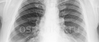
Polysegmental viral pneumonia
If a CT scan reveals that inflammatory foci and infiltrates are present not in one segment of the lung, but in several, such pneumonia is called polysegmental.
More about polysegmental pneumonia
The lungs are usually visually divided into 21 segments - 11 on the right side and 10 on the left side. Bilateral pneumonia of viral origin is the most common. For example, “ground glass” in coronavirus pneumonia is usually localized symmetrically on both sides around the bronchi or in the lateral parts of the lungs. In Pneumocystis pneumonia and influenza pneumonia, they are diffusely located on both sides.
Treatment of idiopathic interstitial pneumonia
Drug therapy
The basis of treatment for most subtypes of IIP (with the exception of primary fibrosing ones - idiopathic pulmonary fibrosis and pleuroparenchymal fibroelastosis) is the administration of immunosuppressive therapy, including:
- glucocorticoids (prednisolone, methylprednisolone): the first line of therapy, the advantages of which include the rapid development of a therapeutic effect;
- cytostatic drugs (mycophenolate mofetil, methotrexate, azathioprine, cyclophosphamide): used in cases of relapse after discontinuation of glucocorticoids, steroid dependence, and the presence of contraindications to the use of glucocorticoids (the advantages of this type of treatment include the absence of pronounced metabolic side effects).
Patients with primary fibrosing variants of IIP (primarily idiopathic pulmonary fibrosis), as well as with the development of progressive pulmonary fibrosis in all variants of IIP, are recommended to take antifibrotic therapy (nintedanib, pirfenidone), which has a proven effect of slowing the progression of respiratory failure and reducing the risk of exacerbations in this cohort sick.
The determining role in the effectiveness of all types of drug therapy is played by its earliest possible administration, with the assessment of clinical, laboratory and radiological parameters over time.
Prevention of exacerbations
Considering the significant contribution of respiratory infections to the development of exacerbation of IIP, vaccination against influenza, pneumococcus, SARS-Cov-2 (the causative agent of COVID-19) is recommended for all patients (in the absence of absolute contraindications).
Also, patients with PPIs, especially those receiving active immunosuppressive therapy, have a wider range of indications for antibiotic therapy, including prophylactic therapy.
Treatment and prevention of concomitant diseases
Taking into account the increased risk of developing cardiovascular complications in patients with IIP, it is important to promptly correct modifiable risk factors - prescribe antihypertensive, lipid-lowering, antiplatelet and anticoagulant therapy.
Due to the increased incidence of lung cancer (in particular, in patients with idiopathic pulmonary fibrosis), targeted screening of this pathology using chest CT is recommended.
In order to reduce the intensity of cough, all patients with IIP are recommended to be screened and, if indicated, treated for gastroesophageal reflux disease.
Non-drug therapy
With the development of respiratory failure and a decrease in blood oxygen saturation in air at rest below 90%, it is recommended to begin continuous oxygen therapy at home using an oxygen concentrator.
All patients with IIP are recommended to eat a balanced high-protein diet, which compensates for energy losses due to additional work of the respiratory muscles. To train the respiratory muscles, reduce shortness of breath, and increase tolerance to physical activity, moderate aerobic physical activity and breathing exercises, including the use of breathing simulators, are recommended for patients with IIP.
How is idiopathic interstitial pneumonia treated in the Rassvet clinic?
At the Rassvet clinic, patients diagnosed with IIP undergo a comprehensive examination to determine the optimal treatment tactics - both the underlying disease and concomitant pathology.
Based on the diagnostic results, a treatment plan is developed, including both drug therapy and dietary recommendations, breathing exercises, and a physical activity plan. During treatment, regular assessment of its effectiveness is carried out with the study of clinical, laboratory and radiological data.
Author:
Akulkina Larisa Anatolyevna pulmonologist
Pneumonia in children - treatment at the RebenOK clinic in Moscow
Our medical center employs pediatricians who have extensive practical experience. Treatment of pneumonia in children is carried out after confirming the diagnosis and determining the type of pathogen, which allows you to get a quick result. Competent specialists determine the symptoms of latent pneumonia in children, which occurs without fever and wheezing.
If signs of a cold appear, it is recommended to consult a pediatrician to rule out the presence of pneumonia or prevent the development of the disease. We use modern diagnostic equipment and prescribe treatment in accordance with international protocols, taking into account the individual characteristics of the patient.
Literature:
1. Erzhanova G.E. Pneumonia in children // Bulletin of the Kazakh National Medical Institute, 2014. URL: https://cyberleninka.ru/article/n/pnevmonii-u-detey (access date: 09/02/2021)
2. Sergeeva E.V., Petrova S.I. Community-acquired pneumonia in children. Modern features // Pediatrician Magazine, 2021. URL: https://cyberleninka.ru/article/n/vnebolnichnaya-pnevmoniya-u-detey-sovremennye-osobennosti (access date: 09/02/2021)
3. Geppe N.A., Kozlova L.V., Kondurina E.G. Community-acquired pneumonia in children // Clinical manual "Moscow Society of Children's Doctors", 2021. URL: https://pulmodeti.ru/wp-content/uploads/Vnebolnichnaya.pdf (access date: 09/02/2021)
4. Petchenko A.I., Luchaninova V.N., Knysh S.V.
Age-related features of the course of community-acquired pneumonia in children // Journal “Fundamental Research” No. 2, 2014. URL: https://fundamental-research.ru/ru/article/view?id=33563 (access date: 09/02/2021) 5. Kakeeva A.A., Bokonbaeva S.D., Dzhanabilova G.A. Etiological structure of community-acquired pneumonia in young children // Journal “Modern Problems of Science and Education” No. 3, 2021. URL: https://science-education.ru/ru/article/view?id=30897 (access date: 02.09. 2021)
General information
According to statistics, in Russia pathology is recorded:
- in preschool children – up to 500 cases per 100 thousand people;
- in adults up to 800 cases per 100 thousand people.
Polysegmental pneumonia is a multicomponent disease that develops due to infection of the body by harmful microorganisms (fungi, viruses, parasites, bacteria) against the background of a sharp decrease in immunity.
Typical side processes affect the lymph nodes and provoke oxygen and heart failure. Due to anatomical features, the right lung in segments II, VI, X and the left lung in VI, VIII, IX, X are most often affected.
80% of patients do not require hospitalization, 20% are treated in a hospital department, patients with an aggressive course of the disease are admitted to intensive care, and the mortality rate is 5%.
Viral pneumonia COVID-19
All known coronaviruses are characterized by rapid (up to 14 days) development of acute respiratory failure. The virus quickly and aggressively infects the lungs, causing not only extensive inflammation, but also associated complications: swelling of the respiratory organ, fibrosis (scarring of the lungs), acute heart failure, myocarditis.
The first coronavirus type B (SARS-CoV) was reported in 2002 and its primary hosts are believed to be horseshoe bats. In 2012, the world was gripped by an epidemic of coronavirus type C MERS-CoV (Middle East respiratory syndrome). Finally, in 2019, there was an outbreak of the new coronavirus type B COVID-19 (or SARS-Cov2). What they have in common is that new viruses are stable, easily attach to the lung parenchyma with protein spikes, and in a short time provoke extensive acute inflammation. The fatality rate is about 10%.
However, pneumonia is not always the cause of death from these viruses. For example, if the patient's medical history is complicated by atherosclerosis or myocarditis, the virus primarily affects the cardiovascular system. In general, the coronavirus family includes about 46 types of virions.
Read more about pneumonia associated with COVID-19 in our articleWhat does a CT scan of the lungs show for coronavirus?
Lung damage due to viral pneumonia
Lung infiltration in viral pneumonia on 3D reconstruction of the respiratory tract (CT-3)
For comparison, this is what normal lungs look like
Ground glass symptom (infiltrates) in viral pneumonia on cross-sectional CT scans
Coronavirus stages: CT-1, CT-2, CT-3, CT-4
Pulmonary fibrosis
Viral-bacterial pneumonia
In some cases, viral pneumonia is complicated by the addition of a secondary bacterial infection. This happens, for example, with respiratory syncytial virus and coronavirus (manifests on days 6-7 and is accompanied by more pronounced swelling of the lung tissue). Treatment of viral-bacterial pneumonia is carried out using a course of antibiotics, which are selected only after accurately determining the type of bacterial infection.