The calendar shows the 35th obstetric week, the 33rd from conception, the 31st from delayed menstruation. There are only 5 weeks left until the expected date of birth (EDD).
- How do mother and fetus feel at this stage?
- What to expect from an appointment with a gynecologist?
- What dietary and behavioral features need to be taken into account?
What should alert a pregnant woman, read the article.
- Physical sensations at 35 weeks
- Ultrasound
- Vitamins
- Sex
Belly size at 35 weeks
The abdominal circumference at 35 weeks of pregnancy is approximately 88-92 cm.
The stomach has already become very large and causes a lot of inconvenience.
At 35 weeks of pregnancy, the uterus became huge, pressing down on the stomach and lungs. At the end of the 35th week, the pregnant woman’s belly will begin to drop and breathing will become easier. If it becomes very difficult for the expectant mother to breathe, it is necessary to perform a special exercise for pregnant women, which will help reduce pressure on the internal organs: get on all fours and smoothly inhale and exhale. You can stand in this position from 5 minutes to half an hour, several times a day.
How does a baby's weight increase at 35 weeks?
A baby at 35 weeks pregnant continues to grow and has already reached the size of a cantaloupe. His weight increases every week by 200-250 g. The fetal movements become more constrained, because there is practically no free space in the mother’s belly. Now he doesn’t push so much as he rolls, sticking out different parts of his body. At this stage, the intrauterine development of the fetus is already quite high, the work of its internal organs and reflexes has been established. If an expectant mother goes into premature labor at 35 weeks of pregnancy, the likelihood of having a healthy baby is very high. However, experts agree that to ensure the survival of the child and its further development, pregnancy must be maintained until the expected date of birth.
Weight gain at 35 weeks of pregnancy ranges from 7.6 kg (with a BMI of more than 26) to 13 kg (with a BMI of less than 19.8). To calculate your individual weight gain at 35 weeks, use the pregnancy weight gain calculator.
Weight
At the 35th week, the weight gained from the moment of registration of a pregnant woman can range from 8 to 14.5 kg. In some cases, deviations from these standards may be greater or lesser. Deciding how much the gained kilograms correspond to the term, whether there is a potentially dangerous underweight or overweight is the task of the gynecologist monitoring the development of pregnancy.
If a deviation from the calculated norm for a particular woman is detected, the doctor may recommend changes to the diet or suggest hospitalization.
Dimensions of the baby at 35 weeks of pregnancy
At 35 weeks, the baby is about the size of a pineapple.
At week 35, the Baby’s height is 43-44 cm, and his weight is 2100-2300 g. At the beginning of the tenth lunar month, the following happens:
- the amount of lubricant on the skin begins to gradually decrease;
- the marigolds almost protrude above the phalanges of the fingers;
- muscles become stronger;
- fat continues to accumulate under the skin, which will provide thermoregulation after birth;
- gaining weight by about 30 g per day, your Baby becomes plumper and cheekier;
- in boys, the testicles descend into the scrotum;
- the child may hiccup and cough, but the hiccups do not cause him any discomfort, even if it continues for a considerable time;
- your Baby begins to recognize changes in lighting. Bright light, for example from a flashlight, can penetrate the skin of the abdomen - this is a new experience for the Baby, to which he can react with a rapid heartbeat.
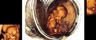
Ultrasound photo at 35 weeks of pregnancy
Belly shape
There is a myth that the gender of the child can be determined by the shape of the expectant mother’s belly: if the belly is wider and rounder, it means there will be a girl; if the stomach is narrower, but protrudes forward more, it’s a boy.
It is impossible to determine the gender of a child by the shape of the belly.
However, this is not so: the shape of the abdomen depends on the individual characteristics of the woman and on the presentation of the fetus. The most reliable way to find out the sex of the baby is an ultrasound.
The shape of the abdomen depends on the individual characteristics of the mother and the presentation of the fetus
Useful advice for future parents
The image of the Kid is rarely clear.
And if it does happen, it is most often tied to the moment of his birth. And there is a simple explanation for this: the image must be holistic, and until the moment of birth the Baby is constantly changing, and it is simply impossible to imagine what he is like at the moment. Ultrasound helps this a little, although the resulting pictures are more likely a positive fact of the child’s existence for you than a portrait that you want to hang on the wall. And yet, you will need to work on the image of the Baby: the more distinct it turns out, the warmer and more active your relationship with the baby will be even before birth. The image of the Kid consists of several components:
- visual image. Someone in their imagination makes the Baby look like toddlers from magazine pages, or familiar children, or older children (if there are any in the family), or even like the parents themselves in infancy. And some people develop an image that is independent of anything. There are also those (and perhaps most of them) who do not have a visual image at all. Be that as it may, the visual image is far from the main component of your Baby’s image;
- emotional image. It is connected with the feelings that you have for your baby. Your love, tenderness, optimism will allow you to imagine a joyful, active, sunny Baby. Mothers experiencing a decline in mood and physical activity begin to develop images of capricious, sad, empathizing Kids. The emotional image of your Baby is one of the most powerful;
- tactile image. It manifests itself in ideas about physical contact with the Baby: how you will feed him, rock him, stroke his head, tummy, back, bend and straighten his arms and legs, etc. Many mothers, without realizing it, actively contribute to the formation of a tactile image: they fuss with babies, sleep cuddled with toys, and if there are cats or dogs in the house, then they will have to try themselves in the role of a Baby who is cuddled and raised. In conjunction with the emotional image, the tactile image creates an almost tangible idea of the Baby.
Information about what he is like in each specific period before birth, information from the leading doctor about the size of his body, how his heart beats, how he has grown over the week (month) and what position he occupies in your tummy helps to create the image of the Baby.
The image of the Baby is closely related to your activities and self-development. So, if you are active, generally satisfied with life, you have many plans for life, and have favorite hobbies, then you will see your Baby as well-rounded, cheerful and talented.
A significant role in shaping the image of the Kid belongs to his dad. Very often, even before birth, fathers boldly create specific images: “When he grows up, we’ll ride bikes with him,” “My daughter won’t be afraid of anything,” “I’ll walk with my son, even in the rain, even in the snow, let him harden himself,” “ Finally, I will have a real rival in computer games,” “When a girl is born, we will grow our braids down to our toes.”
Whatever the image of your Baby, it is needed so that you feel him physically and mentally, strive to communicate with him, feel proud of his success in growth and development, and also love him very much.
Tests and studies during pregnancy
Ultrasound (ultrasound examination) - 35-36 weeks after the first day of the last menstruation.
The condition of the placenta, the height and weight of the child, its position in the uterus, and the quantity and quality of amniotic fluid are assessed. Blood test for AIDS (HIV), syphilis (RW) - 35-36 weeks after the first day of the last menstruation. These diseases can complicate pregnancy and childbirth.
Biochemical blood test - 35-36 weeks after the first day of the last menstruation. Gives an idea of the general health of the pregnant woman.
Vaginal smear - 35-36 weeks after the first day of the last menstrual period. Assessment of microbial flora before the baby passes through the birth canal.
Visiting a doctor monitoring pregnancy: once a week. Weighing, measuring blood pressure, measuring the height of the uterine fundus, listening to the fetal heartbeat.
General urine test - before each visit to the doctor. Indicates the quality of kidney function.
Dopplerography (a study that allows you to evaluate blood flow in the vessels of the uterus, placenta and main vessels of the child) - 33-34 weeks after the first day of the last menstruation, according to indications. The study allows you to find out whether the child is getting enough oxygen and nutrients.
Cardiotocography (CTG, synchronous recording of fetal heartbeats and uterine contractions) - 33-34 weeks after the first day of the last menstruation, according to indications. The child’s condition is assessed and intrauterine hypoxia is excluded.
Receive an exchange card with the results of tests and examinations - 30 weeks after the first day of the last menstruation. It is recommended that you always carry this document with you, which is required for admission to the maternity hospital.
Registration of maternity leave for workers - 30 weeks after the first day of the last menstruation.
Visiting an obstetrician-gynecologist
Medical examination at the 35th week of pregnancy
At the 35th week of pregnancy, it is possible to schedule another scheduled appointment with a gynecologist. The day before, you need to take tests, directions for which were issued during your last visit to the doctor.
Women who have already completed the necessary studies for 3 screening usually undergo only general blood and urine tests, and, if necessary, a test for sugar levels and antibodies (if the pregnant woman has a negative Rh factor). Expectant mothers who, for some reason, have not previously been tested for biochemistry, HIV (AIDS), RW, hepatitis and hormone levels, need to do this before their appointment this week.
It is also worthwhile to undergo fetal cardiotocography (CTG) before appearing. Bring the results to your doctor for evaluation.
During the meeting, the gynecologist will conduct the usual measurements and surveys:
- will inquire about your well-being, ask about the emergence of new sensations;
- measure blood pressure, body temperature, heart rate of the mother (on the arm) and fetus (using a stethoscope);
- weigh the woman and calculate the weight gained since registration;
- will measure the abdominal circumference, the height of the uterine fundus, and compare the results obtained with the norm and past indicators of a particular pregnant woman.
At the end of the appointment, he will give recommendations (if necessary), set a date for the next meeting, and issue directions for blood and urine tests.
Ultrasound
At the 35th week of pregnancy, routine ultrasound is not provided as part of screening. It is carried out from 30 to 34 weeks. But, in some cases, if a pregnant woman has not been examined within the prescribed time frame, a referral may be issued. The study will establish:
- the condition of the fetus, its correspondence to the gestational age;
- position of the baby in the uterus (head, foot or transverse presentation);
- condition of the uterus, its cervix, placenta;
- presence/absence of visible external and internal pathologies of the fetus.
A woman who has undergone a screening ultrasound within the prescribed time frame can undergo an ultrasound examination on her own initiative for a fee.
Uterus and belly
At the 35th week of pregnancy, the fundus of the uterus is at a height of about 35-36 cm from the symphysis pubis, 15 cm above the navel. This week, the woman feels training contractions, during which the walls of the uterus contract, but do not cause pain. If the expectant mother feels pain during muscle contraction, she urgently needs to go to the maternity hospital.
The abdomen at this stage has a large volume and greatly affects the capabilities of the pregnant woman. It becomes uncomfortable to walk and move, and it is almost impossible to put on your shoes on your own. Many pregnant women experience severe itching over the entire surface of the abdomen, caused by skin tension. You should continue to actively care for your skin, using oils or a stretch mark cream recommended by your gynecologist.
This week, the expectant mother may notice that her belly begins to drop a little, her navel, which already protrudes well above her belly, begins to slowly move, looking not up, but forward. This event brings relief to the pregnant woman’s life: it becomes easier to breathe and eat. But the pressure on the bladder increases, and the number of visits to the toilet may become more frequent.
Pain in the abdomen and other parts of the body
Painful sensations at the 35th week in different parts of the body may appear within the normal range or indicate a possible pathology of pregnancy. The expectant mother should clearly understand what kind of pain should prompt her to seek medical help as soon as possible.
Signal for an emergency consultation with a gynecologist:
- pain in the kidney area when urinating;
- painful sensations in the legs not associated with walking or physical fatigue;
- headache, dizziness, heaviness in the back of the head;
- exacerbation or appearance of hemorrhoids.
Signal to call an ambulance or go to the maternity hospital:
- backache. The sensation is pronounced, the pain increases, does not go away at rest;
- abdominal pain. Drawing, cramping pain that increases regardless of body position;
- pain or pressing sensations in the perineum/thighs.
Discharge, bleeding, menstruation
Normally, vaginal discharge should have a slight odor, light or milky color, and an even consistency. At the 35th week, mucous lumps of an even structure may appear.
If the color of the discharge changes (yellow, green, gray), smell (pungent, unpleasant), consistency (flaky, curd-like), you should consult a gynecologist for advice.
If you experience red or brown vaginal discharge, you should immediately seek medical help. There should be no bleeding or menstruation normally during this period.
Excessively thin discharge is a cause for concern. If the expectant mother notes that the discharge has a watery structure, it is worth contacting a gynecologist as soon as possible or heading to the pregnancy pathology department.
Good to know
Childbirth with a scar on the uterus
What awaits a premature baby immediately after birth in the hospital?
How to choose a nursing bra
Is a pregnant woman beautiful?
What worries a woman during pregnancy?
What are the methods of preparing for childbirth?
All texts for pages about mother and baby were kindly provided by RAMA Publishing - these are chapters from the book by Svetlana Klaas “Your Favorite Little Man from Conception to Birth”, reviewer Irina Nikolaevna Kononova, Candidate of Medical Sciences, Associate Professor of the Department of Obstetrics and Gynecology of the Ural State Medical Academy (Ekaterinburg).
Changes in a woman's body
- The uterus puts more and more pressure on the lungs. This makes breathing more difficult. A woman often experiences pain under her ribs.
- At this stage, a slight prolapse of the uterus is already possible, this makes it easier for the woman to breathe.
- Due to the intense increase in the weight of the fetus, the expectant mother finds it increasingly difficult to move, and she quickly gets tired. Despite this, even during such a period, walks in the fresh air should not be neglected.
- Frequent urination continues to torment the woman, which negatively affects the quality of sleep.
- Ligaments become more elastic. This explains the possible change in the woman's gait.
- The birth canal is increasingly preparing for childbirth.
- A woman’s body weight should already be 13 kilograms higher than her original weight.