Posted by Andro-gynecology clinic on 2017-11-03. Published: Women, Diseases
Endometrial polyp is a focal (limited in area) hyperplasia of the tissue of the mucous membrane of the uterine cavity . Hyperplasia is an excessive growth of endometrial tissue, when foci of hyperplasia there is a lot of talk about endometrial polyposis. It can be suspected by ultrasound, removed by hysteroscopy, the final diagnosis is made after histological examination. Article provided by the site “Minimally invasive gynecology in Krasnoyarsk” - original article.
Types of polyps
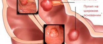
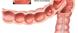
A polyp is a benign formation that has a round shape and sizes from 2 mm to several centimeters. With the help of a stalk, the body of the polyp is attached to the base located under the surface of the mucous membrane. Polyps are overgrown endometrium and, like the uterine mucosa, consist of glands and connective tissue. Depending on the predominance of one of these elements, the following are distinguished:
- Glandular polyps of the endometrium, which consist of stromal cells (the structure of the ovarian stroma) and glands. Most often detected in women of reproductive age.
- Fibrous endometrial polyps, which consist predominantly of connective tissue; on the contrary, there are very few glands. This type of polyp is rarely detected, usually in postmenopausal women.
- Glandular fibrous endometrial polyps, respectively, consist of glands and connective tissue. They can appear at any age.
- Adenomatous endometrial polyps consist of glandular tissue, which shows signs of structural changes. This type of polyp has a high probability of malignancy. Usually detected in postmenopausal patients.
Polyposis is said to occur if a single polyp re-forms or several growths are detected.
Diagnosis of polyps of the uterine cavity
A gynecological examination on a chair is informative only when the formation is large and extends from the uterine cavity into the vagina.
The diagnostic standard is transvaginal ultrasound of the pelvic organs and diagnostic hysteroscopy.
Ultrasonography
Transvaginal ultrasound of the pelvic organs on days 6-7 of the menstrual cycle can identify dense fibrous and glandular-fibrous endometrial polyps.
Glandular polyps are similar in structure to normal endometrium, have a soft consistency, are compressed by the walls of the uterus and are not distinguishable when small in size.
If there are symptoms and no signs of endometrial pathology on ultrasound, it is recommended to conduct a repeat ultrasound examination on days 12-14 of the menstrual cycle, by which time the glandular polyp will have grown, increased in size and become visible on ultrasound.
A more informative ultrasound examination is echohysterosalpingoscopy (ECGS), an ultrasound examination with contrast of the uterine cavity. The uterine cavity is filled with saline, it expands, the polyp takes its natural shape and is contoured against the background of a contrast liquid.
Ultrasound conclusion: suspicion of focal endometrial hyperplasia is an indication for hysteroscopy.
Hysteroscopy
First, diagnostic hysteroscopy is performed - an endoscopic examination of the uterine cavity. Diagnostic hysteroscopy allows you to visually confirm or exclude a uterine polyp.
Video selection of hysteroscopic examinations
If an endometrial polyp is detected during hysteroscopy, it is removed - hysteroresectoscopy. If hysteroscopy does not confirm a uterine polyp, a targeted biopsy of the endometrium is performed in the location described in the ultrasound protocol.
Ultrasound and hysteroscopic findings are not a diagnosis. The final diagnosis is made only after removal of the formation and clarification of its histological structure.
Back to contents
Causes of endometrial polyps
The main causes of endometrial polyposis include the following:
- Ovarian dysfunction caused by increased estrogen levels or decreased progesterone production
- Endocrinopathies
- Surgery in the uterine cavity
- Endometritis
Negative predisposing factors include poor environmental conditions in the region of residence, the use of an IUD, obesity, and decreased immunity.
Despite the fact that endometrial polyps can form at any age, the likelihood of their occurrence increases in women over 35 years of age.
Symptoms of endometrial polyp in the uterus
The pathology is characterized by:
- Menstrual irregularities such as hypermenorrhea
- Intermenstrual bleeding
- Uterine bleeding in the postmenopausal period
- Dyspareunia
- Pain when urinating
- Infertility
- Cramping pain in the lower abdomen
In some cases, there are no obvious signs of endometrial polyposis. It is characteristic that the intensity of clinical manifestations of endometrial polyposis increases in proportion to the woman’s age.
Diagnostics
- Gynecological examination, during which a polyp protruding from the cervical canal may be detected
- Ultrasound of the pelvic organs, during which thickening of the endometrial layer of a local nature is revealed.
- Hysteroscopy with histological examination of the endometrium, during which it is possible not only to detect polyps, but also to remove them.
Endometrial hyperplasia - treatment:
- hysteroscopy;
- hormonal conservative therapy after hysteroscopy;
- removal of the uterus in the presence of recurrent hyperplasia, hormonal ineffectiveness or endometrial hyperplasia with atypia.
Treatment of endometrial hyperplasia begins with surgical removal of the endometrium followed by histological examination. Once an accurate diagnosis has been established, it is possible to determine further management tactics. It is determined both by the histological type of pathology and by a number of other factors: interest in pregnancy, age, the presence of concomitant diseases and many others.
Thus, the main method for diagnosing and treating endometrial hyperplasia is hysteroscopy. Only after the diagnosis has been established is it possible to choose further management tactics.
Treatment of endometrial polyps
A procedure such as removal of a polyp in the uterus is resorted to in most cases after confirmation of the appropriate diagnosis. Conservative therapy in most cases does not have any effect, that is, the hyperplastic formation in the uterine cavity remains.
Treatment involves the elimination of the polyp as a foreign body of the uterus and cervical canal, as well as subsequent histological examination of the removed tissue. This is necessary to prevent the development of oncological pathology against the background of hyperplastic processes of the cervix and endometrium.
Indications and contraindications
Removal of a cervical polyp is carried out only if there are certain indications. These include:
- large polyp size (over 10 millimeters in diameter);
- rapid progression of pathology according to control ultrasound examination;
- presence of intermenstrual bleeding;
- infertility. Problems with conception and a history of spontaneous abortion due to an endometrial polyp are an indication for surgical treatment;
- the presence of an adenomatous polyp (precancerous condition);
- malignant growth of formation.
The procedure of polypectomy (removal of a polyp) is contraindicated in the following conditions:
- with pathology of the cervix (cicatricial deformation, lack of patency of the cervical canal, malignant process);
- acute stage or exacerbation of chronic inflammatory process of the cervix (exocervicitis, endocervicitis);
- presence of uterine bleeding;
- colpitis (inflammatory processes in the vagina of various etiologies).
Preparation for surgery
If an endometrial polyp is detected in the uterus, treatment should begin as early as possible. The procedure to remove such a neoplasm of the cavity or cervix requires preparation.
Before undergoing a polypectomy, a woman undergoes an examination, including:
- blood tests: clinical, biochemical, coagulogram (blood clotting test), HIV, syphilis, and hepatitis markers;
- Analysis of urine;
- vaginal smear for flora and atypical cells;
- colposcopy;
- electrocardiography;
- fluorographic examination.
Since the manipulations are performed under anesthesia, the patient must obtain the opinion of such specialists as a therapist, anesthesiologist, or cardiologist. This is necessary to exclude contraindications for surgical removal of the polyp.
In addition to mandatory examinations, when preparing for polyp removal, a woman must adhere to some rules:
- seven days before the expected date of surgery, you should stop drinking alcoholic beverages;
- three days before the intervention, it is worth excluding foods that promote gas formation from the diet;
- You can eat food no later than 10 hours before the operation;
- on the day of surgery you should not eat or drink;
If a woman is taking any medications, this information must be reported to the attending physician/
Hyperplastic processes of the endometrium (hyperplasia, polyps)
Primary appointment/consultation with a gynecologist—RUB 1,700. Repeated appointment/consultation with a gynecologist—RUB 1,200. See all prices
+7 (499) 400-47-33
Hyperplasia is an increase in the number of cells in any tissue (except tumor) or organ, resulting in an increase in the volume of this anatomical formation or organ. Histologically (according to cellular composition), several types are distinguished: glandular hyperplasia of the endometrium; glandular cystic hyperplasia; atypical endometrial hyperplasia (synonym - adenomatosis, adenomatous hyperplasia); endometrial polyps.
According to most authors, the first two types of hyperplasia are not a precancerous disease. The third type, atypical hyperplasia, is a precancerous disease. If present, the risk of degeneration into a malignant tumor (endometrial cancer) in the absence of therapy ranges from 1 to 14% and is most often observed during menopause (cessation of menstrual function due to age).
Precancerous hyperplastic processes develop into endometrial cancer in approximately 10% of patients (according to various authors, from 2 to 50%), they often persist for a long time, and sometimes undergo reverse development. However, taking into account the real threat of the process progressing to endometrial cancer, a doctor’s attentive attitude towards patients with endometrial adenomatosis and adenomatous polyps is necessary. There are opinions about the possibility of considering glandular hyperplasia and hyperplastic processes that arise again (recur) after endometrial curettage or are not amenable to hormone therapy as precancer of the endometrium. The risk of malignancy (malignancy) of hyperplastic processes increases with metabolic disorders caused by extragenital disease (obesity, impaired carbohydrate and lipid metabolism, dysfunction of the hepatobiliary system and gastrointestinal tract), concomitant with the development of endometrial pathology.
A localized, localized form of endometrial hyperplasia is called an endometrial polyp. Histologically, they are also divided into several types depending on the cells that predominate in their structure: glandular; glandular fibrous and fibrous polyps.
Cause and mechanism of development of the disease
Risk factors for the occurrence of this pathology are:
1. The occurrence of hyperplastic processes in the endometrium is facilitated by hereditary burden (uterine fibroids, genital and breast cancer, hypertension and other diseases), damaging effects during intrauterine life, diseases during puberty and associated disorders of menstrual and subsequently reproductive function . In mature women, the appearance of hyperplastic processes is often preceded by gynecological diseases and surgical interventions on the genital organs.
2. Hyperplastic processes of the endometrium are often accompanied by obesity, hypertension, hyperglycemia (this triad of symptoms is especially often combined with atypical and hyperplastic processes recognized as precancer of the endometrium), uterine fibroids, mastopathy, endometriosis, which are largely hormonal-dependent diseases, as well as dysfunction of the liver, responsible for the metabolism of hormones. The leading factor in the cause of occurrence and further development belongs to hyperestogenia - an increase in the level of the hormone estrogen.
Clinical picture
In the clinical picture of the pathology, the following symptoms are most often noted:
1. Uterine bleeding that occurs after a delay in the next menstruation. They can be either long in duration with moderate blood loss, or short in time but abundant;
2. Bloody intermenstrual discharge;
3. Primary or secondary infertility, which is based on the process of lack of formation of a normal egg;
4. The most common, almost constant symptom of endometrial polyps is menstrual irregularities. With polyps against the background of a normal functioning endometrium, women of reproductive age experience scant intermenstrual and premenstrual blood discharge with a preserved menstrual cycle, as well as an increase in menstrual blood loss.
5. In women of reproductive age, glandular fibrous and fibrous polyps can cause menorrhagia. With anovulatory cycles and the presence of polyps of the glandular structure against the background of hyperplastic processes of the endometrium, metrorrhagia is observed, the cause of which is not polyps, but dystrophic, necrotic changes in the endometrium and hormonal disorders. Such clinical manifestations are more typical for premenopausal women (after 45 years).
Diagnostics
1. Ultrasound using a transvaginal sensor;
2. For diagnostic purposes, diagnostic curettage of the mucous membrane of the uterine body and subsequent histological examination of the resulting material are widely used.
It is recommended to scrape the endometrium on the eve of the expected menstruation or at the very beginning of the appearance of bleeding. In this case, it is necessary to remove the entire mucous membrane, including the area of the uterine fundus and fallopian tube angles, where foci of adenomatosis and polyps are often located. For this purpose, endometrial curettage is performed using hysteroscopy. The removed mucous membrane is sent for histological examination.
Hysteroscopy is the most informative method and allows not only to diagnose endometrial polyps with a high degree of accuracy, but also to specifically remove them, and to monitor the bed of the polyp after its removal. With hysteroscopy, polyps have an oblong or round shape, pale pink, yellowish or dark purple (poor blood circulation) color. Polyps can be single or multiple, often located in the fundus or tubal corners of the uterus and, unlike stationary myomatous nodes, fluctuate in the stream of washing fluid.
The so-called recurrent endometrial polyps are of interest to clinicians. With the introduction of control hysteroscopy after curettage and removal of polyps, there was a firm belief that the “relapse” is the unremoved parts of the polyps.
Histological examination is the most reliable method for diagnosing hyperplastic processes and determining the nature of this pathology (glandular cystic hyperplasia, atypical hyperplasia - diffuse, focal adenomatosis, polyps - glandular, adenomatous, fibrous).
It should be especially noted that separate (separately - the cervical canal, separately - the uterine cavity) curettage of the uterine mucosa is the first stage of treatment for the hyperplastic process of the endometrium. It allows you to obtain a sample of endometrial tissue and make an accurate diagnosis: what kind of hyperplasia, and whether there are any precancerous changes. Further treatment depends on this. The changed endometrium can only be removed mechanically.
Treatment
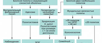
Treatment of endometrial hyperplastic processes is carried out taking into account numerous factors - the patient’s age, the causes of hyperplasia and the nature of this pathology, clinical manifestations, contraindications to a particular treatment method, tolerability of medications, concomitant extragenital and gynecological diseases. The main treatment is hormonal (selected individually). When prescribing hormone therapy, certain conditions and strict consideration of contraindications are required. After 3 and 6 months, a control ultrasound is indicated. For hyperplasia associated with polycystic ovary syndrome, the first stage of treatment is wedge resection of the gonads. This operation is especially indicated for recurrent hyperplasia, which is alarming for endometrial precancer.
If the clinical effect of ovarian resection is insufficient (control - biopsy and histological examination of the endometrium), hormone therapy is carried out according to the guidelines adopted for various types of endometrial hyperplastic processes.
Surgical methods are preferable for recurrent glandular cystic hyperplasia that has developed against the background of diseases of the endocrine glands (diabetes, prediabetes, etc.), obesity, hypertension, liver and vein diseases. Surgical treatment is indicated for precancer (adenomatosis, adenomatous polyps) of the endometrium, especially when this endometrial pathology is combined with adenomyosis and uterine fibroids, and pathological processes in the ovaries.
Patients with fibrous polyps are not subject to hormonal therapy.
Women of reproductive and especially premenopausal age who have glandular and glandular-fibrous polyps against the background of endometrial hyperplastic processes are advised to remove the polyp followed by hormonal therapy.
For adenomatous polyps in premenopausal women with metabolic and endocrine disorders, an alternative is removal of the uterus with a thorough revision of the ovaries (hyperplasia of the tissue body, the presence of hormonally active tumors). Adenomatous polyps in postmenopausal women are an indication for removal of the uterus and appendages.
Polyps in the uterus: surgical treatment methods
In modern gynecological practice, there are several ways to remove polyps of the cervix and uterine cavity.
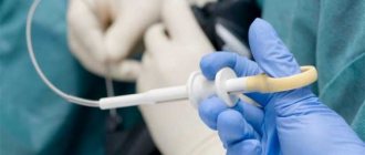
Hysteroresectoscopy
Hysteroresectoscopy is a modern technique that involves inserting an endoscope equipped with a video camera into the uterine cavity.
When performing this procedure, the doctor has the opportunity to assess the condition of the uterine cavity, visualize the polyp itself, and also remove it using mechanical, electrosurgical or laser instruments. Then a histological examination of the formation is carried out.
Advantages of hysteroresectoscopy:
- removal occurs under visual control and not blindly;
- the polyp is removed along with its stem, which prevents its re-formation in this particular place;
- there is no negative or harsh effect on the uterine mucosa, which is extremely important for women planning a pregnancy;
- the risks of trauma to the uterus and the addition of an infectious process are minimized;
- after the operation, the woman recovers quickly and can be discharged from the hospital in the near future;
- there is no pain;
- This minimally invasive intervention allows us to minimize risks for patients with somatic pathologies.
Fractional diagnostic curettage
This surgical intervention involves blind removal of a uterine polyp by curettage of the uterine cavity and cervical canal.
While conducting this study, the doctor is not able to exercise visual control over what is happening in the uterine cavity.
After separate curettage of the canal and the cervical cavity, the resulting material is also sent for histological examination.
According to statistics, after the procedure of curettage of the uterine cavity, recurrence of polyps occurs in more than 50% of cases. That is why in modern gynecology, if there is a diagnosis of “uterine cavity polyp” or “cervical polyp,” treatment involves hysteroresectoscopy of the polyp.
Causes and symptoms of polyps and hyperplasia
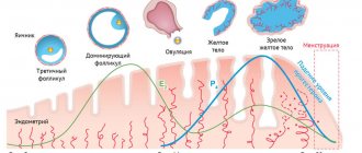
The process of endometrial rejection and renewal is regulated by female sex hormones - estrogens. Accordingly, when hormone balance is disturbed, cell growth processes change. In addition, the following factors can provoke the development of polyps and hyperplasia:
- Inflammatory processes of the genital organs.
- Concomitant gynecological diseases (polycystic ovary syndrome, fibroids).
- Some gynecological procedures (curettage, abortion).
- Diabetes.
- Diseases of internal organs.
- Heredity.
As a result, the endometrium begins to grow pathologically. This can be either a uniform thickening or a combination with cysts or polyps. These diseases are benign, but this does not mean that they do not require treatment.
With a long course, the risk of serious complications increases significantly, up to the transformation of a benign process into a malignant one. In addition, due to dysfunction of the endometrium, it becomes impossible for the egg to attach and, accordingly, pregnancy. That is why, if a woman is diagnosed with infertility, an experienced doctor does not exclude the presence of endometrial hyperplasia and conducts a diagnostic search for this disease.
Pregnancy with endometrial polyposis
Patients with endometrial polyposis may develop infertility. This is due to changes in hormonal levels and difficulties that arise during the implantation of a fertilized egg into the wall of the uterus. According to statistics, removal of polyps significantly increases the chances of both a natural pregnancy and IVF success.
If you have any questions related to polyposis, you can ask them to the doctors at Nova Clinic. You can make an appointment with a doctor by calling the number listed on the website or using the booking button.
