What is the essence of this survey?
Hysterosalpingography is a method of visualizing the fallopian tubes and the uterine cavity, based on filling their lumen with a specially injected fluid. The doctor records the nature of its distribution using equipment, and then analyzes the resulting images.
HSG provides the specialist with reliable information about the patency, location and size of the fallopian tubes. Various deformations, adhesions and neoplasms in the uterine cavity can also be detected. In this case, the patient will be recommended for additional targeted examination in the clinic.
Previously, X-rays with additional contrast enhancement were used for tubal HSG. This method is still used today, although it has a number of disadvantages. Therefore, currently in St. Petersburg, preference is given to an alternative safe technique - contrast ultrasonic hysterosalpingoscopy (CUUSGSS).
X-ray contrast hysterosalpingography is associated with additional (albeit small) irradiation of the patient’s pelvic organs, so it is advisable for her to refrain from becoming pregnant for the next 3 cycles. In addition, the drug used may be toxic or cause an allergic reaction. The ultrasonic method does not have such disadvantages. After all, in this case, the uterine cavity and fallopian tubes are filled with a sterile saline solution that is neutral for the woman’s body, and the ultrasonic waves emitted by the sensor are completely safe and do not cause discomfort.
What methods exist for checking the patency of the fallopian tubes?
- Echosalpingography (sonohysterosalpingography)
- Hysterosalpingography (also known as metrosalpingography)
- Laparoscopy (including transvaginal laparoscopy)
Echosalpingography
Echosalpingography is one of the simplest research methods, performed using ultrasound. A thin rubber catheter is inserted into the uterine cavity, and a saline solution is injected through it using a syringe. Using an ultrasound, the doctor sees the fluid in the uterine cavity, and how it passes through the fallopian tubes - how quickly it fills them and pours into the abdominal cavity. The procedure usually takes no more than 10–15 minutes, is practically painless, but can sometimes be accompanied by slight aching pain in the lower abdomen.
Discomfort often occurs in the presence of an adhesive process; in this case, a solution under pressure can separate thin adhesions. Sometimes this even helps to improve the patency of the tubes, and it is not uncommon for a woman to have a long-awaited pregnancy after this study. But you still shouldn’t rely on echosalpingography as a method of therapy in the presence of obstruction of the fallopian tubes; more often than not, “blowing the tubes” in this way turns out to be at least simply ineffective, and is often associated with the risk of developing an ectopic pregnancy.
X-ray examination of fallopian tube patency
X-ray examination of the patency of the fallopian tubes (hysterosalpingography or metrosalpingography) consists of introducing a water-soluble radiopaque substance into the uterine cavity, followed by an x-ray that evaluates its distribution in the pelvic cavity. There are many types of solutions, all of them containing iodine, so this procedure is possible only in the absence of an allergic reaction to iodine preparations.
The technique of hysterosalpingography is as follows: the procedure is performed without anesthesia in a specially equipped room. A metal catheter is inserted into the woman’s cervical canal, through which 2 ml of a contrast agent is poured into the uterine cavity, after which an X-ray is taken. Then an additional few ml of contrast is injected and a repeat image is taken.
On average, the entire procedure takes about 30 minutes. The image shows the uterine cavity and fallopian tubes along their entire length, filled with contrast. Based on the shape of the fallopian tubes, their filling, and the location of the contrast in the pelvic cavity, it is possible to determine the presence of an adhesive process, and if there is obstruction, in which part of the fallopian tube it occurred. The advantages of such a study are its significantly greater accuracy, compared to ultrasound salpingography, and the fact that you still have pictures of your pipes in your hands, which can be viewed by any specialist even in a few years. The disadvantages include the fact that it is associated with more pronounced discomfort due to the technical features of the procedure.
Laparoscopy
Laparoscopy is the most accurate method for examining the patency of the fallopian tubes, the most valuable factor of which is that it is possible to immediately eliminate almost all defects that are identified, from adhesions to foci of endometriosis. In addition, there is no moment of spasm of the smooth muscles of the tubes in response to a painful stimulus, which can simulate tubal obstruction during echosalpingography or HSG, since laparoscopy is performed under anesthesia and the muscles are completely relaxed. But this is already a full-fledged surgical intervention, so it will be discussed separately.
Indications for such diagnostics
The main purpose of hysterosalpinography is to check the patency of the fallopian tubes. Therefore, this procedure is mainly performed on women of reproductive age who have already encountered problems with natural conception and pregnancy or who are at risk for developing tubal infertility.
Indications for HSG include:
- Marital infertility of unknown origin (not associated with hormonal imbalance and male factor).
- Previously diagnosed tubal infertility, to monitor the results of treatment.
- Planning pregnancy in women who have had ectopic pregnancies and surgical interventions on the appendages in the past.
- Abnormalities of the internal genital organs identified in a woman.
- The woman has a history of diseases that can lead to the formation of tubal adhesions. These include chronic salpingoophoritis, genital tuberculosis, some STDs and a number of other conditions.
- Preparation for fertilization using the AI method (artificial insemination)
Sometimes HSG is also prescribed for examining patients with uterine pathology (polyps, adhesions, fibroids, endometriosis, etc.) and isthmic-cervical insufficiency in a previous pregnancy. But still, in such cases, preference is usually given to other diagnostic methods that provide more information about the condition of the uterine cavity.
When should it be done?
In the absence of pregnancy, within one year of regular sexual activity without contraception, in order to diagnose the causes of infertility, to exclude the tuboperitoneal factor. True, it is important to understand that it is definitely not worth starting a woman’s examination with this procedure. If only because this in itself is not the most comfortable procedure, it can be accompanied by quite intense pain in the lower abdomen, for example. Therefore, the study should be done after all other factors have been excluded (male, ovarian (hormonal levels have been studied) and others).
This procedure is mandatory before planning intrauterine insemination (with the sperm of the husband or donor, it doesn’t matter). This is to ensure that the treatment will be effective and that there are no adhesions that could lead to an ectopic pregnancy.
After surgical interventions on the fallopian tubes (especially for ectopic pregnancy) after 6–12 months to ensure that their patency is not impaired, as well as after excision of adhesions to monitor the effectiveness of the operation.
Contraindications
Checking the fallopian tubes using the GHA method is not carried out during acute infectious diseases, during exacerbation of the patient’s infectious and inflammatory processes of any localization, or with diagnosed and untreated STDs. The procedure is postponed if the patient is diagnosed with colpitis, vulvitis, bartholinitis, acute cervicitis, or if chronic salpingoophoritis is activated.
Contraindications also include somatic and neuropsychiatric diseases in the stage of decompensation. But in many cases, after the woman’s condition improves and stabilizes, she can undergo the necessary examination.
HSG cannot be performed during pregnancy, as well as during menstruation and dysfunctional uterine bleeding.
Indications and contraindications for hysterosalpingography
Typically, an examination is prescribed to diagnose the causes of infertility.
An HSG can be done in a clinic, having previously received a referral from a gynecologist. Typically, an examination is prescribed to diagnose the causes of infertility. The procedure is performed if, in the absence of contraception, a woman under 35 years of age for a year or over 35 years of age for six months cannot become pregnant.
Diagnostics is also necessary if you suspect:
- disorders of the anatomical structure of the uterus and appendages
- fibroids
- adhesions
- polyps and other pathologies
Indications for fallopian tube HSG are determined by the doctor as part of a survey and examination of the patient, as well as after other examinations.
In some cases, no diagnosis is made.
The procedure should be postponed if:
- Pregnancy. It is necessary to limit sexual contact or use contraception for a month before the procedure
- Allergies to contrast agents. During diagnosis, iodine-containing substances are used. They can provoke an acute reaction from the woman’s body
- Bacterial and infectious diseases
- Inflammatory processes (especially in the acute stage)
The final decision on the advisability of fallopian tube HSG is made by the doctor. This allows the procedure to be as safe as possible.
Diagnosis is also refused for uterine bleeding, thrombophlebitis and some other pathologies.
Important! The final decision on the advisability of fallopian tube HSG is made by the doctor. This allows the procedure to be as safe as possible.
Preparation for the procedure
Before checking the fallopian tubes, a woman is prescribed an examination to exclude possible contraindications:
- General clinical tests (CBC, OAM, biochemical blood test).
- ECG.
- Examination by a gynecologist, taking smears to determine the degree of cleanliness of the vagina and from the cervix for oncocytology.
- Examination to exclude major infections (STDs, tuberculosis, viral hepatitis).
- If necessary, consultations with narrow specialists to obtain an opinion on the possibility of carrying out the manipulation.
Direct preparation for hysterosalpingography includes the abolition of any vaginal medications and douching and abstinence from intimate life 7 days before the procedure. During the current cycle, the woman must use adequate protection, and during an X-ray contrast study, she should refrain from becoming pregnant for another 3 months.
Before the procedure, it is necessary to shave the hair in the intimate area and thoroughly empty the intestines (using an enema or medications). You should refrain from eating for 12 hours. When using ultrasound, you need to drink plenty of fluids, but before an x-ray examination, you can drink water no later than 1.5-2 hours before your visit to the clinic.
In consultation with the doctor, the day before HSG, medications with a mild sedative (calming) and antispasmodic effect can be started to reduce the severity of possible discomfort during the procedure.
Sterilization operation
Sterilization of the fallopian tubes, according to L.V. Adamyan (2000), PJ Taylor (1995), is one of the most common gynecological procedures. Indications for surgical sterilization:
- the couple's desire for complete fertility prevention;
- intolerance to other methods of contraception;
- medical indications - diseases of the cardiovascular, endocrine, musculoskeletal systems, mental disorders, hereditary diseases in which pregnancy and childbirth are absolutely contraindicated.
Before the operation, it is necessary to obtain the patient’s personal signature agreeing to sterilization. It is necessary to explain to her that restoration of patency of the fallopian tubes after any method of sterilization is not guaranteed.
With a normal menstrual cycle, surgery should be performed in the early proliferative phase. For women using an IUD, it is advisable to perform it 1 cycle after removal of the intrauterine contraceptive device and prophylactic antibacterial treatment. For patients taking oral contraceptives, sterilization can be performed at any time. The combination of artificial termination of pregnancy and sterilization, in our opinion, is inappropriate.
Currently, there are several options for tubal occlusion. Surgical intervention is performed through 2 approaches: a 5-mm trocar - paraumbilical, 5-mm - in the left (right) iliac region or in the midline above the pubic symphysis. If there is an adhesive process or other complicating issues, a third trocar is introduced.
After fixing the fallopian tube with a clamp, it is monopolarly coagulated over a length of 2 cm, 2-3 cm from the angle of the uterus, followed by dissection with endoscissors to the mesosalpinx, which significantly reduces the risk of recanalization. Coagulation can also be performed using a bipolar electrode in 2 or 3 places.
It is possible to excise a section of the fallopian tube 1-2 cm long after preliminary bi- or monopolar coagulation at a distance of 2 cm from the angle of the uterus.
Fimbriectomy involves coagulation and division of the distal portion of the mesosalpins near the infundibulopelvic ligament and the infundibulum of the fallopian tube.
According to the results of studies by Gomel V., Winston L., J. Hulka J., applying Hulka-Clemens clips to the isthmic part of the tube strictly in a perpendicular direction is a method that ensures the highest efficiency of the refertilization operation. In this regard, when deciding whether to perform sterilization, the patient's future need for a procedure to restore patency of the fallopian tubes should be taken into account.
Yun's ring method involves applying silicone rings using a special tool. The fallopian tube is captured by jaws at the junction of the isthmus and the ampulla, pulled into the lumen of the instrument, the ring is put on the loop of the tube and compresses it. Due to ischemic damage to the tube over a long distance, the likelihood of reversibility of this intervention is very low.
LITERATURE on the topic “Laparoscopic operations on the fallopian tubes”
- Puchkov K.V., Politova A.K., Polyakova O.V. Surgical treatment of infertility using minimally invasive technologies // Treatment of infertility: unresolved problems. – Saratov, 2001. – P. 129-130.
- Puchkov K.V., Politova A.K., Polyakova O.V., Tyurina A.A., Pkhitikova B.Kh. Surgical treatment of female infertility using minimally invasive technologies: method. recommendations. - Ryazan: RyazGMU, 2002. - 37 p.
- Puchkov K.V., Politova A.K., Kozlachkova O.P., Polyakova O.V., Pkhitikova B.Kh. Results of surgical treatment of female infertility using minimally invasive technologies // Laparoscopy and hysteroscopy in gynecology and obstetrics / ed. IN AND. Kulakova, L.V. Adamyan. – M.: PANTORI, 2002. – P.273-274.
- Puchkov K.V., Politova A.K., Polyakova O.V., Ruchkina E.N. Correction of female infertility using minimally invasive technologies // Endoscopic surgery. – 2002. – T.8, No. 3. – P.49.
- Puchkov K.V., Politova A.K., Ivanov V.V., Martynova G.V. Clinical effectiveness of organ-preserving operations in the complex treatment of inflammatory diseases of the uterine appendages // New technologies in gynecology / ed. IN AND. Kulakova, L.V. Adamyan. – PANTORI. – M., 2003. – P.187-188.
- Puchkov K.V., Martynova G.V., Politova A.K., Ivanov V.V. Clinical effectiveness of organ-preserving operations in the complex treatment of inflammatory diseases of the uterine appendages // Current issues of health of the population of the center of Russia / ed. M.F. Sautkina, O.E. Konovalova. – Ryazan, 2003. – P. 72-74. – (Collected scientific works / Ryazan State Medical University named after I.P. Pavlov; Issue III).
- Puchkov K.V., Kozlachkova O.P., Politova A.K., Polyakova O.V., Ivanov V.V. Surgical aspects of the treatment of tubo-peritoneal infertility // Current problems of pelvic surgery. - M., 2003., pp. 70–73.
- Puchkov K.V., Martynova G.V., Ivanov V.V., Polyakova O.V. Organ-preserving operations in the complex treatment of inflammatory diseases of the uterine appendages: assessment of clinical effectiveness // X Russian-Japanese Med. symposium, Yakutsk, August 22 – 25, 2003: abstract. report - Yakutsk, 2003. - P. 629.
- Puchkov K.V., Pkhitikova B.Kh., Shavaeva V.A. Immune status of patients with tubal-peritoneal infertility, the effectiveness of its correction using roncoleukin // Current issues in obstetrics, gynecology and perinatology: materials of the 4th interregion. scientific-practical Conf. - Nalchik, 2004.- P.26-28.
- Puchkov K.V., Ivanov V.V.. Technology of dosed ligating electrothermal effects at the stages of laparoscopic operations: Monograph. - M.: ID MEDPRACTIKA - M. - 2005. - 176 p.
- Puchkov K.V., Politova A.K.. Laparoscopic operations in gynecology: Monograph.- M.: MEDPRACTIKA - M.- 2005.- 212 p.
- Puchkov K.V., Polyakova O.V., Martynova G.V. Surgical treatment of tubo-peritoneal infertility using minimally invasive technologies // Current issues of modern surgery. Regional (Southern Federal District) scientific-practical. conf. surgical doctors, Nalchik, May 26-27, 2006 - Nalchik, 2006.- P. 231-232.
- Puchkov K.V., Polyakova O.V., Martynova G.V. Possibility of increasing the effectiveness of surgical treatment of female tuboperitoneal infertility // Current issues of modern surgery. Regional (Southern Federal District) scientific-practical. conf. surgical doctors, Nalchik, May 26-27, 2006 - Nalchik, 2006.- pp. 232-234.
- Puchkov K.V., Pkhitikova B.Kh., Shavaeva V.A. Immune status of patients with tubo-peritoneal infertility, the effectiveness of its correction using roncoleukin // Current issues of modern surgery. Regional (Southern Federal District) scientific-practical. conf. surgical doctors, Nalchik, May 26-27, 2006 - Nalchik, 2006. - P. 239-241.
- Puchkov K.V., Ivanov V.V., Polyakova O.V. Ways to increase the effectiveness of surgical treatment of female tuboperitoneal infertility // Journal. obstetrics and women's diseases. - 2006. - T. 55 (special issue) - P.34-35.
- Puchkov K.V., Pkhitikova B.Kh., Ivanov V.V. Results of complex treatment of tubo-peritoneal infertility using minimally invasive surgical technologies and immunocorrective therapy of recombinant human interleukin-2 // Endoscopic surgery. – 2007. – T.13, No. 4. – P.36-39.
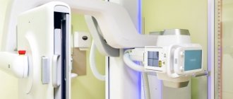
How is it carried out?
HSG is an outpatient procedure that does not require prior hospitalization of the patient. It is performed without the use of general anesthesia (anesthesia). The sensations experienced by a woman resemble mild menstrual discomfort and usually go away quickly on their own. Local anesthesia is only sometimes used to relieve a woman of discomfort during transcervical administration of contrast fluid.
The patient is positioned on a gynecological chair. The doctor disinfects her genitals, after which a catheter is carefully inserted into the cervical canal. Through it, the uterus is partially filled with a warm contrast solution or saline solution, depending on the type of diagnostic used. At the same time, the doctor takes pictures, recording the nature of the filling of the uterine cavity. Then a second portion of the solution is injected to contrast the fallopian tubes, which is also recorded on the images. The entire procedure usually takes no more than 15–30 minutes.
Checking the uterine tubes is not a painful or dangerous type of diagnosis; after some time, if necessary, it can be repeated.
Reasons for the formation of adhesions in the fallopian tubes
Before considering the causes of adhesions in the fallopian tubes, you should familiarize yourself with the mechanism of development of the adhesions process. The fallopian tubes are externally covered with visceral peritoneum, and the abdominal cavity itself is lined internally with parietal tissue. These layers have a smooth surface and secrete a certain amount of fluid, which allows the organs to move freely relative to each other.
If reasons arise that lead to the formation of adhesions, then the fallopian tubes and other organs involved in the pathological process become swollen. A sticky fibrin coating appears on their surface. It is he who contributes to the fact that organs located next to each other are connected to each other. When the inflammatory process has a long course or becomes chronic, adhesive cords form in place of the glued surfaces. In this way, the body responds to inflammation and prevents its further spread. However, the formation of adhesions cannot be considered a normal physiological phenomenon, since it is both pathological and protective in nature.
When the infection penetrates the fallopian tubes, exudate begins to accumulate in them. With adequate therapy, it resolves and does not lead to the formation of adhesions. However, situations are not uncommon when this exudate becomes purulent and spreads throughout the pipe. It is capable of pouring into the peritoneal cavity with fibrin loss. This provokes blockage of the abdominal opening of the fallopian tube by adhesions.
Further progression of the pathological process leads to the fusion of the opposite surfaces of the pipe with each other through partitions. Often, adhesions glue the fallopian tubes to the uterus, oviduct, ovary, intestines, and omentum.
So, doctors consider the inflammatory process to be the main reason for the formation of adhesions in the fallopian tubes, although other factors also influence the growth of strands, including:
- Endometriosis.
This pathological process is characterized by the growth of the endometrium in atypical places, including in the fallopian tubes. (Read more: What is endometriosis? Causes and symptoms of endometriosis).
- Mechanical effects on the fallopian tubes, namely surgical interventions.
In this regard, surgical abortion, ovarian resection, myomectomy, hysteroscopy, intrauterine contraception, IVF attempts, etc. are dangerous. Statistics show that 50% of women who have undergone gynecological operations develop adhesions. Factors such as heavy blood loss, the addition of a purulent infection, and a prolonged course of the operation increase the risk of their formation.
- Undergoing laparoscopy of the genital organs.
No matter how carefully the procedure is performed, it is almost impossible to avoid trauma to the serous membrane of the peritoneum. Therefore, even this operation can trigger the formation of adhesions, which can ultimately block the lumen of the fallopian tubes.
- Difficult childbirth can result in the formation of adhesions
. In this case, the connecting cords grow in the uterine cavity, reach the fallopian tubes and block the entrance to them. A cesarean section, as well as multiple ruptures during the delivery process, are the main factors that lead to the proliferation of connective tissue cords.
- Injuries to the pelvic area accompanied by rupture of an ovarian cyst are dangerous.
In this case, external adhesions are able to attach to the wall of the fallopian tube and block its lumen.
- Infections play a role in the pathogenesis of adhesions formation
sexually transmitted diseases: syphilis, gonorrhea, mycoplasmosis, chlamydia, etc. No less dangerous is genital tuberculosis, which leads to deformation of the tissue structure and the formation of obstruction of the fallopian tubes due to adhesions and scars.
- Inflammatory diseases of the reproductive system
non-infectious nature, for example, salpingitis is another cause of tubal obstruction.
- Reasons that less often lead to the formation of adhesions
– these are hormonal imbalances and irradiation of the genital organs for the treatment of cancer.
The number of inflammatory processes experienced directly affects the risk of developing adhesions in the fallopian tubes.
The statistics are as follows:
- Single inflammation of the fallopian tubes – the risk of formation of adhesions is 12%.
- Having had inflammation twice – the risk increases to 35%.
- Three episodes of inflammation of the appendages - the risk of formation of adhesions in the fallopian tubes is 75%.
What to do after diagnosis
The liquid introduced during HSG is excreted independently through the vagina, so after the procedure it is advisable for a woman to use a pad. For several days, a woman may experience moderate stretching and heaviness in the lower abdomen and in the projection of the ovaries; this is acceptable and does not require urgent consultation with a doctor. To reduce such sensations, it is usually sufficient to take antispasmodics and non-narcotic analgesics (painkillers); this point must be clarified in advance with a gynecologist.
For 3 days after the examination, it is not recommended to use tampons and vaginal devices for collecting secretions (menstrual cups). You should also refrain from douching, intimate relationships, bathing, visiting baths and saunas.
Symptoms of adhesions in the fallopian tubes
The presence of connecting cords in the fallopian tubes may not give themselves away. The fact is that this pathological process does not affect the woman’s well-being. It is possible to suspect adhesive disease of the fallopian tubes only when pregnancy does not occur for a long time (a whole year) in the absence of contraception. Although, with partial patency of the fallopian tubes, conception can still occur.
If the patient suffers from acute or chronic inflammation of the appendages, then the clinical signs of the existing disease come to the fore. They can be diverse and depend on the specific pathology. However, the inflammatory process against the background of formed adhesions will always be reflected by painful sensations in the lower abdomen. The pain can be either moderate or quite severe. They tend to intensify during intimacy and physical activity.
A change in the nature of vaginal discharge is possible. They may contain pus and mucus. The volume of discharge, even without pathological impurities, increases.
Other signs indirectly indicating the adhesive process:
- Periodically occurring pain in the lower abdomen;
- Exacerbation of discomfort due to mechanical irritation of the uterus during regular menstruation;
- Increased volume of vaginal discharge;
- Hypomenstrual syndrome with scanty discharge, which is observed for a long time;
- The menstrual cycle may become unstable, periods will be absent for a long time;
- All symptoms occur against the background of normal body temperature.
An ectopic pregnancy can also be an indirect sign of adhesive disease of the fallopian tubes. Therefore, after the first such incident, it is imperative to check the remaining pipe for patency.
Where can I get an HSG done?
You can check pipes for permeability in St. Petersburg in many institutions that have the necessary equipment and certificates. But when choosing a clinic, you should take into account that modern ultrasound hysterosalpinoscopy is not used everywhere. And the reliability of the results largely depends on the qualifications of the doctor and the quality of the equipment. Therefore, whenever possible, preference should be given to modern, well-equipped medical centers.
In St. Petersburg, the necessary examination can be completed at the ICLINIC Reproductive Medicine Clinic. We specialize in the diagnosis and treatment of various forms of marital infertility, using modern, highly effective and proven methods and high-class equipment.
Testing of the fallopian tubes in ICLINIC is carried out using contrast ultrasonic hysterosalpingoscopy (CUUSH). This is an important modern diagnostic tool that allows you to quickly, safely and with a minimal degree of invasiveness assess the patency of the tubes and the likelihood of a natural successful conception.
After examination at ICLINIC, the patient receives a qualified expert opinion and has the opportunity to consult with experienced reproductive specialists. Our doctors will help you undergo the necessary examination and choose the optimal infertility treatment regimen.
Laparoscopic surgery - salpingo-ovariolysis
The purpose of the operation is to restore normal topographic relationships by cutting the adhesions around the fallopian tube and ovary, isolating them from each other. Salpingo-ovariolysis is performed either as an independent intervention or as a preparatory step for surgery on the fallopian tubes. The fallopian tube (ovary) is picked up with atraumatic forceps and moved upward if possible. The adhesions are cut with endoscissors after their preliminary coagulation. Rough adhesions are excised and removed from the abdominal cavity. Special precautions are necessary when working near the intestines, ureters, and large vessels. The risk of their damage can be reduced by observing the following conditions: the operation is performed under general anesthesia with sufficient relaxation, the absence of defects in the insulating braid of the instruments, short-term switching on of the electrosurgical unit. After complete release of the fallopian tube from adhesions along its entire length, ovariolysis is performed. In this case, it is imperative to lift the ovary and inspect its surface facing the broad uterine ligament, since adhesions can often be localized there.
Complications of the adhesive process of the fallopian tubes
Reproductive dysfunction is the main complication of the presence of adhesions in the fallopian tubes. A woman may become infertile or unable to bear a child. The basal layer of the endometrium becomes damaged, which will prevent fertilization of the egg or cause difficulties with implantation of the embryo.
A third of patients, even with successful conception and gestation, face certain problems during the delivery process. These difficulties are associated with the development of bleeding in the postpartum period.
Failures after IVF can also be caused by the presence of adhesions in the tubes and uterus.
Another complication of adhesions in the fallopian tubes is the occurrence of chronic pelvic pain, which worsens a woman’s quality of life and contributes to the development of neuroses.
Organ structure
The inside of the fallopian tube is lined with ciliated epithelium. The organ wall consists of four layers:
- Serous - comes from the peritoneum;
- Subserous - loose tissue with blood and lymphatic vessels, smooth muscles;
- Lamellar – connective tissue;
- Epithelial.
The shell contains the most ciliated cells; the growth of villi on them is activated by extragen. Sensory cells contain granules that produce fluid. It serves as a source of nutrients for the egg and sperm. The number of such cells is influenced by progesterone.