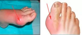
If a person feels pain in the jaw, radiating to the ear and temple, we may be talking about arthritis of the mandibular joint. The pathology is not just painful, but also dangerous. If not diagnosed in a timely manner, it can lead to severe complications, including paralysis of the facial nerves. Therefore, the first thing to do when such symptoms appear is to consult a specialist. In 10-18% of cases, diagnosis and treatment of TMJ arthritis is the responsibility of dentists and occurs in people from 25 to 45 years old.
Anatomical description and location
The blue triangle delimits the temporal region of the head.
The temporal region occupies the lateral sections of the cranial vault and is limited by the zygomatic arch along the lower edge, the outer part of the orbit from the anterior edge, the linear elevation of the frontal bone from the upper edge, the temporal bone and the auricle along the posterior edge. The anatomy of the temple has a rather complex structure:
- Skin. Deepening above the zygomatic arch, they become thinner and separated from fatty (lipid) deposits. Cover the thin terminal branch of the external carotid artery (Arteria temporalis superficialis), which passes in front of the cartilaginous protrusion of the auricle.
- Fat deposits are almost invisible. They contain superficial blood vessels and nerves.
- The superficial fascia is a continuation of the tendon helmet.
- The temporal fascia consists of two dense plates, between which the interaponeurotic space is enclosed.
- The temporal muscle (musculus temporalis), covering the temporal fossa, in the thickness of which there are deep arteries and nerves, as well as lymphatic vessels.
- The periosteum is quite thin and firmly attached to the bone.
The bone base of the temple is the temporal fossa, formed by the parietal squama, the greater wing of the sphenoid and the process of the frontal bone. The bone structures are thin, covered from the inside by grooves of branches of the middle meningeal artery. The temporal region is separated from the infratemporal fossa by the body of the infratemporal crest of the sphenoid bone.
Anatomy of the temporal region
In the subcutaneous layers of tissue of the temporal region, between the plates of the tendon helmet, together with the blood vessels, lies the auriculotemporal nerve, which provides innervation to the temple. This nerve originates from the third branch of the trigeminal nerve. It surrounds it with its endings near the oval opening of the cranium, through which the meningeal branch of the maxillary artery runs, and, bypassing the temporomandibular joint, goes to the temple in the area of the external auditory canal and the superficial artery of the same name.
The zygomaticotemporal nerve passes through the cellular spaces from the second branch of the trigeminal nerve. It enters the temple area through the hole of the same name, which penetrates the outer orbital wall. Near the tragus (the protruding cartilage of the auricle) in the direction of the outer corner of the eye, the zygomatic and temporal branches of the facial nerve pass, providing motor innervation to the muscle fibers of the visual organ, and sometimes to the ear muscles.
In the area of the zygomatic arch lies Arteria temporalis superficialis, which divides into frontal and parietal branches. It is accompanied by veins of the same name, formed from the saphenous veins of the cranial vault, uniting through the parietal openings with the venous sinus of the dura mater of the brain. Together with the maxillary vein, they form the premaxillary vein, which originates in the parotid salivary gland.
Diseases, injuries of the temporal region
Diseases
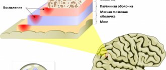
Among V.'s diseases. There are acute and chronic, inflammatory, including specific diseases, tumors of various tissues, fibrous osteitis. Of the acute inflammatory diseases of soft tissues, in addition to erysipelas, the edges usually spread from other parts of the scalp; the most common is deep phlegmon of V. o. Due to its anatomical features, pus accumulates in a closed space between the bone and fascia and hangs over the zygomatic arch, without a tendency to spread to the cranial vault. A breakthrough of pus into the infratemporal, pterygopalatine fossa and to the base of the skull is possible. The opening of the phlegmon is made by two incisions: the front one - slightly inclined anteriorly and the back one - vertical, opening the front and back parts of the fascial sac; if there is pus between the bone and the muscle, the latter must be carefully peeled away from the bone. A transverse incision should not be used, since this can cross vertically running vessels and nerves.
Osteomyelitis of the temporal bone is rare. More often it occurs as a result of the transition of the inflammatory process from soft tissues. Damage to the inner plate of the temporal bone is dangerous due to the possibility of pus breaking into the cranial cavity. There is a high probability of a purulent process transferring to the membranes of the brain in osteomyelitis after open fractures of the temporal bone. Treatment should be active - trepanation with removal of the affected bone, constant aspiration of pus (see Osteomyelitis).
Hron, specific inflammatory diseases (tuberculosis, syphilis) affect V. o. rarely.
Of the diseases of V. o. Non-inflammatory osteitis fibrous, fibromas, angiomas deserve attention. Fibrous osteitis of the Recklinghausen type (see Parathyroid osteodystrophy) occurs in two forms - generalized with multiple lesions of the skeletal bones and limited, in which only the bones of the skull are affected. The disease is rare. Fibrous osteitis of the skull is most often localized in the temporal bone, moving from there to the parietal and zygomatic bones. In V. o. a uniform swelling of bone consistency appears. The bone is sharply changed, thickened, turned into a spongy mass, external and internal plasticity are indistinguishable. This bone structure gives a characteristic x-ray picture with a mesh cellular pattern. The disease must be differentiated from sarcoma, which is characterized by faster growth. With a limited form of fibrous osteitis, surgical removal of the affected area of bone with plastic replacement of the defect can give a favorable result. With extensive lesions, the prognosis is unfavorable.
Rice. 2. Hanging fibroma of the temporoparietal region.
In V. o. Hanging fibroma (fibroma pendula) is relatively common. Fibromas are localized in the thickness of the skin or grow under its surface in the form of warty soft formations (Fig. 2). Treatment boils down to removing individual tumors within healthy tissue.
Of the angiomas of the integument of the skull, cavernous angiomas, which grow in the V. of the lake, are of greatest practical importance. not only the skin, but also the subcutaneous tissue, temporal muscle and can spread to the bone. In the latter case, the angioma communicates with the vessels of the cranial cavity and the venous sinuses of the dura mater. Angiomas are easily traumatized, resulting in bleeding, ulceration, and inflammation. The prognosis is often unfavorable. Treatment Ch. arr. surgical. Angiography helps to plan the upcoming operation (see). For small-sized cavernous angiomas, radiation treatment, injection of 96% alcohol into the angioma, and cryotherapy are used with good results (see Hemangioma).
Cirsoid arterial angioma is a vascular tumor consisting of dilated, tortuous nodular arterial trunks that directly extend into dilated venous collectors. It should be considered characteristic of the presence of pulsation and vascular noises: blowing - venous and systolic - arterial. These angiomas develop in the area of the branching of the temporal artery and other branches of the carotid artery and sometimes occupy almost half of the head. They cause pain of a neuralgic nature (damage to the auriculotemporalis) and constant pulsating noises that are very painful for patients. Treatment is surgical in combination with radiation therapy. The prognosis without surgery is unfavorable (risk of bleeding).
Rice. 3. Fracture (crack) of the scales of the temporal bone with transition to the parieto-occipital region. Rice. 4. Fracture (light oblique line at the top) of the pyramid of the temporal bone (x-ray, special placement).
Cellular space
In the temple area there are two fascia: superficial (in the form of a thin plate running across the entire temple) and proper. When they split, they form an interfascial fiber gap filled with fatty tissue, penetrated by many fibrous bridges.
The cellular spaces of the temporal region consist of the following structures:
- Subcutaneous tissue lying between the skin and the superficial fascia. This is a kind of continuation to the temple area. It contains blood vessels, nerve and muscle fibers.
- Interponeurotic tissue located between the superficial and deep plates of the temporal fascia.
- Subgaleal tissue enclosed between the temporal fascia and musculus temporalis. It contains fat and veins.
The last, deep layer is located between the temporalis muscle and the periosteum. It runs along the blood vessels and nerves that supply muscle tissue.
Treatment
At the moment, there are two directions in the treatment of temporal arteritis: therapeutic and surgical . Surgical methods are resorted to in the event of the development of complications such as aortic aneurysm and thrombosis of blood vessels, especially those supplying blood to the eyeball.
The basis of treatment for the disease, without which it is impossible to achieve positive results, is the prescription of steroid hormones (prednisolone). There is no alternative to glucocorticoids. They are prescribed as early as possible and taken for a long time. Doses and regimen are selected individually, under constant laboratory monitoring of clinical and biochemical blood parameters. A combination of hormones and drugs that suppress immune responses is possible. Symptomatic treatment is also carried out using anticoagulants, agents that improve microcirculation, and vitamins.
Functional meaning
The temple, like the entire cranial vault, performs a protective function, preventing mechanical damage to the brain. This area contains brain regions responsible for the following functions:
- visual and long-term memory;
- processing of sensory information entering the central nervous system;
- acoustic perception with receiving and processing of sound signals;
- visual perception.
The temporal region is penetrated by the most important nerves and blood vessels that supply the brain, organs of hearing and vision.
Diagnostics
The diagnosis of arteritis can be established by performing a histological examination of a section of the superficial temporal artery obtained by biopsy. The sample collection is carried out under local anesthesia and is not difficult. The detection of granulomatous inflammation of the vascular wall with the presence of giant cells is indisputable evidence of this pathology.
But histologists are able to identify typical changes only in half of the cases. The fragmentary nature of the lesion does not always allow for successful selection of the segment for biopsy. However, a negative result does not mean the absence of the disease, since the main criterion for diagnosing Horton’s disease is the totality of clinical manifestations.
Criteria have been formulated and generally accepted to recognize temporal arteritis. The diagnosis is reliable if three or more of the following factors are present:
• age over 50 years;
• headaches with severe intensity;
• vision problems;
• presence of complaints characteristic of polymyalgia rheumatica;
• decrease in the number of red blood cells and hemoglobin level in the blood, increase in ESR.
Sphygmography, rheovasography, and Dopplerography of the affected arteries are of auxiliary value for differential diagnosis. For the same purpose, the presence of C-reactive protein and the level of sialic acid and fibrinogen in the blood are determined.