Photo of worms in the human body. People and animals are attacked by these creatures; the infection is easily transmitted to each other through contaminated food, water and dirty hands. Prevention rules and careful personal hygiene will help prevent the appearance of parasites in the body, but even these measures do not guarantee one hundred percent protection. Helminths that inhabit the human body can live in various organs and differ in appearance, size, and the degree of harm caused to humans.
Parasites in the human body
How many situations are there when a person goes to doctors for years and cannot get rid of allergies, treats asthma, takes glucose-lowering drugs and all to no avail? Each of us has such acquaintances who have spent huge sums on the treatment of a variety of diseases, and have not received any results.
Only in rare cases, when the doctor turns out to be either very smart or very responsible, does he refer such a patient for a routine stool analysis and then... Then parasites are discovered in the human body, which are the cause of dozens of diseases, and with which no one can treat these pathologies doesn't fight.
Photo of worms. Contrary to popular belief, worms are not necessarily “prescribed” in the intestines and can be detected by a simple stool test. Many parasites thrive in the lungs, heart, muscles, even in the brain and eyes.
The well-known malaria, which was once defeated on the territory of the USSR, has returned again and this is the most dangerous parasitic disease, according to WHO experts. Malarial plasmodium lives exclusively in the blood and not every doctor will be able to recognize this disease with sufficient confidence.
Diagnostics
Identifying parasites can be difficult, especially if the disease has just begun. Typically, the first tests a patient is sent for are stool, blood and saliva tests. Diagnosis and treatment should be carried out by a parasitologist or infectious disease specialist.
To accurately confirm the presence of pests in the body and determine their type, they need to be seen. Most often, the diagnosis is confirmed after a stool examination. If worms have affected the organs of the digestive system, then it will contain either whole individuals or their eggs. This way you can detect most types of parasites.
If the body is affected by those pests that live in the organs of the non-digestive system and blood, the diagnosis can be confirmed by examining the blood. Considering that they can circulate through the bloodstream at different times, tests are usually taken several times: in the morning, during the day, in the evening, and at night.
As an additional diagnosis, especially if the suspected parasites are large or there are many of them, ultrasound is prescribed.
Blood for toxocariasis
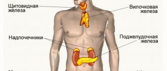
Typically, examination of the following organs is required:
- Gastrointestinal tract (stomach, rectum and others);
- liver;
- gallbladder with ducts;
- muscles;
- lungs.
If the helminthic infestation is in an advanced form, the doctor may prescribe a brain diagnosis. Due to the long life of parasites, neoplasms can be diagnosed, including in the brain. Thanks to ultrasound, it is possible to assess the scale of the negative impact on the body.
How parasites enter the human body
They enter the human body in different ways, most often through the consumption of contaminated water and food.
Pinworm eggs remain viable for up to 6 months and enter the body through toys, carpets, underwear and bedding. Ascaris eggs get inside us through poorly washed vegetables and fruits. Shish kebab or homemade lard is a 95% guarantee of infection with trichinosis.
Parasites penetrate inside us through insect bites, when swimming in freshwater bodies, through the air, and through dust, which carries eggs.
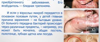
Salted fish, stroganina or caviar are the cause of infection with a tapeworm, the length of which reaches 12 meters and which can live in your body for up to 25 years. Cases of infants becoming infected with parasites in the womb have become more frequent. Dogs and cats, through their moist breath, can disperse parasite eggs at a distance of up to 5 meters.
You can become infected through dirty hands, not only your own, but also sellers, cooks, waiters; parasite eggs travel on money and handrails of public transport. A high concentration of parasite eggs is observed in products such as: bacon, smoked sausage, ham, sausages, pork of any form, beef, chicken, lamb, and even chicken eggs are very often contaminated with them.
Epidemiologists around the world are trying to fight this scourge. In the USA, for example, 1 pig carcass out of every thousand is destroyed to test for helminthiasis. This adds up to multimillion-dollar losses, but there is no other way.
There are no absolutely reliable methods for disinfecting meat, and ordinary cooking does not destroy the larvae. You cannot guarantee the purity of your food by boiling or frying meat; a huge number of parasite larvae still penetrate your body.
Prevention of infection
Often, parasite infection can be prevented; to do this, it is recommended to follow these rules:
- wash your hands with soap before eating, after walking outside, after each contact with pets;
- monitor the hygiene and cleanliness of pets;
- refuse contact with stray animals;
- thoroughly wash vegetables and fruits, soak greens in a vinegar solution;
- drink only purified water (at least boiled);
- clean the house daily, wipe the dust;
- Do a monthly general cleaning of your house or apartment.
By following these simple recommendations, you can protect yourself from not only helminth infection, but also many other diseases.
It is important to know what parasites are, what consequences their presence in the body can have and how to recognize them. The main method of controlling helminths is drug therapy, which includes the use of drugs with a broad spectrum of action. In addition, traditional medicine can be used. The sooner an infestation by parasites can be recognized, the greater the undesirable consequences that can be avoided.
In which human organs can parasites live?
Photo of worms. Helminthic parasites are divided into two categories, which correspond to the location of activity in the donor’s body:
- cavitary : worms that live in various parts of the gastrointestinal tract. There are about 100 species of intestinal parasites, and for every part of the intestine there are a couple of dozen species. The small intestine is ready to accept roundworms, antelostomas, broad tapeworms and other less common “brethren”. The small intestine will “share living space” with pinworms, dwarf tapeworms and others. The medical literature describes cases where one person was infected simultaneously with several types of parasites;
- tissue: worms localized in organs, tissues and even in the blood. Modern medicine successfully copes with paragonimiasis (lungs), cysticercosis (brain), echinococcosis (liver) and filariasis (lymphatic vessels). Some worm larvae move throughout the body through the circulatory system and randomly attach to any organ. If many eggs are introduced, the entire body can be infected.
Possible complications
All parasites in the human intestine are divided into two types:
- Cavity. They affect the organs of the digestive system and live in different parts of the gastrointestinal tract. Complications that arise from cavity pests mainly affect only this department.
- Fabric. They affect various internal organs, tissues and blood. Through the bloodstream, their eggs are quickly transported throughout the body, so complications from the life of tissue parasites usually affect the entire body.
As microorganisms parasitize the internal organs of a person, concomitant diseases develop. These include such serious pathologies as bronchial asthma, diabetes mellitus, and oncology. This is explained by the fact that constant intoxication occurs inside the body, which intensifies as the parasites multiply.
And also undesirable consequences may be associated with a deficiency of beneficial nutrients, which occurs due to the fact that they are absorbed by worms. For example, due to silicon deficiency, osteochondrosis may occur. If the patient does not undergo a comprehensive diagnosis and, without knowing the root cause, treats this pathology, there will be no result.
A connection has been established between parasitic invasion of the body and mental disorders such as chronic fatigue syndrome, depression, and other conditions associated with loss of strength and apathy.
Pinworms - what they look like in the human body (Photo of worms)
Pinworms are one of the most common human parasites, which are a roundworm (nematode). Most often, pinworm infection occurs in children, but it also occurs in adults.
The pinworm is a white parasite, small in size and round in shape. Female specimens measure 8-13 mm in length, 0.5 mm in thickness, oblong in shape and have a straight tail, pointed at the end.
This feature of the tail of the female parasite explains its name - “pinworm”, from the word “sharp”. The male pinworm is much smaller: its length is 2-5 mm, its thickness is 0.2 mm, its tail is curved, unlike the female pinworm.
Photo of worms. Infestation of humans with pinworms is called enterobiasis, and occurs mainly due to non-compliance with personal hygiene rules (insufficient hand washing). Pinworms primarily live in the small intestine and upper part of the large intestine, but in some cases they can also migrate to other organs and organ systems.
The female helminth, having entered the human body orally and mated with the male representative of the nematode, migrates to the large intestine, where she receives the necessary nutrients for life and the maturation of eggs from undigested food debris.
After 4 weeks, the female pinworm begins migrating into the rectum at a speed of 12 cm per hour, crawls out of the anus and lays about 5,000-15,000 eggs in the perianal area, which after 4-6 hours are fully mature and ready for the further life cycle.
This process may be accompanied by itching, which prompts the infected person to scratch the anus, and thus contribute to the further spread of parasites that fall from under the nails into food onto the hands of other people (especially for children who are in very close contact with each other and not always observing the rules of personal hygiene).
Pinworm infection occurs from person to person, through dust with parasite eggs, or objects touched by the patient. Pinworm eggs can also be transferred to food by cockroaches and flies.
Eggs also remain on linens, clothes, and beds, which explains their rapid spread. Due to the fact that the life cycle of the worm is very fleeting, and infection occurs from person to person, it is quite difficult to get rid of parasites, since in addition to taking anthelmintic drugs, it is necessary to treat the patient’s personal belongings and isolate him from other carriers of the nematode.
Treatment
It is impossible to get rid of the infection without drug treatment, so the basis of therapy is always taking medications. If the patient's condition is severe, hospitalization and constant monitoring by the attending physician may be required. Folk remedies will be effective only as an addition to drug therapy.
It may take several years to completely remove parasites from the body, it all depends on their type and the timeliness of seeking medical attention.
Medication
Previously, choosing a medicine was not easy, since most antiparasitic drugs had a narrow spectrum of action, that is, they were effective in the fight against a certain type of helminth. Today there are remedies used for any helminthic infestations.
Most antiparasitic drugs are contraindicated during pregnancy and lactation. For women in such conditions, therapy is selected strictly by a doctor; herbal remedies are often prescribed.
The following drugs are considered the most effective:
- Albendazole - there are also several drugs in which albendazole is an active ingredient: Nemozol, Gelmodol, others. The products are available in the form of tablets and suspensions.
- Mebendazole - there are also several drugs in which mebendazole is an active ingredient: Vormin, Vermox. It affects the metabolic processes inside the parasites, disrupts them, which causes the death of the pests.
- Levamisole or Decaris. Often used either for prevention or for minor infections. The package contains 1 tablet of the drug, it is taken before bedtime. If nausea, vomiting, and other signs of intoxication appear in the morning, this means that more serious antiparasitic therapy is required.
And also general strengthening, painkillers, and other groups of drugs can be prescribed, but the prescription is strictly made by the attending physician individually.
Roundworms - what they look like in the human body (Photo of worms)
Ascaris is a large, spindle-shaped, red-yellow parasite, reaching 40 cm (females) and 15-25 cm (males) in adulthood. Without suction cups or other fastening devices, the roundworm is able to move independently towards food masses. The eggs laid by the female parasite are excreted in the feces.
Infection with ascariasis occurs when mature eggs are ingested along with water or unwashed vegetables and fruits that have soil particles on them. After the eggs penetrate the intestines, mature larvae emerge from them.
Then, penetrating into the intestinal wall, they reach the heart through the bloodstream, and from there they enter the lungs. Through the pulmonary alveoli, the roundworm larva again enters the oral cavity through the respiratory tract.
Photo of worms. In the intestinal phase of their existence, roundworms, endowed with the ability to spiral movements, can penetrate even the narrowest openings. This feature of the parasite often leads to the development of quite serious complications (obstructive jaundice or pancreatitis)
Once ingested again, the parasite reaches the small intestine where it develops into an adult. The worm lives for 12 months, then dies and is excreted in the feces. One or several hundred individuals can live in the intestines of one host.
Allergens released by roundworms can provoke severe allergic reactions. A large number of adults can cause intestinal obstruction, and worms that enter the respiratory tract sometimes cause suffocation.
Whipworms - what they look like in the human body
This type of parasite is quite rare in central Russia. Whipworms often live in the southern regions, since the eggs of this worm love warmth. Most infections are observed in rural areas. Whipworm eggs live in the soil.
Infestation occurs through hands, contaminated soil particles, and poorly washed vegetables and fruits. As a result of infection, a disease occurs - trichocephalosis. Whipworm parasitizes the intestines. This worm causes anemia, as it feeds on human blood, and severe abdominal pain.
To diagnose trichuriasis, the rectum and sigmoid colon are examined with a special device (sigmoidoscopy). In this way, accumulations of parasites in the intestines are detected. Treatment of the infestation takes a long time, since the whipworm eggs are protected by a dense shell.
The eggs of the parasite are excreted in the feces, but they are very small and cannot always be seen even under a microscope. Only with very severe infestation is it possible to detect eggs in a stool test. They are shaped like a barrel and have a brownish-yellow color.
There are holes on both sides of the egg. What do worms look like in feces? They are very difficult to detect alive in feces, since whipworms cannot live long outside the human body. Only with anthelmintic therapy can you notice dead white worms in the feces.
Laboratory tests to identify parasites - tests for worms
Stool examination.
The main laboratory test is examination of stool under a microscope, which allows the identification of cysts, eggs and larvae of parasites. It is very important to do this before starting treatment.
To assess the effectiveness of antiparasitic drugs, a stool sample should be collected 1-3 weeks after the end of therapy. It is recommended to collect the test material three times with an interval of 2-3 days for 10 days.
For patients returning from tropical countries, 4 samples are recommended (last test after challenge with cleaning products). This increases the chance of detecting infection because some forms appear cyclically in the stool.
Increasingly, stool testing is carried out using the Parasep SF system, which is a disposable equipment used for the initial preparation of the test material. This sample preparation allows for more thorough cleaning before microscopic examination, increases the efficiency of infection detection and reduces examination time.
However, it is impossible to detect pinworms using standard methods. They are diagnosed not by examining the feces, but by using sticky tape around the anus.
In addition to microscopic examination of stool, there are also enzyme immunoassays that can detect specific parasite antigens in the stool. They are most often used to confirm infection with parasites such as intestinal giardia, amoebiasis or cryptosporidium. These tests are less important than a basic microscopic examination of the stool; they complement the diagnosis.
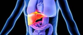
Liver fluke - what it looks like in the human body
The parasite that causes opisthorchiasis is a flatworm that reaches a length of 7-20 mm. It should be noted that more than 50% of cases of infection with liver fluke (also called cat fluke) occur among residents of Russia.
In the acute phase of helminthiasis, the patient experiences pain in the upper abdomen, body temperature rises, nausea, muscle pain develops, possible diarrhea, skin rashes. The larvae of the parasite begin to develop after the eggs enter fresh water (from the snails that swallowed them). Then they penetrate the body of the fish (carp, crucian carp, bream, roach).
Photo of worms. Human infection occurs by eating contaminated fish meat that has not undergone sufficient heat treatment. The larva of the liver fluke from the small intestine penetrates the bile ducts and the gallbladder, fixing there with the help of two suction cups.
The chronic course of opisthorchiasis is manifested by symptoms of hepatitis, inflammation of the bile ducts, cholecystitis, disruption of the digestive tract, nervous disorders, weakness and increased fatigue. The parasite leads to the development of irreversible changes, and even after its expulsion, the patient continues to experience chronic inflammatory processes and functional disorders.
Helminthiasis
Parasitic worms found in the body of humans, animals and plants cause diseases called helminthiasis. Human parasitic worms disrupt the functioning of internal organs and the entire body as a whole, causing poor absorption of nutrients, weakness and diseases of the host body. There are three groups of worms that are classified as helminths:
- Nematodes or roundworms (pinworms, roundworms, Trichinella, Filaria, Intestinal eel);
- Trematodes or flatworms (Opisthorchis, Chinese fluke, Paragonimus, Schistosomes);
- Cestodes or tapeworms (Bovine and pork tapeworms, broad tapeworms, echinococcus, alveococcus).
Helminths have a similar morphology and are multicellular organisms that are visible to the naked eye. In their life cycle, helminths necessarily go through a number of stages: egg, larva and adult. During their life cycle, some worms change hosts. Such helminths are called biohelminths. Geohelminths are parasitic worms that do not require a change of hosts, and their life cycle occurs in the body of a single host. NEMATODOSIS Ascariasis Etiology, pathogenesis .
The causative agent is roundworm, which parasitizes in the adult stage in the small intestine. The lifespan of roundworms is about a year. In the migratory stage (the first 6–8 weeks after infection), roundworm larvae have a mechanical and sensitizing effect, causing hemorrhages and eosinophilic infiltrates in the tissues of various organs. In the intestinal phase (8 weeks after infection), adult roundworms cause general toxicoallergic and neuroreflex reactions of the body and a variety of local mechanical effects.
Symptoms, course
. The migration phase often occurs under the guise of acute respiratory infections and bronchitis; volatile eosinophilic infiltrates in the lungs are possible. In the intestinal phase, a gastrointestinal form is distinguished (salivation, nausea, loss of appetite, cramping pain around the navel, sometimes disorders of stool and gastric secretion); hypotonic (decreased blood pressure, weakness) and neurological (dizziness, headache, fatigue, sleep disturbance, vegetative-vascular disorders) forms.
Complications
. Ascariasis intestinal obstruction, a characteristic feature of which is the release of roundworms with vomit and palpation of a moving “tumor” - a ball of helminths; ascariasis appendicitis; perforated peritonitis; ascariasis of the liver with the development of jaundice, purulent angiocholitis, liver abscess, subphrenic abscess; ascariasis of the pancreas with symptoms of acute pancreatitis; crawling of roundworms into the respiratory tract with the development of asphyxia.
Diagnostics.
The diagnosis of the early phase of ascariasis is based on the detection of nematode larvae in the sputum and antibodies in the blood, and the late intestinal phase - ascaris eggs in the feces.
Prevention
.
Mass examination of the population and treatment of all those infected with ascariasis. Protection of the soil of vegetable gardens, orchards, and berry fields from contamination with feces. Thorough washing and scalding of vegetables and fruits with boiling water. Personal hygiene measures .
Hookworm disease (hookworm and necatoriasis)
Etiology, pathogenesis.
The causative agents are hookworm and necator, parasitic in the small intestines of humans, most often in the duodenum. Infection occurs when the larvae actively penetrate the skin or when they are ingested with contaminated vegetables, fruits, or water. The larvae migrate through the systemic and pulmonary circulation, lasting 7–10 days. In the small intestine, the larvae turn into sexually mature individuals and after 4–6 weeks begin to lay eggs. The lifespan of hookworms ranges from several months to 20 years. During the migration period, the larvae cause toxic-allergic phenomena. Adult helminths are hematophagous. When fixed to the intestinal mucosa, they injure tissue, lead to the formation of hemorrhage, erosion, cause bleeding, anemia, support the state of allergies, gastrointestinal dyskinesia and dyspepsia.
Symptoms, course
. Soon after infection, skin itching and burning, urticarial rashes, asthmatic phenomena, fever, and eosinophilia occur. In the late stage, nausea, drooling, vomiting, abdominal pain, intestinal dysfunction (constipation or diarrhea), and bloating appear. Sometimes pseudoulcer syndrome occurs (hunger pain in the epigastric region, hidden bleeding). The development of hypochromic iron deficiency anemia is characteristic; its severity depends on the intensity of invasion, nutritional deficiency, and individual characteristics of the organism. With low intensity, the invasion can occur subclinically, manifesting itself as eoeinophilia.
Diagnostics
. The diagnosis is confirmed by the detection of eggs in the stool and occasionally in the duodenal contents.
Prevention
. In areas of hookworm infection, you should not walk barefoot or lie on the ground without bedding. It is necessary to thoroughly wash and scald fruits, vegetables, and berries with boiling water; before eating them, you should not drink unboiled water.
Strongyloidiasis
Etiology, pathogenesis.
The causative agent is the intestinal eel, parasitizing in the intestinal wall (mainly the duodenum), sometimes in the ducts of the liver and pancreas, and during the migration period - in the bronchi and lung tissue. The penetration of Strongyloides larvae into the human body can occur actively through the skin (when walking barefoot, etc.) and through the mouth (when eating contaminated fruits, vegetables, and also when drinking water). Adult parasites, localized in the intestinal wall, injure the intestinal crypts (Lieberkühn's glands), solitary follicles and contribute to ulceration of the mucous membrane, and the larvae, migrating, damage the tissue of the liver, lungs and other organs. Of great importance is the toxic-allergic effect of adult parasites and their larvae on the human body, as well as secondary infection.
Symptoms, course
. Signs of allergy often occur: skin itching, burning, urticaria, eosinophilia, sometimes “volatile infiltrates” in the lungs, etc. In the late stage, the symptoms of gastroduodenitis, enterocolitis, and sometimes cholecystitis predominate. The typical triad is chronic recurrent urticaria, persistent enterocolitis and long-term high eosinophilia. Dizziness, headache, insomnia, and increased excitability are common. Sometimes the disease manifests itself only as eosinophilia and skin itching. The course is long, many years (until specific treatment), since with this helminthiasis (especially with constipation), autoreinvasion is possible. There are indications of a connection between hyperinvasion and the use of corticosteroids and immunosuppressive drugs. In severe forms of the disease, ulcerative enterocolitis develops with dehydration, cachexia, anemia and death.
Diagnostics
. The diagnosis is based on clinical data and the detection of acne larvae in feces (when examined using the Berman method) and in duodenal contents, less often in sputum and urine.
Trichocephalosis
Etiology, pathogenesis.
The causative agent is the whipworm, which parasitizes the human colon. The lifespan of the parasite is about 5 years. Whipworms injure the intestinal mucosa, are hematophagous and promote inoculation of microflora, causing reflex reactions in other organs of the abdominal cavity. Their metabolic products sensitize the body.
Symptoms, course
. Worries include drooling, decreased (less often increased) appetite, pain in the right half of the abdomen and epigastrium, nausea, constipation or diarrhea. There may be inflammatory phenomena in the intestines. Headache, dizziness, restless sleep, irritability are often observed; moderate hypochromic anemia and slight leukocytosis are possible. With intense invasion in children, cases of rectal prolapse (due to persistent diarrhea), epileptiform seizures, and Meniere's syndrome have been described. At low intensity, whipworm infestation is asymptomatic.
Diagnostics.
The diagnosis is made when whipworm eggs are found in the stool.
Prevention
. Mass examination of the population and treatment of all infested forms. Protection of the soil of vegetable gardens, orchards, and berry fields from contamination with feces. Thorough washing and scalding of vegetables and fruits with boiling water. Personal hygiene measures.
Trichostrongylidosis
The causative agents are small helminths from the family Trichostrongylidae. The main source of invasion is sheep, goats, and cattle. A person becomes infected through contaminated food or water.
Symptoms, course
. Characterized by nausea, abdominal pain, unstable stools, dizziness, and headache. Sometimes hypochromic or normochromic anemia and loss of strength develop. The diagnosis is based on the detection of trichostrongylid eggs in the stool.
Prevention.
Compliance with personal hygiene measures.
Trichinosis (trichinosis)
Etiology, pathogenesis
. The causative agent is Trichinella. In the larval stage, it enters the human body with the meat of pigs or wild animals (boar, bear, badger, etc.). After digestion of meat, muscle Trichinella are released from capsules, penetrate into the villi tissue, quickly grow, develop, and already 80–90 hours after infection, females lay numerous larvae, which are spread throughout the body. The place of further development of young Trichinella are the striated muscles, especially the intercostal, masticatory, muscles of the diaphragm, tongue, pharynx, and eyes. Here, after 2–3 weeks, trichinella coil up, become encapsulated, and some of them become calcified. The duration of the intestinal stage of Trichinella is about 2 months. Muscular trichinella remain viable for up to 20 years or more. The breakdown and metabolic products of Trichinella sensitize the body, causing eosinophilia, capillaropathy, muscle infiltrates, edema and other painful phenomena. The mechanical effects of adult Trichinella in the intestines and their larvae in muscles and other tissues, as well as secondary infection, are important.
Symptoms, course
. The incubation period is from 3 to 45 days. Early symptoms are characteristic: facial puffiness, accompanied by conjunctivitis, fever, muscle pain, eosinophilia. Various skin rashes are common, as well as gastrointestinal disorders, hypotension, muffled heart sounds, palpitations, headache, and insomnia. In mild forms, only eyelid swelling and eosinophilia are noted. The duration of the disease is from 2–3 days to 8 weeks or more. Sometimes a recurrent course of trichinosis is observed. Complications: myocarditis, meningoencephalitis, thrombosis of arteries and veins, pneumonia, nephritis, etc.
Diagnostics.
The diagnosis is based on epidemiological (consumption of undercooked pork and other meat, group diseases), characteristic clinical data (eosinophilia, swelling of the eyelids, fever, muscle pain). Serological reactions with trichinosis antigen are used. Confirmation of the diagnosis can be the detection of Trichinella and characteristic infiltrates in biopsied pieces of muscle (trapezius, deltoid, gastrocnemius) or in the remains of meat that caused the disease. A biopsy is performed on the 9th–10th day of illness.
Prevention.
You should not eat raw or undercooked pork, as well as the meat of wild boar, bear, badger, etc.
TREMATODOSIS Clonorchiasis
Clonorchiasis is a helminthiasis of the liver and pancreas caused by the fluke fluke. It is found among the population living in the Amur basin. Pathogenesis, clinical picture, diagnosis, treatment and prevention are the same as for opisthorchiasis.
Metagonimiasis
Metagonimiasis is a helminthiasis caused by a small trematode. The pathogen parasitizes in the small intestine of humans, dogs, cats, and pigs. Human infection occurs through consumption of raw fish. In the intestine, larvae hatch from the metacercariae of Metagonimus, which reach sexual maturity in the thickness of the mucous membrane after 2 weeks and emerge into the intestinal lumen. As a result of mechanical and toxicoallergic effects, enteritis develops.
Symptoms, course
. Abdominal pain, drooling, persistent diarrhea. Sometimes it occurs subclinically.
Diagnostics
. The diagnosis is based on the detection of Metagonimus eggs in the stool.
For prevention, see Diphyllobothriasis.
Nanophyetosis
Nanophyetosis is an intestinal helminthiasis that occurs among the population of the Far East. The causative agent is nanophyetus, which parasitizes the intestines of humans, dogs, cats, minks, and badgers. Infection occurs by eating raw fish. Helminths have a mechanical and toxic-allergic effect on intestinal tissue.
Symptoms, course.
Pain and rumbling in the abdomen, diarrhea, constipation, drooling and nausea at night.
Diagnostics
. Diagnosis is made based on the detection of nanophyetus eggs (resembling diphyllobothrid eggs) in the stool.
Treatment
. Male fern extract in a dose of 3–3.5 g.
Prevention
. You should not eat raw, uncooked or insufficiently salted and dried fish, as well as “live” pike caviar.
Opisthorchiasis
Etiology, pathogenesis
. The causative agent is the cat fluke, or Siberian fluke, which parasitizes the bile ducts of the liver, gallbladder and pancreatic ducts of humans, cats, dogs, etc. The parasite lives in the human body for 20–40 years. Infection occurs when eating raw (thawed, frozen), lightly salted and insufficiently fried carp fish (ide, chebak, dace, etc.). Opisthorchises injure the mucous membranes of the pancreatic and bile ducts, create obstacles to the outflow of bile, and contribute to the development of cystic enlargements and liver tumors. They have toxic and neuro-reflex effects. In the early stage, pronounced allergization of the body is observed (eosinophilic-leukemoid blood reactions).
Symptoms, course
. The incubation period is about 2 weeks. In the early period there may be fever, pain in muscles and joints, vomiting, diarrhea, pain and enlargement of the liver, less often the spleen, leukocytosis and high eosinophilia, allergic skin rashes. In the chronic stage, the most common complaints are pain in the epigastric region, right hypochondrium, radiating to the back and left hypochondrium. Sometimes there are attacks of pain such as gall bladder colic. Frequent dizziness and various dyspeptic symptoms. Muscle resistance in the right hypochondrium, liver enlargement, occasionally icteric sclera, enlarged gallbladder, and symptoms of pancreatitis are detected. Most often, with opisthorchiasis, the phenomena of angiocholitis, cholecystitis, biliary dyskinesia, chronic hepatitis and pancreatitis develop, and less often - symptoms of gastroduodenitis and enterocolitis. In some cases, a picture of cholangitic cirrhosis of the liver occurs. Opisthorchiasis can be asymptomatic.
Diagnostics
. The diagnosis is based on the detection of helminth eggs in the stool and duodenal contents.
Prevention
. Explaining to the population the dangers of eating raw, thawed and frozen (stroganina), lightly salted and insufficiently fried fish.
Fascioliasis
Etiology, pathogenesis.
Pathogens: liver fluke and giant fluke. The main source of human invasion is various farm animals. Human infection usually occurs in the warm season when fasciola larvae are ingested in water, sorrel, lettuce and other greens. The lifespan of helminths in the body is about 10 years. Trauma and toxic-allergic damage to the hepatobiliary system are important. Fasciolae can be carried into other tissues and organs.
Symptoms, course
. The disease is characterized by eosinophilia, allergic phenomena, disorders of the liver and gall bladder, reminiscent of the symptoms of opisthorchiasis (jaundice and attacks of gall bladder colic are more common).
Diagnostics
. Diagnosis of the early stage of fascioliasis is difficult, since helminth eggs are released only 3–4 months after infection. Immunological methods are used. In the late stage, the diagnosis is based on the detection of fasciola eggs in the duodenal contents and feces.
Prevention
.
Prohibition of drinking water from stagnant bodies of water; Thoroughly washing and scalding the greens with boiling water. CESTODOSES Alveococcosis Etiology, pathogenesis.
The causative agent is the larval stage of alveococcus. Infection occurs after oncospheres enter the mouth after contact with contaminated skins of foxes, arctic foxes, dogs, with the water of stagnant reservoirs and by eating wild berries collected in endemic areas. Clusters of larvae (usually in the liver) infiltrate and grow into tissues, disrupt the blood supply to organs, cause tissue degeneration and atrophy, and have a toxic-allergenic effect.
Symptoms, course
. The disease develops slowly and remains asymptomatic for a long time. There is a progressive enlargement of the liver, heaviness and pressure appear in the right hypochondrium, and a dull aching pain. After a few years, the liver becomes lumpy and very dense. Jaundice may develop. The spleen often becomes enlarged. Possible ascites. When the nodes disintegrate, body temperature rises, sweating, leukocytosis, eosinophilia, and increased ESR are observed. Hyperproteinemia and hypergammaglobulinemia are characteristic. Necrotization and invasion into the inferior vena cava can lead to profuse bleeding. With metastases to the lungs, symptoms of pneumonia, bronchitis, and hemoptysis may occur. Brain metastases mimic the clinical picture of a brain tumor.
Diagnostics.
Diagnosis is based on clinical data. Serological tests with alveococcal antigen are performed. To clarify the localization, X-ray and ultrasound examination, liver scanning, peritoneoscopy, and computed tomography are used. Test puncture is strictly prohibited due to the risk of contamination of other organs. Differentiate from tumors, echinococcosis and cirrhosis of the liver.
Hymenolepiasis
Etiology, pathogenesis
. The causative agent is the dwarf tapeworm. Infection occurs through ingestion of parasite eggs that come into contact with the patient and with contaminated feces, household items (chamber pots, toilet seats, etc.) and restroom walls. The local effect of larvae and adult forms on the intestinal mucosa is expressed in the destruction of villi, proliferative inflammation (sometimes with ulceration) of the mucous membrane with copious secretion of mucus. Toxic-allergic effects are also observed.
Symptoms, course
. Decreased appetite, nausea, abdominal pain, diarrhea or constipation, dizziness, headache, irritability, increased fatigue, restless sleep. Sometimes weight loss, moderate anemia, and eosinophilia are observed. Hymenolepidosis can be asymptomatic.
Diagnostics _
. The diagnosis is made based on the detection of dwarf tapeworm eggs in the feces.
Prevention
. Maintaining hygiene of the body, clothing, home, and public hygiene. Examination of all members of the patient’s family for helminths and simultaneous treatment of all infested ones.
Diphyllobothriasis
Etiology, pathogenesis
. The causative agent is the broad tapeworm. Its lifespan is tens of years. Human infection occurs when eating fresh, insufficiently salted caviar and raw fish (pike, perch, omul, grayling, etc.). The tapeworm, attaching to the intestinal mucosa with its bothria, injures it. Large accumulations of the parasite can clog the intestinal lumen. Helminth metabolic products sensitize the body. Absorption of vitamin B12 by the broad tapeworm directly from the digestive tract leads to hypovitaminosis B12 and the development of anemia.
Symptoms, course
. Characterized by nausea, unstable stools, and the release of fragments of strobili during defecation. Patients sometimes complain of weakness, dizziness, and abdominal pain. Occasionally, pernicious anemia develops, close to Addison-Birmer anemia.
Diagnostics
. The diagnosis is made by detecting tapeworm eggs and strobila fragments in the feces.
Prevention
. You should not eat raw, uncooked or insufficiently salted and dried fish, as well as “live” pike caviar.
Taeniasis
Etiology. The causative agent is the pork tapeworm, which can parasitize humans not only in the sexually mature stage, but also in the larval stage, causing the disease cysticercosis. The adult helminth parasitizes the small intestine for many years. Humans become infected with taeniasis by eating raw or half-raw meat containing taeniasis.
Symptoms, course.
The same as with teniarhynchosis, however, the segments of the parasite do not actively come out of the anus (occasionally, tender segments come out with feces, which the patient usually does not notice).
Diagnostics
. The diagnosis is made on the basis of repeated examination of stool for the presence of helminth segments and mucus from the perianal folds (by scraping) for the presence of tapeworm eggs.
Prevention.
You should not eat undercooked or undercooked pork.
Trichinella - what it looks like in the human body (Photo of worms)
The causative agent of trichinosis is a small round helminth, reaching 2-5 mm in length. Infection occurs when eating poorly cooked meat (pork, bear, wild boar). Penetrating into the intestines, the parasite larva matures in 3-4 days to the state of a sexually mature individual.
The lifespan of the worm is 40 days, after which the parasite dies. By boring through the intestinal wall, the larvae penetrate the bloodstream and spread to all organs of the human body, settling in the muscles. In this case, the respiratory and facial muscles, as well as the flexor muscles of the limbs, are most often affected.
In the first days after the invasion, patients complain of abdominal pain.
Then, after about 2 weeks, the body temperature rises to 39-40 C, itchy rashes appear on the skin, muscle pain develops, and the face swells.
During this period, in case of massive infection, there is a significant risk of death. After about a month, recovery occurs. The parasite is encapsulated in a spiral form, after which it dies within two years.
Wide tapeworm - what it looks like in the human body
This is one of the largest helminths, reaching a length of 10-20 meters. The disease caused by this parasite is called diphyllobothriasis. The development cycle of the worm begins with freshwater fish or crustaceans.
Photo of worms. Reaching the small intestine, the parasite attaches to its wall and grows to a sexually mature individual within 20-25 days.
The larva enters the human body, which is the definitive host of the broad tapeworm, along with caviar or infected fish fillets.
Diphyllobothriasis occurs against the background of disorders of the digestive tract and B12-deficiency anemia.
Molecular Biology Research
In more complex diagnostic cases, genetic tests from the field of molecular biology can be performed. Indications for such tests:
- suspicion of low intensity infection (cannot be detected using basic tests);
- localization of the parasite;
- difficult to reach organically (sampling may be an invasive procedure);
- difficulties in differentiating species and the need to determine drug sensitivity.
These tests also complement imaging tests (ultrasound) and negative stool microscopic examination results. The material may be blood, feces, or other tissue from the injured person.
Molecular studies use the following methods: PCR (polymerase chain reaction), real-time PCR, nested PCR, loop isothermal amplification method (LAMP) and fluorescence in situ hybridization (FISH).
Echinococcus - what it looks like in the human body
For this parasite, humans are the intermediate host. The worm parasitizes the human body in the form of finna. The definitive host of Echinococcus is a wolf, dog or cat.
Infection occurs through nutritional routes through contact with animals and environmental objects contaminated with Echinococcus eggs. After entering the intestine, oncospheres (six-hooked larvae) develop from them. From the intestines they enter the bloodstream and spread throughout the body.
Photo of worms. The “favorite” places for parasitism of the worm are the liver and lungs. Settling in these organs, the larva turns into a finna (echinococcal cyst), which, gradually increasing in size, begins to destroy nearby tissues.
Often, echinococcosis during the diagnostic process is mistakenly mistaken for a tumor of benign or malignant origin. In addition to mechanical effects (compression of organs and blood vessels), rupture of an echinococcal cyst sometimes occurs. This condition can cause toxic shock or the formation of multiple new cysts.
Alveococcus - what it looks like in the human body
This parasite, considered a type of echinococcus, is the cause of one of the most dangerous helminthiases (alveococcosis), which is similar in severity to cirrhosis and liver cancer. Infection occurs when oncospheres (eggs with mature larvae) penetrate the intestines.
This parasite, considered a type of echinococcus, is the cause of one of the most dangerous helminthiasis (alveococcosis)
There, the embryo emerges from the egg and, penetrating the intestinal walls, penetrates the bloodstream. Further, through the bloodstream, the parasite spreads throughout all tissues and organs of the body (most often localized in the liver). It is there that the main stage of development begins in the larvae (a multi-chambered bladder, laurel cyst, is formed).
Photo of worms. Each chamber contains the embryonic head of the parasite, which continues to gradually develop. Laurocysts are very aggressive formations, constantly growing due to enlarging vesicles, and also have the ability to grow into the liver, like cancer metastases.
Due to disruption of blood vessels, nearby tissues undergo necrotic changes. Spreading to nearby structures, the alveococcus forms fibrous nodes with inclusions of multi-chamber blisters. This condition can last for several years, and therefore requires mandatory surgical intervention.
Schistosoma - what it looks like in the human body (Photo of worms)
Schistosoma is a blood fluke that belongs to the class of trematodes and, depending on the species, causes various schistosomiasis. This is a flat dioecious helminth, reaching 4-20 millimeters in length and 0.25 mm in width. The body of the schistosome is equipped with 2 suckers - oral and abdominal, they are located close to each other. Female schistosomes are longer and thinner than males. The male has a longitudinal groove on his body, with its help he holds the female. Their eggs are 0.1 mm in diameter, oval in shape, and have a large spike on the surface of one of the poles.
Human worms, schistosomes, choose humans as their final host; in their bodies they parasitize in the small veins of the colon, abdominal cavity, uterus, and bladder. Worms feed on blood and partially absorb nutrients through the cuticle. Schistosome eggs are transported to the intestines and bladder, where they mature and are excreted along with feces or urine. In freshwater waters, a larva emerges from the eggs - a miracidium; its intermediate host is mollusks. In the body of a mollusk, metacercariae develop into cercariae in 4-8 weeks.
Pork tapeworm - what it looks like in the human body (Photo of worms)
The pork tapeworm, like the bovine tapeworm, has 4 suckers on its body, but in addition to this, the body of the helminth is also equipped with a double crown of hooks. Strobila reaches two to three meters in length. The pork tapeworm has a three-lobed ovary; the uterus has from 7 to 12 branches on each side. A characteristic feature of this helminth is the ability of the segments to crawl out of the anus. After exiting, their shell becomes dry and bursts, so helminth eggs enter the external environment. The intermediate host of tapeworm can be pigs and humans. Photo of worms.
The main owner is a person. Intestinal parasites in humans include the pork tapeworm, the helminth is located in the intestines of the patient, where it lays its eggs. Infection occurs through consumption of invasive meat.
How can you get infected with them?
The eggs of many types of parasites are so picky that they can survive in any conditions. Each type of pest deposits them in different places: in the human body, in the folds of the anus, in the soil and other conditions. The main routes of infection are considered to be:
- Soil and water. Different types of soil, even sand, are a suitable breeding ground. As soon as a person does not wash his hands after contact with the ground or drinks contaminated water, the eggs quickly penetrate the intestines. Then part of it is excreted from the body naturally and enters the external environment again, ready to infect the next victim.
- Direct contact. Infected pets and people carry helminths that can be transmitted through even minor contact. This could include shaking hands, eating from the same utensils, and other joint activities.
- Animal products. Not all meat and fish are potentially dangerous foods. The risk of infection occurs when consuming dried or lightly processed products. For example, the source of infection could be barbecue, lard, dried fish, or simply an undercooked meat dish.
- Insect bites. Of all the routes of infection, this one is less common than others, but it is no less dangerous. Carriers of helminths can be flies, mosquitoes, housebugs, and other insects.
Worms that are infected through soil and water are called geohelminths, and through meat - biohelminths.
Children are exposed to infection much more often than adults. This is explained by the fact that the child has a lot of contact with the external environment, spending a lot of time outside. And also a child’s body has a weaker immune system, protective functions are just beginning to form, so infection occurs faster.
According to statistics, about 90% of children under the age of 5 were infected with worms.