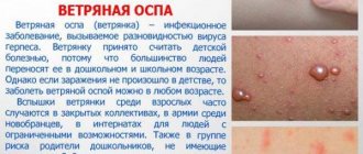
Cutaneous leishmaniasis is a group of diseases that manifest as damage to the mucous membranes, skin and tissues. There are several types of leishmaniasis:
- Late ulcerating. Distributed in cities. Mosquitoes and sick people act as carriers of infection. After recovery, a person develops immunity for life.
- Penda ulcer. Registered in rural areas. The infection is seasonal. Its carriers are mosquitoes and rodents.
Symptoms of the disease
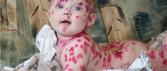
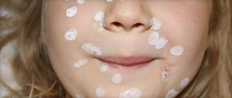
The urban type is characterized by a long incubation period from two months to several years. The disease develops slowly. First, a nodule forms on the skin, sometimes a horny plug can be seen in its center. After 3-5 months, a crust forms on the surface of the nodule. If it is removed, an abrasion opens. After some time it turns into an ulcer. A dark red ridge rises above healthy skin. Serous matter with pus is released from the ulcer. It gradually increases, reaching a size from 2 to 5 centimeters. A scar forms at the healing site.
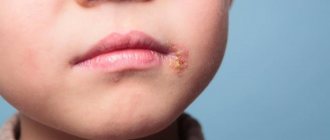
In the case of the rural type, there is a short incubation period (1-5 weeks) and it develops faster. Red-bluish bumps appear on the skin. Gradually they increase and an irregularly shaped ulcer forms. Skin damage in the rural type is much more significant than in the urban type. When healing occurs, an unsightly scar forms.
In children, the disease has a more acute course. Often, cutaneous leishmaniasis is complicated by infection.
Other rare forms of the disease also occur:
- Anthroponotic - reminiscent of lupus. Soft brown bumps form on the scars. In this case, the disease is difficult to treat, and the number of tubercles gradually increases.
- Mucocutaneous - it takes a very long time to develop and healing occurs from one to three years. In the early stages, metastases spread to the mucous membranes. Soft tissue destruction occurs. An infection can enter the body. The process ends with disfigurement.
- Diffuse leishmaniasis spreads to exposed skin. The rashes merge and form lesions. In this case, the disease does not manifest itself on the mucous membranes.
Characteristic signs of leishmaniasis
The clinical picture of the disease, depending on its form, may vary. Small formations in the form of ulcers appear on the patient’s skin and mucous membranes. The edges of the ulcers look blurry, they often appear on the face and look in the form of edematous growths, with the separation of exudative effusion.
Treatment of leishmaniasis is complex, especially when it comes to severe systemic infections. In addition, during their existence, the parasites managed to develop resistance to a number of drugs.
Treatment and prevention
Treatment of cutaneous leishmaniasis is carried out in medical institutions under the strict supervision of doctors. If any signs are detected, you should immediately contact a specialist. If treatment was carried out in a timely manner, then complete recovery occurs; otherwise, the person may die if cachexia develops or another infection occurs.
For prevention in areas where leishmaniasis is common, rodents and mosquitoes are controlled, and vaccination is required 3 months before visiting the focal area.
References
- Potekaev, N.S. Zoonotic cutaneous leishmaniasis: historical background and clinical observation. Wedge. dermatology and venereology, - 2015. - No. 5. - P. 41-50.
- Ponirovsky, E.N. Leishmaniasis: features of epidemiology, clinical picture, diagnosis, treatment, prevention. Quality of life, 2005. - No. 1(8). — P. 58-56.
- Tumolskaya, N.I., Zavoykin, V.D., Mazmanyan, M.V. and others. Tourism, imported parasitoses and their prevention. Honey. Parasitol., 2012. - No. 4. - P. 3-7.
- Control of the leishmaniases. World Health Organ. Tech.Rep. Ser., 2010. - Vol. 949. - R. 202.
- Handler, Z. Cutaneous and mucocutaneous leishmaniasis: Differential diagnosis, diagnosis, histopathology, and management. J. Am. Acad. Dermatol., 2015. - Vol. 73(6). - R.911-926.
- Bari, A., Rahman, B. Many faces of cutaneous leishmaniasis. Indian J. Dermatol. Venereol. Leprol., - 2008. - Vol. 74(1). — P. 23-27.
Some statistics
Every year, more than 11-12 million people in the world become ill with leishmaniasis. Of these, about 1.5 million people get sick for the first time in their lives. The practice of international tourism and constant military operations in a number of countries have led to periodic outbreaks of infection in recent years.
Risk groups for leishmaniasis include:
- HIV-infected;
- AIDS patients;
- tourists vacationing in epidemiologically dangerous regions;
- “disadvantaged” citizens;
- indigenous people of developing countries.
There is no officially approved vaccination against the disease yet.
LEISCHMANIASIS
Leishmaniasis is a disease caused by parasitic protozoa of the genus Leischmania. The first clinical descriptions of the disease appeared in European medical literature in the middle of the 18th century. In 1898, the Russian military doctor P.F. Borovsky discovered the causative agent of cutaneous leishmaniasis (Borovsky's disease), in 1900 Leishman, and in 1903 Donovan discovered the causative agent of visceral leishmaniasis (kala-azar). The spread of leishmaniasis by mosquitoes was established in 1921. Every year, at least 60 million people in the world suffer from leishmaniasis.
Etiology of leishmaniasis. The causative agent of cutaneous leishmaniasis is Leishmania tropica. There are two varieties of the causative agent of cutaneous leishmaniasis: L. tropica minor, which causes cutaneous leishmaniasis of the anthroponotic (urban) type, and L. tropica major, which causes cutaneous leishmaniasis of the zoonotic (rural) type. The causative agent of Mediterranean visceral leishmaniasis is L. donovani infantum. Leishmania is a type of protozoa. The development cycle of Leishmania is associated with a change of hosts - a vertebrate animal (human), in whose body the parasites are in a flagellated form, and a blood-sucking insect (mosquito), where Leishmania transforms into a flagellated form. Mosquitoes become infected by sucking the blood or interstitial fluid of animals (humans) infected with leishmaniasis. Leishmania multiplies in the mosquito's body, and after 6-8 days the parasites accumulate in the mosquito's throat, from where, when bitten by a person, they penetrate into the wound and penetrate into the cells of the skin or internal organs (depending on the type of leishmania).
Epidemiology . Mediterranean-Central Asian leishmaniasis is a zoonotic disease. There are three types of foci: 1. natural foci in which the pathogen circulates among wild animals (jackals, foxes, gophers and other rodents), which are the source of invasion; 2. rural foci in which the pathogen circulates in dogs, the main sources of infection, as well as in wild animals; 3. urban (synanthropic) foci, in which the main source of invasion is dogs and synanthropic rats. Carriers are various types of mosquitoes - phlebotomuses. Mostly children aged 1 to 5 years are affected, as well as adults - visitors from non-endemic areas. The incidence is sporadic; outbreaks are possible in cities. The infection season is summer, and the morbidity season is the fall of this year or the spring of next year. Outbreaks are recorded in the Mediterranean countries, China, the Middle East, and Latin America. Sporadic cases are recorded in Central Asia and Transcaucasia: Turkmenistan, southern Kazakhstan, Georgia, Armenia, Azerbaijan.
Zoonotic cutaneous leishmaniasis . Sources of infection are animals: gerbils, thin-toed ground squirrels, house mice, hedgehogs. Human infection occurs in late summer - early autumn - during the period of maximum mosquito population. The incubation period is from 2 to 6 weeks, then a small tubercle forms at the site of entry of the pathogen, which subsequently ulcerates, and subsequently the ulcers scar. There may be several such formations. In uncomplicated cases, the disease lasts 2-4 months. After the disease, strong immunity remains. This is a natural focal infection, the foci are located in rural areas. Among the local population, children mainly become ill, since adults have previously suffered from this disease. There may be widespread cases of cutaneous leishmaniasis among visitors and soldiers. For example, in 1885, in a detachment of Russian troops that arrived in the Merv oasis, 85% of the personnel fell ill with cutaneous leishmaniasis.
Anthroponotic (urban) cutaneous leishmaniasis. The incubation period is from 2 months to 2 years. The tubercle and infiltrate stage lasts 4-6 months, the ulcer stage - 6 months, the scarring period - 1-2 months. The total duration of the disease is up to 1 year. There are a lot of Leishmania at the affected area. Immunogenesis is slowed down. Both variants of cutaneous leishmaniasis provide cross-immunity. The source of infection is humans, but the role of dogs and rodents cannot be ruled out. The incidence is of a pronounced focal nature: residents of certain neighborhoods and houses are affected. Seasonality is not expressed. Currently, this disease has been practically eliminated from our country.
Mediterranean visceral leishmaniasis The duration of the incubation period ranges from 10-20 days to 1 year. The disease begins with the appearance of a papule at the site of a mosquito bite. From here, Leishmania hematogenously enters the cells of the reticuloendothelial system. Developing systemic parasitic reticuloendotheliosis leads to enlargement of the liver, spleen, lymph nodes and disruption of their function. There is general intoxication, impaired hematopoiesis and allergization of the body. The disease occurs against a background of fever. Untreated visceral leishmaniasis is often fatal. In cities, the sources of infection are dogs, and in rural areas - jackals, foxes, and porcupines. Infections occur in the summer, but due to the length of the incubation period, diseases more often occur in the cold season. Among the cases, 80-90% are children under 3 years of age.
Complications of cutaneous leishmaniasis It is possible that ulcers become infected with secondary bacterial flora, which delays recovery and can lead to the development of erysipelas and an abscess. The prognosis is favorable, cosmetic defects are possible.
Diagnosis of cutaneous leishmaniasis The main symptoms of the clinical diagnosis of cutaneous leishmaniasis are the formation of papules at the site of a mosquito bite with subsequent transformation into leishmania, an ulcer with undermined edges and subsequent scarring. Epidemiological data - stay in endemic regions in the last two months - are of great importance.
Specific diagnosis of cutaneous leishmaniasis: Microscopy of the contents of ulcers and marginal infiltrate is carried out. Since there are few leishmania in the material, microscopic examination is carried out several times. A Montenegro skin allergy test with leishmanin (the surface liquid of leishmania killed by phenol) is used. A biological test is also used on white mice and hamsters, which are infected intradermally with material from the patient’s ulcers.
Differential diagnosis of cutaneous leishmaniasis Differential diagnosis is carried out with furunculosis, leprosy, syphilis, epithelioma, trophic ulcer, anthrax, etc. Treatment of cutaneous leishmaniasis Treatment is predominantly local. Apply lotions with furatsilin, gramicidin, ointments: 0.5% akrikhinova, monomycinova, Vishnevsky. In the early stages of leishmanioma disease, it is recommended to sprinkle or inject intradermally with quinine, monomycin, berberine sulfate or hexamethylenetetramine. In recent years, laser therapy has been used, the advantage of which is the healing of ulcers without scarring. In the case of multiple ulcers with significant infiltrates, parenteral monomycin is prescribed 0.25 g 3 times a day for 10-14 days, aminohiol 0.2 g 3 times a day (for a course of 10-12 g). Stimulating and restorative treatment, multivitamin preparations, etc. are widely used. Prevention of leishmaniasis In case of zoonotic cutaneous leishmaniasis, deratization measures are necessary, directed against the main source of infection - great gerbils. Events are held within a radius of 2.5-3 km around the locality. In the anthroponotic form of cutaneous leishmaniasis (the source of infection is humans), patients are hospitalized in areas inaccessible to mosquitoes; Protect skin lesions with a bandage from mosquito bites. With visceral leishmaniasis, sick dogs (with skin lesions, emaciated, bald) are destroyed and stray dogs are caught. Measures to prevent transmission of the disease are the same for all forms of leishmaniasis, since in all cases mosquitoes are carriers. In populated areas, insecticides are used to treat mosquito breeding sites, as well as their roosting areas. Individual protection measures against mosquitoes - the use of canopies, nets, and repellents - are of particular importance. In natural foci of leishmaniasis, children and adults arriving for work and permanent residence are given artificial immunization by intradermal injection of a live culture of L. tropica major. A benign local process occurs, ending in scarring. Since the culture is injected into areas of the body covered by clothing, the cosmetic defect is negligible. Immunity is developed 3 months after vaccination.