Syphilis in development
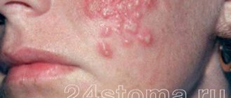
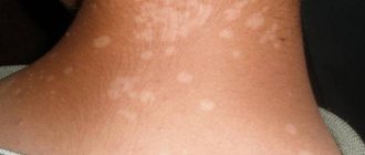
The disappearance of the spots of syphilis shown in the photo indicates the development of a secondary stage of the disease in the patient. From the second stage, a person begins to have problems with internal organs; the rash becomes pale. After 3-5 years from the moment of infection, tertiary syphilis develops, which is characterized by ulcers, scars, and total infection of internal organs and systems. Treatment at this stage is very difficult and often does not save from imminent death.
If the symptoms are similar to a cold, how can you find out about the disease before your nose drops?
With the help of testing, of course!
To test for syphilis, you need to do two tests. The first is the RPR test (synonyms: nonspecific antiphospholipid test, cardiolipin test, modified Wasserman reaction (RW)). It is used to determine the presence in the patient’s blood of antibodies to cardiolipin, a special phospholipid, antibodies to which appear during infection with Treponema pallidum and some other diseases. The cardiolipin test is good for its simplicity, low cost and high speed - a positive result will be within 7-10 days after the appearance of the chancre, so it is used for primary screening for syphilis. The disadvantage of the method is that a positive result does not necessarily mean syphilis, because antibodies are determined to cardiolipin, and not to the treponema itself, and the result may be false positive. This test is also used to monitor the effectiveness of syphilis treatment. If the treatment was successful, the titer (concentration) of antibodies to cardiolipin will decrease by 4 or more times or they will disappear completely.
To confirm syphilis, in addition to the cardiolipin test, a test for total antibodies (IgM + IgG) to treponema pallidum antigens is done. This test has high specificity, since antibodies to the causative agent of the disease are determined. The problem is that IgG antibodies can remain in the blood even after successful treatment of syphilis. Therefore, only positive results of two tests - cardiolipin and total antibodies to Treponema pallidum - will indicate an active infection.
If the test for antibodies to Treponema pallidum is positive and the cardiolipin test is negative, this indicates that syphilis has been suffered and treated in the past. A positive cardiolipin test with a negative total antibody test may indicate either a very early stage of syphilis (in this case, the test for antibodies to Treponema pallidum is repeated after 10–14 days), or another disease that will also be characterized by the presence of antibodies to cardiolipin. Both negative tests will indicate the absence of the disease or its very early stage.
To determine the stage of syphilis, you can separately do an IgM test for Treponema pallidum. This class of antibodies predominates at the onset of the disease. Therefore, a positive result for IgM (with a positive cardiolipin test) will indicate a “fresh” infection that has not yet entered the asymptomatic latent phase. In later stages of infection, the IgM test will be negative with positive cardiolipin and positive IgG. The disappearance of IgM does not say anything about the effectiveness of treatment - these antibodies will disappear on their own over time, even if the disease is not treated at all. Effective treatment will be indicated only by a decrease in titer in the cardiolipin test.
Treponema pallidum can also be detected in smears under a microscope and using a PCR reaction. But these methods are used less frequently and only as an auxiliary one, since in the early stages of the disease (before the onset of secondary syphilis) there will not yet be a pathogen in the blood and it is also not always possible to detect it in a chancre smear.
Sexual infection with syphilis in women goes unnoticed
During sexual infection, chancre sometimes goes unnoticed by a woman. This happens in cases where the ulcer occurs on the inner surface of the labia, in the clitoris, external opening of the urethra, on the vaginal mucosa or on the cervix.
In all cases of ulceration, you should immediately consult a doctor. If a woman is not treated, she develops multiple enlargement of the lymph nodes, and then a rash on the skin and mucous membranes (secondary syphilis). This rash subsequently disappears, but then may appear again (recurrent syphilis).
Hard chancroid and soft chancroid are different diseases
In addition to gonorrhea and syphilis, venereal diseases include chancroid. This disease is transmitted from one person to another almost exclusively through sexual contact.
Chancroid is caused by a microbe that is shaped like rods, arranged one after the other in a chain. This microbe enters the body through cracks or abrasions in the skin and mucous membranes.
A few days (most often 3-4) after infection, an ulcer appears in the place where the microbe penetrated. This ulcer is covered with pus, has a round or oval shape with uneven edges and bottom; its size is 1-2 cm, sometimes more, it seems soft to the touch, in contrast to a dense syphilitic ulcer.
The ulcer is painful; More often there is not one, but several ulcers located on the mucous membrane of the vagina, the skin of the external genitalia, the perineum in the anus and on the thighs.
If a woman with chancroid is not treated, the ulcers heal very slowly (after 1-2 months, and sometimes after longer periods). In all cases, when a woman notices the appearance of an ulcer in the external genital area or increased vaginal discharge, she should consult a doctor to determine the nature of the disease and prescribe the necessary treatment.
In addition to the formation of ulcerations, with chancre, in some cases, inflammation of the inguinal lymph nodes is also observed. This complication can develop when the patient does not carry out the treatment prescribed to her or does not follow the correct regimen recommended by the doctor.
How is it transmitted?
Syphilis is caused by Treponema pallidum, which lives in the external environment for only 3 minutes. Therefore, the main route of transmission of the disease is sexual. Infection of the fetus is possible in utero (vertical route) or intrapartum, when the child passes through the mother’s birth canal.
The household route of transmission is uncommon; infection is possible from persons with the tertiary stage of syphilis, when treponema pallidum gets on dishes, linen, towels, etc. from decaying gums. Hematogenous transmission of syphilis through blood transfusion cannot be ruled out.
Cases of infection of medical workers through contact with the blood of a patient are not that rare. It is possible to become infected through “bloody” objects: a shared toothbrush, razor, manicure set, etc.
Characteristics of the pathogen: external structure
Treponema pallidum resembles a corkscrew in appearance. It has 8 - 14 equal-sized curls, the height of which decreases at the ends. The spiral shape of the pathogen is preserved in all cases and under all conditions. The length of the microorganism is from 5 to 15 microns, the width is 0.2 microns.
Rice. 4. The photo shows the causative agent of syphilis Treponema pallidum (view under an electron microscope).
"End" formations
The ends of most treponemes are pointed. They contain disc-shaped outgrowths (blepharoplasts) with 10-11 fibrils attached to them.
The fibrils stretch along the body of the treponema and wrap around it, providing the bacteria with a spiral shape. From each end there are 2 independent bundles of fibrils. They are located under the outer wall, passing above the cytoplasmic membrane. Fibrils were also found under the cytoplasmic membrane. They are thinner and more numerous. The fibrils of the external bundle ensure movement of the treponema; they are twice as thick. They are long tubes consisting of flagellin protein, which is quite resistant to the action of a number of enzymes. The fibrils of the inner layer play the role of a framework.
At one end of the bacterium there are two round structures (corpus spongiosum). It ensures active penetration of bacteria into host cells.
Treponema pallidum can perform translational (back and forth), rotational, flexion, wave-like (convulsive) and helical movements.
Rice. 5. The photo shows treponema pallidum (cultured form).
Rice. 6. The photo shows pale treponema with a magnification of 3000 times (dark-field microscopy). This type of research allows you to record the shape and movement of living bacteria.
Stability of pathogens in the external environment
- Treponema pallidum is resistant to low temperatures. Can withstand freezing for up to one year. Pathogenic strains of pathogens are stored in an oxygen-free environment at low temperatures (20 - 70 ° C) or dried from a frozen state.
- Treponemas retain their virulence on environmental objects until they dry out. At temperatures up to 42°C, the activity of bacteria first increases, and then they die. When heated to 60°C, treponemes remain active for 15 minutes. For more than 3 days, syphilis pathogens retain their pathogenic properties in cadaveric material.
- Outside the human body, bacteria quickly die. At a temperature of 100°C they die instantly. Treponema pallidum is sensitive to disinfectants and some antibiotics.
Rice. 2. To identify bacteria, an immunofluorescence reaction is used.
Internal structure of Treponema pallidum (ultrastructure)
Mucoprotein "case"
The body of the bacterium is surrounded by a mucus-like structureless substance (microcapsule). This mucopolysaccharide substance protects treponemes from phagocytes and antibodies. The capsular substance is produced by the treponema itself.
Cytoplasmic membrane
The cytoplasmic membrane of bacteria performs a number of vital functions: transport, protective, is the site of localization of antigens and enzymes, it takes an active part in cell division, L-transformation and sporulation. The cytoplasmic membrane has a three-layer structure. Its inner layer forms numerous outgrowths in the protoplasmic cylinder, due to which the active transfer of nutrients from the outside occurs. The vital activity of bacteria depends on the state of the cytoplasmic membrane.
Protoplasmic cylinder
The protoplasmic cylinder is located under the outer wall. The structure of the cytoplasm of Treponema pallidum is finely granular. Ribosome granules and many lamellar structures are immersed in the transparent hyaloplasm. Ribosomes provide protein synthesis in the bacterial cell. The cytoplasm also contains a nucleotide that does not have a limiting membrane. It stretches along the entire length of the protoplasmic cylinder.
Mesosomes
Mesosomes are derivatives of the cytoplasmic membrane. They occupy half or the entire axis of the diameter of the treponema. Mesosomes supply energy to the bacterial cell at points of increased growth during sporulation and division. Their function is similar to mitochondria. Mesosomes differ in the nature of their structure, but their number is always very large.
Rice. 7. Ultrastructure of Treponema pallidum. Mucoprotein cover on top, then a three-layer cell wall, inside there is a cytoplasmic membrane and a cylinder with nucleotide, mesosomes, ribosomes and other inclusions. The photo clearly shows the fibrils running along the body of the bacterium.
Possible consequences
Pathology in both sexes and all ages is associated with serious consequences:
- failure or deformation of internal organs;
- internal hemorrhages;
- irreversible changes in appearance;
- death.
In some cases, syphilis may appear after treatment: due to re-infection or unscrupulous therapy.
To avoid contracting these unpleasant diseases, choose protected sex. Do not neglect routine examinations so that a diagnosis can be made at an early stage or a hidden form of the disease can be identified. If trouble has already happened, do not despair and go to an appointment - the doctor will help you.
This article has been verified by a current qualified physician, Victoria Druzhikina, and can be considered a reliable source of information for site users.

