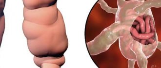
Harlequin ichthyosis is a skin disease. It is diagnosed when the fetus is still in the womb. The disease spreads throughout the body and is incurable.
This is a hereditary disease, very rare, and belongs to the group of genodermatoses. Ichthyosis harlequin anencephaly is formed in the womb and is the most severe form of the disease. The entire body of the sufferer is covered with formations in the form of geometric patterns, like a harlequin. That is why the disease got its name.
Causes of harlequin ichthyosis
The disease is transmitted in an autosomal recessive manner. As a result of biochemical processes triggered by the mutating gene, protein function is lost. The ABCA11 DNA polypeptide chain becomes shorter, and fat and protein metabolism is disrupted. A large amount of amino acids accumulate in the skin.
The granular membranes do not line up as they should. Lipids (fats) are unable to form a healthy stratum corneum. The gene that transports lipid granules into keratinocyte membranes loses memory. As a result of the mutation, a disease develops. In addition, lipids (fats) undergo increased secretion.
Find out more
Diagnosis of the disease
Using an ultrasound machine, Harlequin ichthyosis can be suspected in utero at 4-5 months of pregnancy.
To confirm the diagnosis, amniocentesis is prescribed - analysis of amniotic fluid.
If the result is positive, mothers explain the symptoms of the disease in detail and offer an abortion. The decision always remains with the woman.
A diagnosis of “Harlequin ichthyosis” can be made to an already born baby based on an examination. This can be done by a neonatologist or dermatologist. If necessary, the doctor takes a piece of skin for analysis. Diagnostics is carried out by electron microscopy.
In this case, you cannot do without consulting a geneticist who conducts molecular genetic analysis.
Additionally, the patient is prescribed a general and biochemical blood test, and a general urine test. Once every 2 weeks, they must take a culture from the skin folds for the presence of pathogenic microflora.
Differential diagnosis is carried out with desquamative erythroderma, Ritter's dermatitis, epidermolysis bullosa, congenital syphilis, etc.
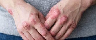
Symptoms of harlequin ichthyosis
- There are gray and beige scales on the skin.
- There are cracks between the scales.
- The lips are twisted.
- The ears are damaged and may be covered with a layer of altered skin.
- The mouth is either stretched or narrowed due to the density of the diseased dermis (babies are fed only through a tube).
- There are membranes between the fingers.
- Lack of nails.
- Skeletal defects.
- There is no hair on the body.
- The eyes do not close, the eyelids are turned out.
- Dehydration of the dermis (water balance in tissues is not regulated).
A very small percentage of babies survive. About 3% of children reach the age of 11-12 years. A few live up to 18 years. They are constantly fighting for life.
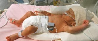
Harlequin ichthyosis in children
One sick child is born in a ratio of 1x1000000. Children have cracks, formations in the form of scales on the skin that fit tightly to each other. The formations tighten the mouth and fingers, and movements are limited. Feedings are only possible through a tube. Because of their clumsy, inverted eyelids, they sleep with their eyes open. Because the plates tighten the abdomen and chest tightly, it may be difficult for the child to breathe.
The skin is pink and reddish in color. It is very dry and requires a constant flow of water. Due to lesions throughout the body, on which there are formations in the form of thick plates, an imbalance in water balance occurs. Most children do not live to see 10-12 years of age. They require careful care, constant care and attention.
Children with this disease are transferred to intensive care wards immediately after birth. The thick, lamellar skin soon begins to peel off. It peels off centimeter by centimeter. The process takes several weeks.
Children are given antibiotics to prevent infections. When the thick skin completely peels off, the baby is left with red, dry skin. Thin plates may remain on it. After bathing, the dermis is lubricated with ointments and emollient creams. During the first weeks of life, suffering babies require constant care. You can't leave them for a minute.
Types of pathology
More than 90% of patients develop ichthyosis vulgaris, a disease that first appears in the second or third month of life. Depending on the nature of the skin lesion, the following clinical forms are distinguished:
- xeroderma, or abortive form, is the mildest variant of the disease, in which the skin becomes dry and rough, but there are no scaly layers;
- a simple shape with small scales, thin at the edges and with a dense center, which cover the entire body up to the scalp;
- shiny shape with transparent scales located in the form of a mosaic mainly on the legs;
- white form with white or slightly yellowish scales, similar in appearance to asbestos fibers;
- serpentine in shape with rough keratinizations of brown, brown or gray color, reminiscent of snake scales.
Much less common is the lamellar form of the disease (collodion fetus), fetal (Harlequin fetus), linear, etc. Some of them are incompatible with life - for example, fetal ichthyosis develops in the second trimester of intrauterine development, and the child dies in the first days of life or is born already dead.
Treatment of harlequin ichthyosis
This is done by a group of doctors, which includes:
- Dermatologist (main treatment).
- Therapist, pediatrician, family doctor (increasing the body’s protective properties, general strengthening).
- Psychologist (relieving stress, depression, breakdowns, increasing self-esteem).
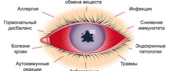
- Oculist (observes vision, advises).
- Otorhinolaryngologist (ENT) (observes the ears, checks hearing).
- Gastroenterologist (observes how the body works during nutrition).
- Nutritionist (proper nutrition).
- Cardiologist (monitoring the work of the heart).
Traditional treatment (drugs)
The complex of drugs includes creams, ointments, gels. They should be selected so that the components contain the necessary substances. It is imperative to look at antimicrobial and antibacterial properties. The preparations must contain urea. The patient will have to constantly nourish and moisturize the dermis, so it is necessary to select moisturizing and nourishing preparations.
Narcotic painkillers are introduced into therapy. Keratolytics will help get rid of scales.
Vitamin and mineral complexes
Treatment is aimed at increasing immunity. Patients feel better. The courses last a month, then are repeated after some time. The body receives all the necessary vitamins and minerals. Doctors assign a special role to taking vitamins of groups A, B, C, E, and nicotinic acid.
Vitamin A and its analogues, when used internally, help get rid of scales. However, the oral dose is prescribed only by a doctor, since retinoids cause side effects and are toxic. They come in the form of tablets, capsules, injections, droppers.
Hydrotherapy
Patients with the disease constantly take baths, as their skin is dehydrated. In the morning you need to saturate your skin with moisture for at least two hours. Herbal baths with the addition of string, chamomile, and straw provide a good anti-inflammatory effect. Water with borax softens the dermis. Patients can simply bathe in ordinary water, but for at least 2 hours. After bathing, lubricate the skin with Vaseline.
Spa treatment
Consists of a complex of therapeutic measures. They are aimed at cleansing the skin and getting rid of scales. Separation of the scales is painful, so they are first softened. This process is helped by vitamin U and other drugs. Patients are treated by bathing in sulfide and carbon dioxide baths.
Heliotherapy
Sun treatment can be offered to patients in precise dosages. A beam of infrared radiation is directed to the affected skin and lingers on one of the areas. After a short period of time, the sheaf of light is transferred to another zone. It also lingers here, saturating the stratum corneum with particles.
Thalassotherapy
This is one of the methods of climatotherapy, which is a treatment by the sea. Those suffering from the disease are recommended to take sea bathing, wrapping themselves with seaweed and estuary mud. It is beneficial for patients to breathe sea air; a large amount of seafood is included in the menu. Sun, sand, and air baths increase immunity, improve the well-being and mood of patients.
Treatment of severe forms
Patients are given plasma transfusions, drugs with a high content of iron and calcium, and aloe extract. All procedures are aimed at stimulating the immune system. Hormone therapy may be prescribed.
ethnoscience
At home, harlequin ichthyosis is treated with herbal baths and wraps. A softening ointment is boiled in lard with herbs. The grass can be changed, take the appropriate action: rowan, motherwort, sea buckthorn, peony, tansy, plantain, celandine, horsetail. Decoctions are prepared and drunk in courses of 30 days. After a break, repeat. Treatment is aimed at relieving pain and symptoms. The disease harlequin ichthyosis is incurable. All healing methods are aimed at alleviating the condition of those suffering.
Diet therapy
The menu of patients should be rich in products with a high content of vitamins of groups A, B, E, C. You can grind and prepare dishes in the form of puree, make juices, compotes, jelly. Vegetables and fruits are required, but in moderation. The diet should include:
- lactic acid products;
- meat (game, chicken, beef, pig);
- porridge (buckwheat, oatmeal, rice, others);
- seafood;
- fruits and vegetables of any kind, but yellow and red ones are especially useful.
To choose the right diet, it is better to consult a professional nutritionist.
Physiotherapy
Physiotherapy procedures help patients well. Applications with emollient and analgesic drugs may be prescribed. Baths with the addition of soda, starch, and sea salt soothe the dermis. UV irradiation activates metabolic processes and improves skin metabolism.
Climatotherapy
Patients are recommended to live in an area with a mild climate. The gifts of the Dead Sea can also be used in the fight against disease. Air, sun, and sand baths have a beneficial effect on the skin. Therapeutic mud saturates the dermis with many useful elements.
Negative impact of the disease on the patient
Ichthyosis causes not only external harm, but also affects the processes and functions of the body:
- the functionality of the pancreas and adrenal glands is inhibited;
- the functioning of the gonads is disrupted;
- there is a lack of cellular immunity;
- vitamin A is not absorbed;
- spastic paralysis;
- oligophrenia;
- the functionality of the glands responsible for sweat production decreases;
- pain when trying to “clean” the skin;
- cardiac dysfunction in the body;
- mental retardation;
- Possible death.
Mortality among children with congenital ichthyosis is highest.
Prevention of harlequin ichthyosis
Unfortunately, the only method here is consultation with a geneticist. Expectant parents can take a special test, the results of which will show whether someone is a carrier of the mutating gene. If there is one in the family, the geneticist calculates the likelihood of having a healthy child.
If the fetus is affected by the disease, a sick child will be born. Parents themselves decide what to do in such a situation. Carriers of the mutating gene do not necessarily get sick themselves, but they can give birth to sick offspring.
Modern medicine does not know how to cope with the disease. Scientists have not yet fully uncovered the processes occurring in the gene, which has forgotten how to build the stratum corneum of the human dermis. According to some hypotheses, the gene builds the dermis for another biological species related to the underwater world, but not for humans.
Forecast
The mortality rate from Harlequin ichthyosis is high, with rates approaching 50% worldwide. A review of 45 cases by Dr Rajpopat et al identified 25 survivors (56%) ranging in age from 10 months to 25 years. Twenty deaths (44%) occurred from days 1 to 52 and were as likely to cause respiratory failure as fulminant sepsis. A Japanese survey of 16 patients reported a survival rate of 81.3% (13 of 16 patients). Respiratory failure, fulminant sepsis, or a combination of both are the most common causes of death in newborns.