The term “ichthyosis” combines a list of pathologies of the human skin that are of genetic origin. One of the most severe cases among such diseases is Harlequin ichthyosis (Harlequin syndrome). Children with this mutation die either immediately after birth or in the first weeks of life. Only 2-3% of children can live to be 10-12 years old; only one child out of several hundred patients manages to survive until adulthood. At the same time, all his life he has been tormented by constant pain, inflammation, problems with internal organs and psychological ones.
General information
The disease is typical for newborns of any sex. In total, less than 100 episodes of Harlequin syndrome in a newborn have been recorded worldwide.
It is assumed that this syndrome is inherited by an autosomal recessive mechanism (that is, both parents have a hidden disease that does not manifest itself, but when they meet, the “dormant” genes of the parents combine, and the child has a congenital pathology).
Doctors described Harlequin syndrome in a newborn boy
Mrs. Esther Booth, née Santlow, dressed as a harlequin / John Ellis, 1719
Doctors have described another case of Harlequin syndrome, in which only one side of the person's body turns red. This time the syndrome occurred in a newborn boy. The child was born prematurely with pathologies of the heart and lungs, but the syndrome appeared independently of them on the third day of life, and then just as suddenly disappeared. It is still unclear what caused it, perhaps due to the medications that were given to the child immediately after birth. Brief report published in The New England Journal of Medicine
.
Harlequin syndrome is an asymmetrical redness of the body: while on one half (right or left) all signs of erythema appear (redness, sometimes beads of sweat), the other remains unchanged. In itself, this condition is considered safe, does not lead to any health consequences and does not require treatment. However, there is still no explanation for this phenomenon.
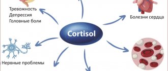
Doctors believe that asymmetrical redness may be related to the sympathetic nervous system, the part of the peripheral nervous system that is responsible for the stress response. Apparently, the different halves of this system work differently in Harlequin syndrome. Moreover, as a rule, the blame is placed on the half that does not show signs of a stress reaction - it is likely that in a given situation it turns out to be inoperative and does not respond to a stressful stimulus.
Gert van den Berg and Hanneke Bakker from the Sofia Children's Hospital in Rotterdam described the development of Harlequin syndrome in the newborn. The child was born prematurely, at the 35th week of pregnancy. Even before giving birth, he was suspected of having a rare heart pathology - Ebstein's anomaly - in which the valve between the right ventricle and the right atrium is displaced towards the ventricle and does not close completely. Because of this, the right atrium increases in size; The oval window, the duct between the right and left atria, also becomes larger. Usually after childbirth it heals, but in such patients this is difficult, so the blood in the atria mixes, and as a result, the body tissues receive less oxygen.
As one would expect, the child’s right atrium turned out to be hypertrophied, and the lungs, on the contrary, were underdeveloped. To compensate for this, immediately after birth he was prescribed artificial ventilation and injections of prostaglandin E1. This is a pro-inflammatory substance that is designed to inhibit the fusion of another germinal duct - the Botallic duct - between the pulmonary artery and the aorta. This duct allows the embryo to dump “extra” blood that the unexpanded lungs are not able to accept. Prostaglandin E1 was supposed to keep the ductus botallus open and relieve the strain on the patient's underdeveloped lungs.
On the third day of life, pronounced erythema appeared on the right half of the child’s body, while the left remained pale. However, no other physiological parameters changed, including vital ones - breathing and blood circulation. Therefore, the doctors did not take any measures, and after a few minutes the Harlequin syndrome disappeared as suddenly as it appeared.
Van Den Berg, Bakker/NEJM, 2020
Share
It can be assumed that uneven skin coloring appeared as a consequence of therapy - for example, because fever is sometimes a side effect of taking prostaglandins. However, the authors of the work note that Harlequin syndrome was previously described both against the background of taking prostaglandins and independently of them, so its true cause remains unknown.
Previously, doctors described the unusual color of the iris, which turned the patient’s eye into the “Eye of Sauron.” You can find out what unusual colors urine can take from our test “For every taste and color.”
Polina Loseva
Pathophysiology
Harlequin ichthyosis is accompanied by disturbances of microcirculation and peripheral blood flow due to neurological disorders, which consist of unilateral damage or asymmetry of the functions of the nervous sympathetic system. And, as mentioned above, it is a pathognomonic sign of asphyxia or TBI in newborns.
There is an assumption that the thickening (induration) of the stratum corneum, characteristic of this pathology, develops as a result of a defect in lipid synthesis or dephosphorylation of proteins, resulting in their secretion into the skin and, as a result, the formation of the latter is disrupted.
Manifestations of the disease
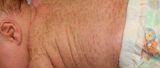
Pathologies are visible to the naked eye at birth. The skin is intensely flattened, there are cracks and characteristic defects on the surface. In some cases, part of the body is covered with bright spots. As the temperature in the room rises, the stains acquire a rich burgundy tint; when the temperature drops, they become blue. The child experiences increased sweating on the affected side. The skin is hot to the touch and red.
Congenital ichthyosis is difficult to confuse with another dermatological disease due to the characteristics of the symptoms:
- The child's lips are stretched, which is why the mouth does not close. This phenomenon gave the disease its name due to its resemblance to the Harlequin mask;
- the eye, nasal slits and mouth have an irregular shape;
- the ears are underdeveloped;
- nose flattened;
- the skin in appearance resembles a scaly shell;
- The child usually has no hair or nails;
- keratinized tissue prevents free flexion and extension of the limbs.
History and clinic
The onset of the disease, that is, pathological thickening of the skin and its cracking with the formation of deep defects, is observed already at birth.
The characteristic symptoms of this syndrome are spots that can completely cover one half of the body, while the baby’s legs and arms are only partially covered with such spots. In case of hyperthermia or overheating of the baby, the spots acquire a bright burgundy hue, and in case of hypothermia, they turn blue. When the child is in normal condition, they are practically invisible.
In addition, Harlequin syndrome is accompanied by hypertrophy of the legs and arms on one side, redness (hyperemia) of one half of the face and increased sweating of the same half.
Local symptoms. Skin manifestations of Harlequin syndrome manifest as thick and rigid skin with the appearance of triangular/hexagonal/polygonal plaques that form when the affected skin cracks. In this case, the skin has a yellow or gray color with plaques and bright red deep cracks on the surface.
In addition, there is pronounced ectropion (that is, eversion) of the eyelids, eclabion (deformation in the form of a fish mouth), flat deformed ears and, in some cases, microcephaly.
General symptoms. The majority of babies with Harlequin syndrome are stillborn or die immediately after birth. Such infants are malnourished and suffer from electrolyte imbalance and heat exchange disorders. Due to skin rigidity, breathing and movement difficulties are noted.
Deep fissures often become infected, which leads to the development of sepsis.
Causes of severe illness
Harlequin ichthyosis is an inherited disorder that occurs at the genetic level. All possible causes of Harlequin ichthyosis have not yet been fully investigated. In the fetus at 4-5 months of intrauterine development, a shortening of the polypeptide chain in the ABCA11 gene occurs. As a result of this process, the protein responsible for the normal development of the skin disappears from the gene.
The mutation leads to the fact that the baby is born with a “shell” of dehydrated and dense skin. Doctors also note significant distortions in the shape of the nose, ears, mouth and eyelids. The limbs of a newborn are often deformed; in addition, neonatologists diagnose abnormal development of important internal organs.
The disease is transmitted to children only in one case - if each of the biological parents is a carrier of the altered gene. If one of the parents is the carrier of this gene, then the unborn child is not in danger. But in such a situation, the baby also becomes a carrier of the mutated gene.
The disease is very rare; statistics indicate one case per 800,000 healthy children born. Ichthyosis affects both sexes equally. It is impossible to examine pathology in the early stages using ultrasound. If parents are aware of their family history, invasive diagnostics should be performed in the first trimester of pregnancy. If the diagnosis is confirmed, the only chance to avoid the birth of a seriously ill baby, for whom life will be short and very painful, is to decide to terminate the pregnancy.
Diagnostics
The diagnosis of “Harlequin syndrome” is made in accordance with the characteristic clinical manifestations and data from neurological studies, which includes MRI of the spinal cord and brain.
Differential diagnosis of this syndrome should be carried out with a milder form of congenital ichthyosis, the so-called “colloidal fetus”.
Histopathology. In the skin biopsy of patients with Harlequin syndrome, there are signs characteristic of this pathology: compact orthokeratosis and thickening of the stratum corneum and absence of the granular layer, obstruction (obstruction) of the ducts of the exocrine (sweat) glands and the presence of hyperkeratotic detritus in the sebaceous glands. In the hair follicles, there is an accumulation of keratin masses localized around the hair shaft, which gives rise to the manifestation of pathological prenatal disorders.
Ultramicroscopy. When examining the granular layer, pathologically transformed lamellar bodies are observed in it, and in the stratum corneum there is a complete absence (lack) of lipid plates and/or lipid inclusions.
Symptoms
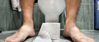
According to some reports, the syndrome may be a consequence of fetal asphyxia or intracranial injury during childbirth. It is also diagnosed in cases of damage to the hypothalamus or medulla oblongata. In any case, a neurologist is immediately called for a consultation. Detecting the disease is quite simple: its main symptom is redness of the skin when the baby lies on its side. In this case, the border of the color change runs exactly along the line of the spine.
Uneven coloring of body parts occurs a few minutes after the baby is laid down. This is how Harlequin syndrome manifests itself. Photos of young children in medical encyclopedias show an unsightly picture - the upper part of their body has a normal color, the underlying part becomes bright red, sometimes with a bluish tint. The phenomenon is noticeable within a few minutes. Some experts say the syndrome may be a sign of prematurity.
Therapy
Treatment of this pathology is carried out by a neurologist.
Increased survival is achieved through the administration of systemic retinoids, such as Acitretin and Isotretinoin, but pathological skin changes persist, which adversely affects the quality of life of such patients.
In addition, systemic intensive supportive treatment is carried out to prevent infectious complications, maintain electrolyte-water balance and normal temperature, and ensure normal nutrition of the infant.
In the case of a mild course of the syndrome, parents are advised to maintain the air temperature at 20 degrees and train the child’s autonomic system and blood vessels, that is, the child’s gradual adaptation to cold and heat.
Features of the disease
Babies do not always die if they have Harlequin pathology. The syndrome is not a death sentence. The disease may not be pronounced; moreover, if it is quickly and correctly diagnosed, the baby’s condition can be alleviated with special medications. In such children, not only heat exchange is disturbed, but also electrolyte balance. Horny skin makes breathing and movement difficult. Large cracks can become infected and develop sepsis.
Fortunately, the disease is not very common. At the same time, the gender of the baby does not matter: both boys and girls are susceptible to pathology. Doctors say the syndrome is often severe. However, the prognosis in most cases is quite unfavorable. The etiology of the syndrome has not yet been established.
Helping a sick person
Symptomatic therapy can alleviate the condition of the newborn. Treatment of this type of ichthyosis comes down to the following measures:
- intensive skin hydration – eliminates cracking, peeling, and soreness;
- increased sterility - necessary to prevent secondary infection. The patient's skin is vulnerable to external influences; coccal infections are especially dangerous for a child;
- restoration of thermoregulation - babies up to six months need comfortable living conditions, and patients with ichthyosis suffer more from heat deficiency.