In this article we will tell you:
- Causes of retinal detachment
- Types of retinal detachment
- Rhegmatogenous retinal detachment (RRD)
- Tractional retinal detachment (TRD)
- Exudative retinal detachment (ERD)
- Symptoms of retinal detachment
- Diagnosis and treatment of retinal detachment
- Prevention of retinal detachment
Retina
- the thinnest multilayer structure of the eye, covering 70% of the area of the inner surface of the eyeball. The retina of the eye is connected to the visual analyzers of the brain, into which it transmits information and is responsible for converting light impulses into nerve impulses.
The inner side of the retina is adjacent to the vitreous body, and the outer side is adjacent to the choroid: the choroid from which it receives nutrients and oxygen. Sometimes, under the influence of one reason or another, part of the retina moves away from the choroid. In this case, cells deprived of nutrition and oxygen quickly die. Without surgical intervention, the process leads to disturbances up to complete loss of vision. This condition is called retinal detachment.
Retinal disinsertion
–
a severe pathological process requiring emergency surgical care, in which the reticular layer of the eyeball is separated from the vascular layer.
The term itself was first proposed at the beginning of the 8th century, but its clinical confirmation was made only in 1851 after the invention of the ophthalmoscope. According to research, the incidence of the disease has increased significantly over the past decade: previously, 1 in 15,000 people were at risk of retinal detachment, now it is 1 in 10,000. Patients with myopia and aphakia often suffer from retinal detachment, especially if part of the vitreous lens was removed during surgery simultaneously with the lens. bodies. In the latter case, the risk of developing pathology increases to 10%.
Retinal functions
So, the main role of the retina is to perceive an image that is focused on it, passing through the cornea and lens, and then convert the light flux into nerve impulses and transmit them to the brain. Already there the image is processed in the necessary way, and we see a clear picture. Thanks to the work of the retina, it is possible to provide central and peripheral vision.
The retina is a very sensitive structure, and any pathology of it, if not treated promptly, will inevitably lead to sad consequences - loss of vision. The most common ailment is retinal detachment, and in the initial stages of the disease a person does not even suspect it. At the end of the article, we will tell you why it is necessary to regularly conduct fundus examinations.
Causes and mechanisms
In fact, retinal detachment is always caused by the same reason: prolonged disruption or interruption of the blood supply. In this case, after some time, dystrophic and atrophic processes develop, organic degeneration of thin retinal tissue begins, often the “spare”, compensatory vascular network grows (the phenomenon of neovascularization), which leads to all kinds of adhesions, tense ties (tractions), hemorrhages, swelling , accumulations of exudative fluid. When exudate penetrates between the neuroepithelium and the choroid, or with constant mechanical “tearing” force from traction, scars and overgrown connective tissue, neovascular vessels that have grown into the vitreous body, the retina is torn away from the original vascular “soil” and becomes functionally untenable: reception and transmission of the visual neurosignal in case of total detachment is excluded, and in case of partial detachment it is extremely poor quality.
As for the immediate and most common causes of such a catastrophic development, they include:
- angiopathy (vascular pathology) and secondary retinopathy caused by them (degenerative changes in the retina) in a number of systemic diseases - diabetes mellitus, blood diseases, arterial hypertension and atherosclerosis, as well as, in some cases, in pregnant women or very premature newborns;
- idiopathic, primary, independent of external causes, degenerative processes in the retina, caused by internal characteristics or conditions of a particular organism;
- eyeball injuries (sometimes chest injuries);
- oncological neoplasms in the visual system;
- pronounced degrees of myopia.
Causes of retinal detachment
Retinal detachment is the process of separation of the retina from the adjacent choroid. In this case, intraocular fluid penetrates and accumulates between the layers. This is a serious disease that requires urgent action.
There are several types of retinal detachment. They are divided depending on the reasons that caused this pathology. Only an accurate diagnosis will allow you to choose the right method of treating the disease. So, let's look at these types:
- Traction. Occurs when there is tension on the part of the vitreous body as a result of various disorders: vascular ingrowth, diabetic retinopathy, retinal vein thrombosis. The second most common cause of retinal detachment.
- Rhegmatogenous. The main provoking factor is thinning of the retinal layer due to various pathologies. A rupture can occur with sudden movements or strong physical stress. The likelihood of occurrence is also aggravated by eye diseases - cataracts, glaucoma. This is the most common type, diagnosed in every tenth of 100 thousand people, and most often in men over the age of 45.
- Traumatic. Its rupture can occur immediately at the time of injury (for example, due to a strong blow to the head) or develop in the subsequent period, which sometimes takes up to several years. Traumatic detachment can also result from surgery.
- Exudative or serous. In this case, the retina itself remains intact, but begins to peel off as a result of the accumulation of fluid under it due to some pathological processes: toxoplasmosis, neoplasms, choroidal melanoma, etc.
- Secondary retinal detachment occurs due to various disorders in the organs of vision: hemorrhages and thrombosis, inflammation of the choroid, etc.
For any reason of detachment, the result is the penetration of intraocular fluid between the layers, as a result of which the retina begins to peel off from the choroid. The risk of this pathology increases in patients at risk:
- with type 2 diabetes mellitus;
- myopia, especially severe degrees;
- retinal dystrophy;
- after eye surgery;
- after injuries to the organs of vision or craniocerebral; vascular diseases.
Only correct and timely determination of the cause of retinal detachment can help eliminate the problem.
Diagnosis of retinal pigment epithelium detachment
Despite the fact that in ophthalmological practice, retinal detachment with its symptoms is an emergency condition, before starting treatment for this condition, it is necessary to conduct a comprehensive medical examination of the patient.
- An examination using an ophthalmoscope will allow you to assess the location, shape and size of the pathological process. The presence of retinal tears is confirmed or denied.
- Conduct research using contact and non-contact lenses.
- In case of chronic retinal detachment, electrophysiological research methods are used to assess the functional capabilities of the eye and at the same time make a prognosis regarding the restoration of vision in the patient.
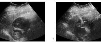
- If there are concomitant eye diseases that make examinations with lenses or an ophthalmoscope difficult, ultrasound examination is used.
- Perimetry and assessment of visual acuity are carried out, which makes a proper contribution to determining the magnitude and localization of the pathological process.
- Additionally, intraocular pressure is measured, which may be reduced in comparison with a healthy eye.
Retinal detachment: main symptoms
At the initial stage of this eye disease, a person has no idea that a pathological process has begun. However, if the slightest symptoms appear that may indicate this, it is necessary to visit an ophthalmologist for a thorough examination of the fundus.
Often the signs of the disease are attributed to fatigue after long visual work, but in the meantime irreversible changes occur in the eyes.
When the symptoms become too obvious and the quality of vision begins to deteriorate, a person visits a doctor, but the disease is diagnosed already at a critical stage. The fact is that during detachment, the cones and rods responsible for vision die, and their regeneration is no longer possible.
So, here are the signs you need to visit a specialist if you find that they recur on an ongoing basis:
- flashes, sparkles in the eyes - photopsia;
- distortion of the shape, size and shade of objects (metamorphopsia);
- “spots” and black dots before the eyes (a sign of damage to the retinal vessel);
- the appearance of a black veil, which covers an increasingly larger area, decreased quality of peripheral vision.
Sometimes patients note that they see better immediately after waking up. However, doctors explain it this way: when the body is in a horizontal position, the retina is adjacent to the choroid, and in a vertical position, when active daytime activity begins, it moves away from it, and vision begins to deteriorate again.
The detachment cannot be cured with medications or drops. Only timely diagnosis and retinal surgery will help restore visual functions.
Reasons why you should contact the Federal Scientific Clinical Center for treatment of this disease
- The Department of Ophthalmology of the Federal Scientific and Clinical Center of the Federal Medical and Biological Agency has the most advanced technologies and extensive experience in the treatment of diseases of the retina;
- Modern surgical equipment: a DORC (Eva) combine is used during operations on the retina, has high accuracy, provides optimal clinical results, and a quick recovery period;
- The extensive clinical experience of ophthalmologists of the Federal Scientific and Clinical Center of the Federal Medical and Biological Agency and first-class technical equipment make it possible to successfully treat various eye diseases even in advanced stages of the disease;
- At the Federal Scientific and Clinical Center of the Federal Medical and Biological Agency, all necessary preoperative preparation is carried out in just one day.
- Taking into account the presence of a multidisciplinary hospital, resuscitation and anesthesiology service, we perform operations on somatically severe patients with a risk of developing cardiovascular complications.
What is a retinal detachment?
Retinoschisis (as this pathology of the retina is called in medicine) is the division of the retinal layers into outer (choroidal) and inner (vitreal). Fluid accumulates between them and cysts form. Retinoschisis occurs as a result of developmental defects or dystrophic changes in the retinal membrane.
In Europe, the incidence of the disease is approximately 5% in the population over 30 years of age. It is also inherited. The main problem of retinoschisis is its asymptomatic course; it is diagnosed accidentally or already in an advanced stage when complications arise.
Contraindications for illness
The most important contraindication for those who suspect retinal detachment: in no case should you delay contacting an ophthalmologist and self-medicate. This disease can progress rapidly. It cannot be treated with traditional methods, and incorrectly selected medications can only aggravate the situation.
After surgery to eliminate detachments and tears of the retina, a certain number of restrictions must be observed. Contraindicated:
- do not sleep on your back unless specifically instructed by your doctor;
- make sudden head movements, bend over, lift weights of more than 5 kg;
- reading for a long time, working on a computer or watching TV - you need to limit eye strain;
- wear contact lenses in the first 1.5 months after surgery;
- go outside without sunglasses.
What forms of retinoschisis are there?
Russian ophthalmologists classify this disease into three types, depending on the cause of retinal separation:
- Hereditary. It develops as a consequence of genetic disorders and is transmitted as an autosomal dominant or recessive trait. Sometimes it is a symptom of genetic disorders such as Goldmann-Favre disease or Wagner syndrome.
- Degenerative. The acquired form usually occurs after the age of 40 due to degenerative changes in the retina.
- Secondary. It develops as a result of vascular diseases of the organs of vision (diabetic retinopathy, thrombosis of the central retinal vein), as well as inflammation of the eyes - uveitis, iridocyclitis in chronic stages, with oncology of the vascular system, after eye injuries.
With the development of retinoschisis, circulatory disturbances are observed in the macular and peripheral areas of the retina. In this case, a large number of compactions - cysts - appear. As they grow, they merge with each other, forming large cavities that stratify the retina along its entire length. This process leads to the development of retinal detachment and death of retinal structures.
Symptoms of retinal dissection include:
- decreased visual acuity;
- narrowing of peripheral fields;
- flashes in the eyes;
- distortion of the outlines of objects.
Usually these signs appear at a fairly advanced stage.
Classification of pathology
Clinical classification T.A. Bagdasarova, distinguishes three stages of retinoschisis:
- First. The retinal split is limited to a separate area, there is no vascular elevation, and there is no intraretinal fluid.
- Second. Retinal splitting progresses without a definite boundary between the affected area and the healthy retina. The formation of microcysts is detected in its layers.
- Third. The lesion with extensive cysts is localized in several quadrants of the retina. Their cavities rupture under the retina. There is a risk of transition of the pathological process to a limited detachment.
Separately, it should be said about central (macular) retinoschisis , which is less common, but has more severe consequences due to damage to the center of the visual field and possible complications - the formation of a macular hole.[4]
Fig.2 Picture of maculoschisis (OCT image - optical coherence tomography)
How is retinal detachment and separation diagnosed?
To verify the expected diagnosis, a specialist will conduct an examination of the visual organs using modern equipment. This procedure includes several stages. The main one is coherence tomography, which allows you to accurately identify changes in the macula of the retina, determine the presence of cysts and detachments in the peripheral area.
Computer perimetry will allow you to determine the boundaries of the patient’s field of vision. In addition to detachment, perimetry will also help to recognize glaucoma, tumors, and pathologies of the optic nerve.
Ophthalmoscopy - examination of the fundus of the eye - allows you to determine the location of ruptures and identify thinned areas where new foci of detachment may appear.
An ophthalmologist can, if necessary, perform viziometry, tonometry, ultrasound of the eyeball, electroretinography, and fluorescein angiography. Unlike detachment, which can only be treated surgically, for retinoschisis, conservative therapy with the help of vitamin preparations and drops can also be prescribed.
Make an appointment and ask a question:
What to do if detachment does begin?
See a doctor immediately. The detachment process occurs according to the following scheme: a gap is formed, vitreous water enters it, and the process of tissue scarring begins. In some cases, during a retinal tear, blood enters the eye and the person sees dark blots floating in the field of vision. We have already said that if you reached a competent ophthalmic surgeon before the center was detached, then with a successful operation, most likely your vision will be completely restored. And if you didn’t have time, then the periphery will be restored as it was, but the center of the retina will not return to its original characteristics. As a rule, it is good if half of what it was before the detachment is restored. Moreover, recovery sometimes takes up to two to three years. And even after a successful operation, curvature of the lines in the central field of vision may remain.
How is surgery to repair retinal detachment performed?
There is only one cause of detachment – rupture of the retinal tissue itself. The retina still remains “alive”, still consists of “pixels” and transmits the image to the brain. But outside of its substrate it does not work well. So the point of the operation is to put the retina in its rightful place. The only question is how to do it.
If the detachment is fresh,
then you can try to quickly close the gap with a simple filling operation, thereby stopping the flow of fluid into it, and after a couple of days all the excess water will be absorbed into the substrate itself, and the retina will return to its place. Sometimes this can even happen within a few hours.
If the patient does not quickly reach the doctor, then the situation is more complicated.
The scarring process has already started, the retina is already curved and stuck in an uneven position to the vitreous body. This means that simply closing the hole will not result in restoration. In this case, the retina is first separated, cleaned of overgrown connective tissue, the hole is closed and the retina is put in place. It is not simple. It happens that the retina is cleaned two or three times. Fortunately, the scarring process is not endless.
The sooner the patient sees a specialist from the start of the detachment process, the easier it will be to restore vision. The longer a person waits, the more his body wrinkles the eye and the more difficult the manipulations for treatment.
How is the retina fixed in its “rightful” place? Some kind of glue?
Such glue has not yet been invented to close tissue gaps; we can only do this with the help of natural scars. To do this, laser microburns are made around the tears so that scars or “biological rivets” are formed at the site of the burns, which will tightly block the tears. But a scar does not form instantly. A full-fledged scar is obtained only after two to three weeks, or even after a month. And all this time, something must hold the retina in place, pressing it against the substrate, the choroid, on which, in fact, the scar forms. This is reminiscent of the process of gluing wallpaper, someone must hold it tightly against the wall until the glue hardens, otherwise there will be no complete fixation. In order to solve the problem of holding the retina in the correct position until fixing scars appear, various gases or silicone oil are used.
Gas is used
, in simpler cases. It is injected into the eye cavity at the end of the operation. There is no need to remove the gas afterwards; it gradually dissolves in the natural fluid of the eye. Various gases are used, each with its own absorption rate. Ordinary air dissolves the fastest, in a week or two. The type of gas is chosen based on how quickly it needs to dissolve.
Silicone
in most cases it holds the retina better than gas. It is not as mobile and is used in more severe cases. There are different types of silicone. The lower parts of the retina are attached to the one that is heavier than water, and the upper parts are attached to the one that is lighter. But silicone does not dissolve on its own; it must subsequently be removed from the eye. This is not difficult, but an additional operation is required, which is usually performed 1.5 months after the main operation.
There is one more nuance with silicone - its structure refracts light differently than the vitreous fluid. And as a rule, the refraction of the eye becomes positive, for example +4 or even +7. You need to understand that this is temporary and after removing the silicone everything returns to normal. However, there are cases when, for some reason, the situation is so complicated that the presence of silicone in the eye is the only chance today to have vision. And then the silicone cannot be removed. This means that a person will have to use plus glasses or lenses. Although this can be solved if you replace the natural lens with an artificial one, and compensate for the new properties of the eye.
Does silicone need to be replaced over time?
In some cases, the silicone gradually emulsifies into microdroplets inside the eye. And there are situations when silicone changes every 8-10 years.
Is the vitreous removed to introduce gas or silicone into the eye?
The vitreous body is 98% water and 2% collagen filaments. And it does not have any particularly important function. But it participates in the processes of inflammation and scarring, which harm the eye by tightening the retina, so yes, it is removed. But after two to three hours, the vitreous body is replaced by liquid, which passed through the eye even before its removal. That is, in essence, the liquid remains as it was, and only the threads are removed.
Sealing
What is retinal filling?
This is another option for closing a tissue tear. No scarring. It is used in uncomplicated cases of retinal detachment in young people. If a person is young and the vitreous body of the eye has not yet broken down into fibers and water, that is, it is still naturally elastic like a gel, then you can create a kind of mound on the side of the break. This gel will press down and the hole will be blocked. But you need to understand that time will pass and the vitreous body will still begin to stratify with age. Therefore, after installing the filling, when the detachment has closed, usually after a month, it is necessary to go through this area with a laser to strengthen the gap.
Retinal detachment and separation: treatment
Surgical treatment of retinoschisis and retinal detachment includes several methods prescribed depending on the stage of the disease and the presence of damage. They can be surgical, combined, or only laser restoration is possible.
Extrascleral ballooning. The purpose of this method is to mechanically press the retina against the choroid in order to restore blood supply to the retina in the area of detachment.
To do this, a balloon is placed behind the eyeball, which the doctor gradually fills with liquid through a catheter. The balloon, increasing in size, begins to put pressure on the sclera, after which the inner shell is soldered in the right place with a laser beam. The operation takes 30-50 minutes and is low-traumatic. However, to consolidate the result and natural fusion of the combined layers, the balloon is removed from the eyes only after a week.
Scleral filling. The surgery treats retinal detachment and separation. Its purpose is to ensure that the exfoliated area adheres to the epithelial layer. To do this, a special filling made of biocompatible elastic material is used, which is applied to the desired segment and then fixed with absorbable sutures.
Vitrectomy. This is the name given to partial or complete removal of the vitreous humor from the eyeball. Instead, silicone oil and special medical polymers are temporarily introduced into the cavity.
After completion of the vitrectomy, the same laser coagulation of the retina is performed, holding the tissue together. This is the most modern and effective way to treat detachment and separation.
Under the influence of a laser beam, partial destruction of the retinal proteins occurs. At the same time, its damaged areas are soldered to the choroid, and the pathologically altered retinal vessels stop growing and bleeding. This helps stop the development of the disease, and in other cases completely cures it. The procedure is completed in 15-20 minutes; if there are no complications, the patient can be discharged from the clinic on the same day.
It is noteworthy that this method is allowed even for pregnant women, while other methods are contraindicated for them. Retinal tearing at the time of childbirth is not uncommon, especially if the woman has a high degree of myopia. Before the invention of the laser, such patients were prohibited from natural childbirth precisely for this reason and a cesarean section was prescribed. Now preventive laser coagulation allows you to give birth naturally.
Cost of retinal detachment surgery:
| № | Service name | Price in rubles | Make an appointment |
| 2011074 | Introduction of drugs of the 3rd degree of complexity into the vitreous cavity | 78000 | Sign up |
| 2011073 | Introduction of drugs of the 2nd degree of complexity into the vitreous cavity | 27000 | Sign up |
| 2011072 | Introduction of drugs of the 1st degree of complexity into the vitreous cavity | 22200 | Sign up |
| 2011071 | Peripheral vitrectomy 3rd category of complexity | 14400 | Sign up |
| 2011070 | Peripheral vitrectomy 2nd category of complexity | 12000 | Sign up |
| 2011069 | Peripheral vitrectomy of 1st category of complexity | 9600 | Sign up |
| 2011068 | Microinvasive revision of the anterior chamber | 14400 | Sign up |
| 2011075 | Intraoperative administration | 7200 | Sign up |
| 2011066 | Reconstruction of the anterior chamber | 7200 | Sign up |
| 2011065 | Removal of perfluoroorganic liquids from the vitreous cavity | 9000 | Sign up |
| 2011064 | Removing liquid silicone from the vitreous cavity | 12000 | Sign up |
| 2011063 | Circular retinotomy or retinectomy | 16800 | Sign up |
| 2011062 | Retinotomy and retinectomy | 9000 | Sign up |
| 2011061 | Injection of gas into the vitreous cavity | 12000 | Sign up |
| 2011060 | Injection of liquid silicone into the vitreous cavity | 18000 | Sign up |
| 2011059 | Introduction of perfluoroorganic liquids into the vitreous cavity | 15000 | Sign up |
| 2011058 | Circular peripheral endolaser coagulation of the retina | 16500 | Sign up |
| 2011057 | Endolaser coagulation of the retina, delimiting (one quadrant) | 7200 | Sign up |
| 2011056 | Endodiathermocoagulation | 9000 | Sign up |
| 2011055 | Removal of epiretinal membranes or posterior hyaloid membrane of the third category of complexity | 57600 | Sign up |
| 2011054 | Removal of epiretinal membranes or posterior hyaloid membrane of the second category of complexity | 47700 | Sign up |
| 2011053 | Removal of epiretinal membranes or posterior hyaloid membrane of the first category of complexity | 36600 | Sign up |
| 2011067 | Endodrainage of subretinal fluid | 7200 | Sign up |
| 2011052 | Unscheduled revision of the vitreal cavity | 42000 | Sign up |
| 2011051 | Revision of the vitreous cavity of the third category of complexity | 33000 | Sign up |
| 2011050 | Revision of the vitreous cavity of the second category of complexity | 28200 | Sign up |
| 2011049 | Revision of the vitreous cavity of the first category of complexity | 20400 | Sign up |
| 2011048 | Vitreoretinal surgery for complicated conditions (2-stage) of the highest category | 210060 | Sign up |
| 2011047 | Vitreoretinal surgery for complicated conditions (2-stage) of the third category | 162060 | Sign up |
| 2011046 | Vitreoretinal surgery for complicated conditions (2-stage) of the second category of complexity | 119940 | Sign up |
| 2011045 | Vitreoretinal surgery for complicated conditions (2-stage) of the first category of complexity | 84420 | Sign up |
| 2011042 | Vitrectomy for complicated conditions of the second category | 90000 | Sign up |
| 2011041 | Vitrectomy for complicated conditions of the first category of complexity | 83400 | Sign up |
| 2011039 | Vitrectomy for uncomplicated hemophthalmos or vitreous opacities of the second category | 64500 | Sign up |
| 2011038 | Vitrectomy for uncomplicated hemophthalmia or vitreous opacities of the first category | 54000 | Sign up |
| 2011037 | Removal of a silicone filling implanted in another medical institution | 24900 | Sign up |
| 2011036 | Removal of a silicone filling within a period of more than 6 months. after the first operation | 18660 | Sign up |
| 2011035 | Pneumoretinopexy for retinal detachment | 22200 | Sign up |
| 2011034 | Additional extrascleral filling in case of detachment | 28860 | Sign up |
| 2011033 | Combined extrascleral filling for detachment | 64800 | Sign up |
| 2011032 | Circular extrascleral filling for detachment | 48420 | Sign up |
| 2011031 | Local extrascleral filling for retinal detachment | 37800 | Sign up |
| 2011030 | Extrascleral ballooning for retinal detachment | 31800 | Sign up |
| 2011028 | Set of disposable consumables for vitreoretinal surgery | 57600 | Sign up |
| 2011027 | Introduction of drugs into the vitreous cavity (Eylea, Lucentis, Ozurdex) | 55200 | Sign up |
| 2011040 | Vitrectomy for uncomplicated hemophthalmia or vitreous opacities of the third category | 78600 | Sign up |
| 2011044 | Vitrectomy for complicated conditions of the third category of complexity | 144900 | Sign up |
Making an appointment Today: 9 registered
Why is retinal detachment dangerous?
The peeling process can last up to several years, and the person will not even suspect it. Phenomena such as “floaters” before the eyes, temporary blurred vision, light flashes are attributed by many to fatigue after work and do not turn to an ophthalmologist. Meanwhile, light-sensitive receptors die off.
In 15% of cases, when detachment occurs, it also occurs in the second eye. And even surgical intervention is not a guarantee of the return of visual functions - this method can only stop the development of detachment or stratification, but not restore lost cells.
In advanced cases, these retinal pathologies result in complete blindness. That is why it is so important not to delay a visit to an ophthalmologist when symptoms appear in order to prevent irreparable consequences.
Symptoms
People who are not even aware of their pathology may experience the following symptoms in the eyes:
- sparks,
- flares,
- unclear vision of objects,
- flickering
- opacity of vision.
If you experience any such sensation, you should immediately consult an ophthalmologist. Only a specialized specialist can diagnose diseases. You won't be able to do this on your own. The study uses specialized instruments. As a result, it is possible to understand not only the degree of development, but also the reasons. This provides an opportunity to choose the most effective treatment.
Prevention of retinoschisis and retinal detachment
Prevention of these diseases consists of regularly visiting a doctor, especially after 40 years, to check the fundus, peripheral vision, etc. If myopia is severe, high physical activity, extreme and traumatic sports should be avoided.
With eye and head injuries, the risk of retinal detachment increases several times, and this may not happen immediately, but over a period of time.
If this happens, then visiting an ophthalmologist for a check-up should become the norm. Patients with glaucoma need to monitor intraocular pressure and take medications on time.
Only a careful and careful attitude to eye health will help maintain good quality vision for many years. Regular visits to the doctor will help to detect many diseases in time and take appropriate measures.
Indications
In the presence of the risk factors mentioned above, competent prevention plays a major role. What is important is regular observation by an ophthalmologist, limiting physical activity, reducing psycho-emotional stress, sufficient supply of eye tissue with vitamins and microelements, etc.
Among the indications of doctors is eye gymnastics. Its goal is to improve the nutrition of eye tissue (including the retina). This “digital gymnastics” is especially recommended for those whose eyes are constantly under strain:
- rub your palms together vigorously;
- close your eyes and cover them with warm palms;
- With your eyes closed and with maximum amplitude, “draw” the numbers from 1 to 10. Do the same in reverse order - from 10 to 1.
Make an appointment and ask a question:
Can detachment spread to the other eye?
If a person’s retina has detached in one eye, it means that there is a predisposition to detachment in the second. It is necessary to be examined and, if necessary, take measures to surround areas of thinning of the retina with laser coagulants.
If the detachment occurs due to injury, then an examination of the second eye is still carried out, but one cannot talk about predisposition.
Complex surgery
How long does surgery take to treat retinal detachment?
This is difficult surgery. It takes about an hour. The operation is performed under local anesthesia or general anesthesia. For the patient, the operation is painless, since there is no nerve tissue in the retina. Therefore, there is no pain when there is a rupture. We can only understand from signs of deterioration in vision that a detachment is occurring.
Is it necessary to perform any manipulations with the lens if surgery is required to treat retinal detachment?
For good visualization and illumination of the posterior chamber of the eye, it is important to have a clean lens, because it is through it that the doctor observes the operation. If the lens is cloudy due to cataracts, then it needs to be replaced. The lens can also become cloudy during surgery if it is touched or if gas gets on it. Therefore, it may be necessary to combine the operation with an operation to replace the lens with an artificial one. This is not a problem, since today artificial lenses have almost surpassed natural lenses in their properties.
Are there any restrictions after surgery?
The operation has only one goal: to close the gap. After a mini-laser burn is made, the patient can even stand on his head, a scar will still form. That is, there are no restrictions. Especially if silicone is used. Within two to three hours after the operation, you just need to make sure that the person has recovered from the anesthesia, and then he can go home.
Naturally, microbes live around us and after surgery there is slight inflammation, so special drops containing anti-inflammatory drugs and antibiotics are prescribed, both before and after surgery.
Then every six months you should undergo a fundus examination to ensure that no new weak areas appear in the second eye. If there was a detachment and surgery, then in the eye that was worked on, all possible weak areas have already been strengthened, so the likelihood of a recurrence of the rupture is small. But in the second eye there is still a predisposition.
Does it happen that you can’t achieve results the first time?
It happens that people come for alterations, yes. Sometimes even people from abroad come to us for repeated surgery. Today, any retina, no matter how long it has been detached, no matter how twisted or scarred it may be, can be cleaned and returned to its anatomical place. The results will be different for everyone, and so will their vision, but it can be done. Several operations may be required. Usually a prognosis is made about what result we expect after successful treatment, and then together with the patient we decide whether it makes sense to operate or not.
How do you treat detachment that occurs in case of injury?
Eye injuries have certain nuances. The blood supply to our head is very active and if something ruptures, it is accompanied by a large amount of blood. And if at this moment you begin to restore the retina, then it will, firstly, be very difficult, and secondly, the results will not be up to par. In case of injuries, the first task is to sew up the torn vessels and allow them to close. It takes 1-3 days for the vessels to stop bleeding.
Of course, injuries are very variable. But usually, when a person with an injury arrives at any hospital, the membranes of his eye are simply sewn up, restoring its integrity. After this is done and anti-inflammatory treatment is prescribed, starting from the second or third day, usually after a week, we can talk about surgical treatment in order to restore the functions of what remains intact. Post-traumatic restoration of the eye and vision is sometimes a complex, multi-stage surgical process. And very often we manage to get excellent results.
When a retinal detachment occurs, the key point remains whether a person sees at least some light or not. If he sees, then the retina channel - the optic nerve - the brain is working and you can and should fight and gain vision.. If the eye does not see light, then modern medicine is powerless for now.
How much does surgery to treat retinal detachment cost in your clinic?
If this is a filling, then the price starts from 90,000 rubles for manipulation on one eye, if vitreoretinal surgery is required, then from 130,000 rubles. If a complex operation with increased complexity is required, the cost can be more than 200,000 rubles.
Oleg Vladimirovich, thank you very much for the detailed coverage of such a serious problem. We wish you success!