Calcium is a vital macronutrient that performs many functions in the human body (providing muscle contraction, blood clotting, conducting nerve impulses). Calcium is also a major component of bone tissue. Calcitriol (vitamin D) and parathyroid hormone take the most important part in the regulation of its metabolism. Hypercalcemia is a fairly common electrolyte disorder, occurring in approximately 0.17 to 3.9 cases per 100 people. Gender differences vary among people of different ages. Young men and women over 45 are more susceptible to this condition.
Causes of hypercalcemia
Hypercalcemia almost always indicates some disease or pathological process. However, sometimes it develops due to physiological reasons (in newborns on the 4th day of life, in adults after eating). The pathological causes of this condition are as follows:
- Hyperparathyroidism.
This is an endocrine disease characterized by hypersecretion of parathyroid hormone (PTH). It is the most common cause of hypercalcemia. Hyperparathyroidism is caused by adenoma, hyperplasia of the parathyroid glands, and renal failure. Sometimes hyperparathyroidism occurs as part of autoimmune polylinglandular syndrome or multiple endocrine neoplasia. - Oncological diseases.
Recognized as the second most common cause of this electrolyte disorder. In cancer, it occurs through two mechanisms. The first is bone destruction by metastases or primary focus (leukemia, lymphoma, myeloma). The second mechanism is the synthesis of PTH-like peptide by cancer cells (lung, breast, bladder cancer). - Granulomatous processes.
Chronic diseases characterized by the formation of cellular granulomas in tissues (primarily in the lungs) can also cause hypercalcemia. These include tuberculosis, sarcoidosis, histoplasmosis. Mononuclear phagocytes that are part of granulomas, due to the expression of 1-alpha hydroxylase, are able to convert vitamin D into the active form (calcitrirol, 1,25OH-D3), which enhances the absorption of calcium ions by the small intestine. - Prolonged immobilization.
As a result of prolonged inactivity, osteoclasts (cells that destroy bone tissue by dissolving mineral compounds) are activated. This leads to the release of calcium ions from the bones. This phenomenon occurs during forced immobilization after injuries, being in conditions of weightlessness (during space flights). - Taking medications.
This primarily applies to vitamin D and calcium supplements. Other medications (thiazide diuretics, theophylline, lithium) can also cause calcium imbalance by increasing osteodestruction or reabsorption processes in the tubules of the nephrons of the kidneys. - Other endocrine disorders.
In addition to the pathology of the parathyroid glands, other endocrine diseases are sometimes the cause of hypercalcemia. For example, an excess of thyroid hormones in hyperthyroidism increases the destruction of bone tissue. With adrenal insufficiency, the inhibitory effect of glucocorticoids on calcium metabolism is reduced.
Hypocalcemia
etiology, pathogenesis Hypocalcemia and hypocalcemic tetany (HT) are a striking metabolic-endocrine syndrome, the leading clinical manifestations of which are paresthesia and generalized, and sometimes local, convulsions associated with a decrease in calcium concentration in the blood. HT is observed in 70–90% of patients in intensive care units due to pancreatitis, sepsis, major operations and injuries, as well as other acute conditions [1]. At the same time, HT is often recorded as the first and even the only manifestation of various chronic or latent pathologies [2, 3]. Numerous etiological and pathogenetic variants of HT are given in the classification. Classification of hypocalcemia: • Dysregulation of calcium-phosphorus-magnesium metabolism. I. Parathyroid hormone deficiency, i.e. hypoparathyroidism (HyPT): 1) removal or damage of the parathyroid glands (PTG) during surgery; 2) radioiodine therapy or radiation therapy for diseases of the neck organs; 3) hemochromatosis; 4) tumor metastases in the parathyroid gland; 5) congenital underdevelopment of the parathyroid gland (idiopathic HyPT); 6) autoimmune destruction of the parathyroid gland. II. Violation of the action of parathyroid hormone: 1) pseudohypoparathyroidism (Phypoparathyroidism) or Martin-Albright syndrome: 1. PhiPT type 1; 2. PGIPT type 2; 2) pseudohypohyperparathyroidism (Costello-Dent syndrome). III. Impaired secretion or action of other hormones that regulate calcium-phosphorus-magnesium metabolism: 1) excessive secretion of calcitonin: 1. medullary thyroid cancer; 2. other apudoms; 2) vitamin D deficiency or impaired action of D-hormone: 1. rickets or osteomalacia in adults; 2. malabsorption syndrome; 3. insensitivity to vitamin D. • Functional hypocalcemia: 1) alkalosis (respiratory, metabolic, iatrogenic); 2) hyperproteinemia (hyperalbuminemia); 3) “hungry bones” syndrome; 4) neonatal hypocalcemia of infants born to mothers with hyperparathyroidism; 5) increased calcium uptake by osteoblastic tumors; 6) chronic renal failure (CRF); 7) endocrinopathies (diabetes mellitus, hypogonadism, hypopituitarism); acute destructive pancreatitis; 9) rhabdomyolysis. • Toxigenic and iatrogenic hypocalcemia: 1) excessive intake of phosphorus into the body; 2) hypomagnesemia; 3) introduction of EDTA; 4) treatment with mithramycin, neomycin, cisplatin; 5) use of phenobarbital, phenytoin, glucagon, laxatives, antacids; 6) massive infusion of citrated blood or extracorporeal hemoperfusion. By definition, the pathobiochemical factor of HT is a low concentration of circulating and cytosolic calcium, which is accompanied by increased excitability of neuromuscular and interneural synapses and, as a consequence, paresthesia, muscle cramps and other neuroregulation disorders. The most common cause of HT is hypoparathyroidism (HyPT), which complicates approximately 8% of surgical operations on the thyroid gland and almost all interventions on the parathyroid gland [4, 5]. Thyroid surgery, even by experienced surgeons, is fraught with the development of hyperthyroidism, since in approximately 2% of people the PTG is located inside the thyroid gland [6]. Irreversible HyPT sometimes occurs after radioiodine therapy for thyrotoxicosis, thyroid cancer, or radiation therapy for other malignant tumors of the neck [2, 3]. Destruction of the parathyroid gland by tumor metastases is considered a casuistic cause of hyperthyroidism and tetany. Meanwhile, such cases are apparently not uncommon, because some authors found metastases in the parathyroid gland in 5–12% of patients with cancer pathology, especially in breast cancer [2, 3, 5]. Autoimmune HyPT as part of autoimmune polyglandular syndrome (APS), like the syndrome itself, is an undoubted rarity, and congenital aplasia or hemochromatosis of the parathyroid gland is simply a casuistry. There is only one report of HyPT due to hemochromatosis of the parathyroid gland after multiple infusions of iron supplements for hypochromic anemia [1, 5]. Idiopathic pseudohypoparathyroidism (PhyPTH) is the insensitivity of target organs to parathyroid hormone (PTH). There are two forms of the syndrome, differing in that with PhiPT-1, the injection of parathyroid hormone does not affect the excretion of cAMP and phosphaturia, and with PhiPT-2, after the administration of PTH, the excretion of cAMP increases, but phosphaturia does not (Table 1). Pseudohypohyperparathyroidism is a variant of PhiPT, in which the kidneys are resistant to PTH, but skeletal PTH receptors are preserved. As a result, a bone form of primary hyperparathyroidism (osteitis fibrosis cystica) is formed, but calcium in the blood is not increased, but decreased [4, 5]. Pseudopseudohypoparathyroidism is another genetically determined syndrome, but here the defect is not in the PTH receptors, but in the PTH molecule [4, 5]. The syndrome is recognized by a test with PTH, during which both cAMP excretion and phosphaturia increase (Table 1). In terms of its effect on calcium-phosphorus-magnesium metabolism, calcitonin (CT) is an antagonist of PHT and D-hormone. In this regard, it is logical to expect that medullary thyroid carcinoma and other APUD cell tumors that secrete a lot of CT should be accompanied by attacks of tetany. However, in practice, hypocalcemia is extremely rare in such patients, which is explained by the powerful effect of PTH and D-hormone, which block the effects of calcitonin. The pathogenesis of tetany with vitamin D deficiency or a defect in D-hormone receptors is close to that with HyPT and PhiPT. This makes their differential diagnosis difficult, which, however, is possible using a PTH test. After eliminating hypocalcemia by administering calcium, patients with D-hormone deficiency give a normal phosphaturic response to the administration of PTH, while in HyPT and PhiPT, parathyrine does not increase phosphaturia. Hyperalbuminemia and alkalosis reduce the level of ionized calcium, which, in combination with other causes, can cause HT. Hungry bone syndrome is the name given to hypocalcemia that often occurs after surgical treatment of severe thyrotoxicosis or hyperparathyroidism. Temporary hypocalcemia in these cases is due to the fact that bone tissue intensively absorbs calcium, which has been “washed out” of the bones by excess thyroid hormones or PTH. Neonatal hypocalcemia of infants born to patients with hyperparathyroidism is explained by transient hypersecretion of CT and suppression of parathyroid function in infants against the background of excess parathyrine and calcium in the blood of the mother. The mechanism of development of hypocalcemia in patients with massive osteoblastocytomas that absorb calcium is obvious. Hypocalcemia in chronic renal failure is associated with increased loss of calcium in the urine due to a decrease in renal Ca2+ reabsorption and insufficient synthesis of D-hormone in the kidneys. Hypocalcemia against the background of some endocrinopathies is explained by a weakening of the permissive or increased opposing effect of systemic hormones in relation to PTH, CT and D-hormone. Finally, hypocalcemia in pancreatitis and rhabdomyolysis is due to chelation, i.e. calcium deposition in areas of massive destruction of adipose and muscle tissue, as well as hyperphosphatemia and hyperkalemia, which increase calcium excretion. In addition, these patients have impaired renal, gastrointestinal and endocrine system function. The phosphate ion (PO3-) is reciprocally bound to the calcium ion, so hyperphosphatemia is necessarily accompanied by hypocalcemia. This, in particular, explains late postnatal tetany and iatrogenic rickets if the child is not breastfed, but cow's milk, which contains a lot of phosphorus. The nature of hypocalcemia against the background of hypomagnesemia is not entirely clear, just as the nature of hypomagnesemia itself is not always clear. It is believed that hypomagnesemia inhibits PTH secretion and receptor binding [7]. The most common causes of hypomagnesemia are alcoholism, cytostatics, diabetes mellitus, parenteral nutrition, and a congenital defect in the intestinal absorption of magnesium. Iatrogenic hypocalcemia from a number of drugs is due to their chelating effect or blockade of vitamin D, or inhibition of intestinal calcium absorption. HT syndrome often complicates massive citrated blood transfusion or cardiopulmonary bypass procedures [3]. The sodium hydrogen citrate contained in preserved blood converts Ca2+ into poorly dissociated calcium hydrogen citrate. Clinical picture and diagnosis of hypocalcemia The most striking manifestation of hypocalcemia is prolonged, usually generalized, tonic convulsions, the so-called tetany. Tetany is painful for the patient, but the danger to life is low. In a few reports, death from tetany in the setting of postoperative hypoparathyroidism has been associated with asphyxia from laryngeal stridor and diaphragmatic contraction or cardiac tetany [1–3]. However, chronic hypocalcemia creates serious problems for the patient's health. It is associated with the development of cataracts, metastatic calcifications in organs and the brain, severe mental disorders with depression and suicide, infertility or miscarriage with fetal death, and finally, against the background of hypocalcemia, a chronic infection is activated. The classic triad of incipient HT is a combination of paresthesia, difficulty breathing and convulsions. Paresthesia begins from the lips and spreads to the hands and feet. Tonic convulsions can be local - a sardonic mask on the face or “carpopedal syndrome”. Almost always, seizures are accompanied by clear neurological symptoms - dysarthria, dysphagia, paresis of cranial nerves, stigmata of cerebrovascular, extrapyramidal and brainstem disorders, spastic paresis of the limbs. Disorders of the somatic nervous system are accompanied by dysfunction of the autonomic nervous system: profuse sweating, bronchospasm, renal or hepatic colic, vomiting, diarrhea. Intracranial hypertension and swelling of the optic nerves are often recorded. Convulsive syndrome in patients with HT is often regarded as epilepsy, although in epilepsy the seizures are usually clonic, with loss of consciousness. However, HT can also occur in the form of clonic convulsions with syncope, i.e. with short-term loss of consciousness. Sometimes HT resembles mental disorders: paranoid or hallucinatory psychosis, depressive-catatonic attack. Manifestations of hypocalcemia such as laryngospasm, tetany of the stomach and heart were mentioned above. In this case, asphyxia or “acute abdomen” syndrome, or an attack of angina pectoris and even small focal myocardial infarction are attributed, because The ECG shows QT prolongation, ST depression, sharpening or inversion of T. It is clear that the listed phantoms can cause therapeutic and diagnostic errors, sometimes dramatic, for example, hospitalization in a psychiatric clinic or surgery. Atypical and latent HT are recognized using tests for spasmophilia: • Khvostek's sign: twitching of the lips or contraction of the orbicularis oris muscle when tapping with a fingertip in front of the tragus of the auricle or between the zygomatic arch and the corner of the mouth. • Weiss's sign: contraction of the facial muscles when tapping on the outer edge of the eye orbit (outer corner of the eye). • Trousseau's symptom: the appearance of the so-called “obstetrician's hand” after compression of the arm with a sphygmomanometer cuff. • Schlesinger's symptom: in a patient lying on his back, with passive flexion in the hip joint, the thigh muscles contract convulsively and supination of the foot is noted. It must be remembered that symptoms of spasmophilia are present in 10–25% of healthy people and in all patients with vegetative dystonia and psychasthenia. At the same time, 30% of patients with hypocalcemia do not have signs of spasmophilia [1, 3]. In the diagnosis of hypocalcemia, anamnesis is important: previously observed convulsive attacks, thyroid surgery, radioiodine therapy or radiation therapy to the neck, chronic renal failure, enterocolitis, frequent fractures. There are specific signs for certain types of chronic hypocalcemia. For example, postoperative hypoparathyroidism, in addition to a scar on the neck, is characterized by eczema and other dermatoses, dry skin, hard, dry and brittle hair, uneven, brittle nails. Cataracts are often detected, which are considered a specific complication of chronic hypocalcemia. Ro-graphy reveals calcification of the basal ganglion, calcifications in the subcutaneous tissue, muscles, internal organs, subperiosteal ossification of tubular bones, diffuse fibrous osteitis [8]. Pseudohypoparathyroidism in 75% of cases has clear morphotypic stigmas: short stature, round face, short neck, brachydactyly, rachitic skeletal deformities, mental retardation [2, 5]. A screening test for hypocalcemia is the determination of calcium in the blood. The lower limit of total calcium is 2.2 mmol/l, ionized calcium is 1 mmol/l. The diagnosis is clarified using additional tests (Table 1). Treatment of hypocalcemic tetany Standard emergency therapy for HT is an intravenous bolus injection of 10–20 ml of a 10% calcium chloride solution. You need to beware of extravasal ingestion of calcium chloride, because it causes massive tissue necrosis. The danger of extravasation is very high, since the injection has to be done against the background of seizures. Instead of calcium chloride, you can use a 10% solution of calcium gluconate or lactate, however, to relieve HT with these solutions, you need to administer at least 20 ml, and preferably 40 ml, since they contain half as much Ca2+ ion per unit volume. It should be remembered that massive extravasation of these drugs also causes tissue necrosis. The most reliable way to prevent such a complication is to administer calcium solutions not as a stream, but by infusion through a cannula in a vein of 50 ml of a 10% CaCL2 solution diluted in 100–200 ml of a 5% glucose solution. If the diagnosis of HT is beyond doubt, and the administration of calcium-containing solutions does not have an effect, you should think about alkalosis or hypomagnesemia. These conditions can be confirmed and eliminated ex curantibus - by intravenously administering 10 ml of a 5% solution of ascorbic acid and 10 ml of a 25% solution of magnesium sulfate. But it is better to clarify the diagnosis by determining the concentration of magnesium and blood pH. The introduction of 10–20 ml of 10% calcium chloride stops tetany for only 2–3 hours. Therefore, it is advisable to immediately administer 40 ml of 10% CaCL2, or better, as mentioned above, to carry out a long intravenous infusion of 50–100 ml of 10% calcium chloride, diluted in 200–400 ml 5% glucose. The algorithm for diagnosing and treating HT is shown in Figures 1 and 2. After stopping an attack of HT, calcium and vitamin D preparations are prescribed orally. Calcium preparations irritate the mucous membrane of the stomach and intestines, so their dose should be as low as possible, and it is advisable to abandon them as quickly as possible. The patient is recommended a diet enriched with calcium, including dairy products, hard cheeses, soy, sesame, almonds, dried apricots, celery, canned fish “with bones”: sardines, sprats, pink salmon. Normally, the daily requirement for Ca2+ is at least 1000 mg. Naturally, in patients with most forms of hypocalcemia, the need for calcium is 2–3 times higher. It is clear that it is almost impossible to provide 2000–3000 mg of Ca2+ from food products. Therefore, the “calcium diet” still has to be supplemented with calcium supplements, preferably twice a day. Of the numerous vitamin D preparations, its active metabolites are preferred: calcitriol and alfacalcidol, prescribed in a dose of 0.25–1.0 mcg 1 or 2 times a day. A calcium-rich milk-vegetable diet contains a lot of phosphorus, which enhances calcium excretion in the urine and its absorption by the skeleton. To bind phosphorus, take 20–40 ml of aluminum hydroxide during meals. If hypocalcemia is not effectively eliminated, magnesium deficiency should be assumed and appropriate medications should be prescribed. These are magnesium sulfate, potassium and magnesium aspartate, magnesium orotate, etc. The adequacy of treatment is monitored clinically (absence of symptoms of spasmophilia) and by monitoring the levels of calcium, phosphorus and magnesium in the blood. In conclusion, it should be noted that it is necessary to focus on the prevention of HT, which consists in the timely diagnosis of hypocalcemia, which is achieved by mandatory determination of calcium in the blood, better ionized, in all outpatient and hospital patients.
Literature 1. Marino P. Intensive care; lane from English M.: GEOTAR-Media, 1998. P. 461–463. 2. Lukyanchikov V.S., Korolevskaya L.I. Hypoparathyroidism and hypocalcemic syndrome. // Clinical medical 2003. No. 1. P. 66–70. 3. Carlstedt F., Lind L. Hypocalcemic Syndrome // Critical. Care Clinics. 2001. Vol. 17. No. 1. P. 139–153. 4. Potts J. Hypocalcemia // Internal diseases (translated from English) / Ed. E. Braunwald et al. M: Medicine, 1997. T. 9. pp. 397–411. 5. Wilson J., Foster D. Hypoparathyroidism. In: Williams Textbook of Endocrinology (8th ed.). . Philad.: WB Sounders, 1992. P. 1456–1476. 6. Wheeler M., Williams E., Path F. et al. The Hyperfunctioning intrathyroidal parathyroid gland // World. J. Surg. 1987. No. l. R. 10–114. 7. Dacey M. Hypomagnesemic disorders // Critical. Care Clinics. 2001. Vol. 17. No. 1. P. 155–179. 8. General Guide to Radiology (translated from English) / Ed. N. Pettersson. M.: RA “Spas”, 1996. T. 2. P. 690–733.
Pathogenesis
An increase in calcium content in the blood changes the membrane potential of cells, which leads to inhibition of neuromuscular conduction in skeletal muscles, myocardium, and the gastrointestinal tract. The pathogenesis of neuropsychiatric symptoms is not completely clear. The role of slowing down the conduction of nerve impulses is assumed. Calcification of blood vessels, internal organs, dystrophy, and tissue shrinkage develops.
Due to hypercalciuria (increased filtration of calcium in the nephron tubules), the risk of nephrolithiasis increases. Calcium inhibits adenylate cyclase, which suppresses the renal effect of antidiuretic hormone. Also, due to the high extracellular concentration of this cation, the secretion of hydrochloric acid by the lining cells of the stomach increases, which leads to the development of peptic ulcers.
What is hypocalcemia, how to recognize it?
The essence of hypocalcemia is a decrease in calcium levels to below normal levels. The disease is serious because it is fraught with serious complications. There are acute and chronic forms of pathology.
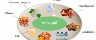
For reference, why does the body need calcium?
Calcium circulates in the blood and is stored in the bones; by the age of 18, about 1 kg of the element accumulates in the bones and teeth. Calcium has many functions, without it the body will not be strong; it is involved in the process of blood clotting, muscle activity, and the functioning of the nervous and immune systems. The body regulates the level of the element, but sometimes its content decreases, which negatively affects health.
Why should hypocalcemia be treated?
The disease provokes serious complications, including osteoporosis (brittle bones), cataracts and other vision problems, disruptions in the cardiovascular system, neuritis, skeletal deformation, etc. People with diabetes have an increased likelihood of pathology.
Symptoms of hypocalcemia
The signs of the disease are similar regardless of the cause of its occurrence:
- spasms and muscle cramps;
- disturbances in the functioning of the heart (since calcium ions are involved in the contraction of muscle fibers);
- peeling and dryness of the skin;
- increased fragility of hair and nails;
- bone pain;
- short temper and sudden mood swings.
Patients often experience body weakness and dizziness. Sleep disturbances and memory problems often occur. In severe cases, patients suffer from depression.
The clinical picture is complemented by symptoms associated with the disease, which has led to a decrease in calcium levels. If this is a pathology of the digestive tract, patients experience abdominal pain, nausea, vomiting, decreased appetite, breathing problems, etc.
Hypocalcemia also affects the condition of the teeth, more precisely the enamel. The risk of developing caries increases, tissues and gums become inflamed. In some cases, tooth loss occurs.
The most pronounced is chronic hypocalcemia, which occurs against the background of rickets, renal failure and other diseases. The main symptom is increased excitability in the neuromuscular system - spasms, convulsions, psychosis, depression, irritability, etc.
Classification
According to the current, chronic and acute hypercalcemia (hypercalcemic crisis) are distinguished. Based on the level of cation (in mmol/l), the following degrees of severity of hypercalcemia are distinguished:
- Easy.
The content of total Ca is less than 3, ionized Ca is less than 1.5. - Moderate.
The level of total Ca is up to 3.5, ionized - up to 1.8. - Heavy.
Total Ca is above 3.5, ionized Ca is more than 1.8.
Pseudohypercalcemia is considered separately. Part of the calcium binds to plasma proteins, therefore diseases such as paraproteinemic hemoblastoses (multiple myeloma), characterized by a high protein content in the blood, are accompanied by an increase in the level of total calcium. Determination of ionized calcium helps to exclude false hypercalcemia.
Rickets
In some cases, differential diagnosis with rickets-like diseases (primary tubulopathies) is necessary. These include renal tubular acidosis, osteogenesis imperfecta, Fanconi syndrome, hydrocephalus, chondrodystrophy, and cerebral palsy. Which doctors should I contact?
First you need to contact.
The doctor’s task is to prescribe a dose of vitamin D appropriate for the child’s age and condition and refer the child to an appointment with specialized pediatric specialists.
Treatment of rickets
First of all, it is necessary to adjust the regimen: walking in the fresh air for at least 2-3 hours a day, observing periods of wakefulness and sleep during the day, organizing deep sleep at night.
In infants with rickets, natural feeding is considered optimal. If the baby is on mixed or artificial feeding, then an alternative to breast milk is adapted milk formulas, which include vitamin D in a prophylactic dose (400 IU per 1 liter), a complex of other vitamins and microelements. It is important to timely introduce fruit and vegetable juices, purees, poultry, and fish into the diet.
2 weeks after the start of drug therapy, complex treatment includes physical therapy and massage for 1.5-2 months.
Therapeutic baths (coniferous and sodium chloride), applications of paraffin and therapeutic mud, ultraviolet irradiation are used, which are prescribed after the end of drug treatment.
Specific therapy for rickets requires the administration of vitamin D in therapeutic doses depending on the severity of the disease. After completing the main course, vitamin D is prescribed in a prophylactic dose. Since polyhypovitaminosis is often observed with rickets, children are advised to take multivitamin complexes, group B drugs, calcium, and phosphorus. The combination with vitamins B2 and C is especially important, since if they are deficient, the effect of treatment with vitamin D may be low.
To eliminate muscle hypotension and improve metabolic processes, carnitine hydrochloride is prescribed; calcium glycerophosphate is used to correct phosphorus metabolism.
A citrate mixture based on citric acid improves the absorption of calcium and phosphorus salts in the intestine.
If the disease is caused by disturbances in the functioning of internal organs, then therapy is first aimed at eliminating this pathology.
It is important to understand that such measures are effective only in the early stages. If rickets is detected at the stage of skeletal deformation, it will not go away without consequences - surgical intervention may be required.
Complications
Rickets is a dangerous disease that can leave a mark for life. If measures are not taken in time, the baby may develop serious pathologies of bone tissue. To prevent this, you need to be attentive to any changes in the child’s behavior and report them to the doctor.
The most severe consequences are associated with changes in the shape of the skeleton:
- curvature of the jaw and, as a result, difficulty chewing, malocclusion, caries and problems with diction;
- deformation of the pelvis, spine, flat feet;
- muscular dystrophy, impaired motility of the gastrointestinal tract, decreased rate of absorption of macro- and microelements;
- mental retardation.
Violation of the secretory activity of the stomach, intestines and pancreas is expressed in flatulence, unstable stools, enlargement of the liver and spleen.
During the acute course of rickets, children may be diagnosed with hypochromic anemia. In rare cases, a severe form of Yaksh Gayema anemia develops.
Children who have had rickets are prone to frequent colds. Rickets causes narrowing of the pelvis in women, which makes normal childbirth difficult in the future.
Prevention of rickets
Prevention of rickets should begin already during pregnancy. The expectant mother should walk every day, eat right, take multivitamins, and follow a daily routine.
Preventive measures for infants include: breastfeeding, daily walks, regular exercise, massage, swimming, and timely introduction of complementary foods.
Children older than 6 months benefit from baths with sea salt.
To prevent rickets in healthy full-term infants, water-soluble vitamin D is used, 1-2 drops daily in the winter during the 3rd year of life.
It should be remembered that vitamin D is found in dairy products, butter, fish oil, egg yolk, and vegetable oil.
Sources:
- Zakharova I.N., Dmitrieva Yu.A., Vasilyeva S.V., Evseeva E.A. What a pediatrician needs to know about vitamin D: new data on its role in the body (part 1). //Pediatrics, 2014, T. 93, No. 3. – P.111-117.
- Prokoptseva N. L. Rickets in children. (Lecture) // Siberian Medical Review, 2013, No. 5. P. 88-98.
- Clinical recommendations. Rickets in children in general medical practice. Moscow. 2014.
IMPORTANT!
The information in this section cannot be used for self-diagnosis and self-treatment. In case of pain or other exacerbation of the disease, diagnostic tests should be prescribed only by the attending physician. To make a diagnosis and properly prescribe treatment, you should contact your doctor.
Symptoms of hypercalcemia
With a mild degree of pathology, there may be no symptoms at all. In moderate to severe cases, muscle weakness appears, sometimes reaching such severity that it is difficult for the patient to get out of bed. Symptoms from the gastrointestinal tract are typical - nausea, vomiting and abdominal pain. Appetite decreases significantly and constipation occurs. Cardiac symptoms (increased blood pressure, tachycardia) are often observed.
Even with hypercalcemia of non-oncological origin due to anorexia and muscular dystrophy, the patient loses a lot of weight, acquires a cachectic appearance, which may give a false impression that he has a malignant neoplasm. The weakening of the effect of antidiuretic hormone on the kidneys causes the appearance of symptoms such as severe thirst, an increase in urine output to 5-6 liters per day.
Neuropsychological symptoms are especially clearly presented. First, emotional instability, impaired concentration, and slight drowsiness occur. In severe cases of pathology, confusion, delirium, and psychosis develop. Hallucinations are possible. With long-term high levels of calcium, it begins to be deposited in the tissues of the joints (chondrocalcinosis), which causes arthralgia.
Signs and symptoms
Purpura
The neuromuscular symptoms of hypocalcemia are caused by a positive bathmotropic effect (ie, increased sensitivity) due to decreased interaction of calcium with sodium channels. Because calcium blocks sodium channels and inhibits depolarization of nerve and muscle fibers, decreased calcium levels lower the threshold for depolarization.[7] About the symptoms, you can recall the mnemonic “numbness of cats” - convulsions, arrhythmias, tetany, numbness of the arms, legs and around the mouth.[ citation needed
]
- Petechiae which appear as on-off spots and then coalesce and appear as purpura (larger areas of bruising, usually in dependent areas of the body).[ citation needed
] - Oral, perioral and acral paresthesia, a tingling or pins-and-needles sensation in and around the mouth and lips, and in the extremities of the arms and legs. This is often the earliest symptom of hypocalcemia.
- Carpopedal and generalized tetany (unrelenting and strong contractions of the arms and large muscles of the rest of the body are observed).
- Hidden tetany Trousseau's sign of latent tetany (detecting carpal spasm by inflating the blood pressure cuff and maintaining the cuff pressure above systolic)
- Chvostek's sign (tapping the lower part of the cheekbone will cause facial spasms)[8]
- Laryngospasm
- Negative chronotropic effect, or decreased heart rate.
- Intermittent Prolongation of the QT interval, or there is intermittent prolongation of the QTc (corrected QT interval) on the ECG (electrocardiogram). The consequences of intermittent QTc prolongation predispose to life-threatening electrical instability of the heart (and therefore is a more critical condition than persistent QTc prolongation). This type of electrical instability puts a person at high risk for pointe tachycardia, a specific type of ventricular tachycardia that appears on an electrocardiogram (or EKG) as something similar to a sine wave with constantly increasing and decreasing amplitude. (Torsades de pointes can cause death if a person fails to undergo cardioversion through medical or electrical means and return the heart to a normal rhythm.)
Complications
Hypercalcemia has a wide range of adverse effects. The most common complications are osteoporosis (due to increased release of calcium ions from bones), pathological fractures, and urolithiasis. Acute pancreatitis and intestinal obstruction occur less frequently. The most life-threatening condition is considered to be hypercalcemic crisis, in which mortality reaches 60%. The cause of death is heart or kidney failure.
Another severe but rare complication is calciphylaxis (calcifying uremic arteriolopathy), characterized by ischemic necrosis of the skin and subcutaneous fat. It develops in patients with end-stage renal failure. A prolonged increase in calcium in the blood can also lead to band keratopathy, calcification of the aorta and heart valves with the formation of heart defects.
Where to get diagnosed and treated for hypocalcemia in Moscow
Make an appointment with an endocrinologist at the El.En clinic. The specialist will conduct an examination and prescribe the necessary diagnostic procedures. Tests can be carried out in the clinic; we have modern equipment for making a diagnosis. When making an appointment, you choose a convenient time.
The disease is treated through drug therapy; the patient is prescribed drugs that compensate for the lack of necessary substances - calcium, vitamin D, magnesium, etc.
At the first consultation you will receive answers to all questions regarding this pathology. Call us or leave a request on the website.
Diagnostics
The profile of a medical specialist supervising a patient with this pathology is determined by the cause that caused this condition. Most often, such patients are seen by endocrinologists, nephrologists, and oncologists. When interviewing the patient, it is necessary to clarify what medications he is taking. During the examination, the doctor pays attention to symptoms such as decreased muscle tone and suppressed tendon reflexes. An additional examination is prescribed, including:
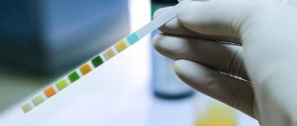
- Laboratory research.
A biochemical blood test measures the level of albumin, urea, and creatinine. Of the electrolytes, in addition to total and ionized Ca, the concentration of phosphorus and chlorides is determined. The content of vitamin D (25OH-D) is studied. PTH, PTH-like peptides. Daily urinary Ca excretion is checked. If thyrotoxicosis or hypocortisolism is suspected, a blood test for hormones (TSH, free T4, cortisol) is performed. - Functional tests.
Special provoking tests provide invaluable assistance for differential diagnosis of the causes of this disorder. These include tests with native vitamin D, thiazide diuretics, and calcitonin. A steroid suppression test with prednisolone can be used to exclude a process not associated with increased PTH secretion. - Instrumental research.
To search for adenoma or hyperplasia of the parathyroid glands, ultrasound, computed tomography, and scintigraphy are performed. Densitometry is performed to determine bone mineral density, and renal ultrasound is performed to diagnose nephrolithiasis. If there are symptoms that raise suspicion of an inflammatory process in the lungs or a malignant neoplasm, radiography, CT scan of the lungs, abdominal organs, and mammography should be prescribed to identify them.
Differential diagnosis must be made based on the predominant symptoms. Insipidal syndrome must be differentiated from diabetes mellitus and diabetes insipidus. Muscle weakness and hypotension should be distinguished from that with muscular dystrophies, myasthenia gravis, and polymyositis. Neuropsychiatric symptoms require the exclusion of psychiatric diseases.
Scintigraphy. Parathyroid adenoma
Recommendations
- ^ a b c d f g gram hour i j
Takeoff, J;
Perkins, G.D.; Abbas, G; Alfonzo, A; Barelli, A; Bierens, J. J.; Brugger, H; Deakin, C. D.; Dunning, J; Georgiou, M; Handley, A. J.; Locky, DJ; Paal, P; Sandroni, C; Thies, K. C.; Zideman D.A.; Nolan, J.P. (October 2010). "European Resuscitation Council Guidelines 2010, section 8. Cardiac arrest in special circumstances: electrolyte abnormalities, poisoning, drowning, accidental hypothermia, hyperthermia, asthma, anaphylaxis, cardiac surgery, trauma, pregnancy, electric shock." Resuscitation
.
81
(10): 1400–33. doi:10.1016/j.resuscitation.2010.08.015. PMID 20956045. - ^ a b c d f g gram h i j k
Fong, J;
Khan, A (February 2012). "Hypocalcemia: Updates in Diagnosis and Management in Primary Care." Canadian family doctor
.
58
(2): 158–62. PMC 3279267. PMID 22439169. - ^ a b
Patey, M. John (2006).
"Appendix 1: Converting SI Units to Standard Units." Principles and Practice of Geriatric Medicine
.
2
(4th ed.). Chichester [ua]: Wiley. p. Appendix. Doi:10.1002/047009057X.app01. ISBN 9780470090558. - ^ a b c
Cooper, MS;
Gittoes, New Jersey (June 7, 2008). "Diagnosis and treatment of hypocalcemia." BMJ (ed. Clinical Research)
.
336
(7656):1298–302. Doi:10.1136/bmj.39582.589433.be. PMC 2413335. PMID 18535072. - Lemone, Priscilla; Burke, Karen; Dwyer, Trudy; Levett-Jones, Tracey; Moxham, Lorna; Reid-Searle, Kerry (2015). Medical-surgical nursing
. Pearson Higher Education, Australia. p. 237. ISBN 9781486014408. In the archive from the original dated 10/02/2016. - Minisola, S; Pepe, J; Piedmonte, S; Cipriani, C (2 June 2015). "Diagnosis and treatment of hypercalcemia." BMJ (ed. Clinical Research)
.
350
: h2723. doi:10.1136/bmj.h2723. PMID 26037642. S2CID 28462200. - Armstrong, C. M.; Cota, Gabriel (1999). "Calcium block of Na+ channels and its effect on the rate of closure". Proceedings of the National Academy of Sciences of the United States of America
.
96
(7):4154–4157. Bibcode:1999PNAS...96.4154A. doi:10.1073/psas.96.7.4154. PMC 22436. PMID 10097179. - Durlach, J; Bac, P; Durlach, V; Bara, M; Guyet-Bara, A (June 1997). "Neurotic, neuromuscular and autonomic nervous forms of magnesium imbalance." Magnesium Research
.
10
(2): 169–95. PMID 9368238. - ^ a b
Nussey, S. S.;
Whitehead, S. A. (2013-04-08). Endocrinology: an integrated approach
. CRC Press. paragraph 194. ISBN 9780203450437. - Bijlani, R. L.; Manjunatha, S. (November 26, 2010). Understanding Medical Physiology: A Textbook for Medical Students
. Published by Jaypee Brothers. item 465. ISBN 9789380704814. - “Hypoparathyroidism. Symptoms and diseases of the parathyroid glands | Patient". Patient
. Retrieved 2015-09-05. - "Hypoparathyroidism." NORD (National Organization for Rare Diseases)
. Retrieved 2019-01-09. These cases may be called autoimmune hypoparathyroidism and develop when the body's own immune system mistakenly attacks parathyroid tissue and results in loss of parathyroid hormone secretion. - ^ a b c
Metheny, Norma (2012).
Fluid and Electrolyte Balance: Care Guidelines
(5th ed.). Sudbury, MA: Jones and Bartlett Learning. paragraph 93. ISBN 978-0-7637-8164-4. Retrieved September 4, 2015. - ^ a b c d
Helms, Richard (2006).
Textbook of therapy: drugs and treatment of diseases
(8th ed.). Philadelphia, PA [ua]: Lippincott Williams and Wilkins. item 1035. ISBN 978-0-7817-5734-8. Retrieved September 4, 2015. - ^ a b c d f f
Fong, Jeremy;
Khan, Aliya (2012). "Hypocalcemia: Updates in Diagnosis and Management in Primary Care." Canadian family doctor
.
58
(2): 158–62. PMC 3279267. PMID 22439169. - Hall, edited by Patrick T. Murray, Hugh R. Brady, Jesse B. (2006). Intensive care in nephrology
. London: Taylor and Francis. paragraph 129. ISBN 978-0-203-02482-9. Retrieved September 4, 2015.CS1 maint: additional text: list of authors (link) - MedlinePlus Encyclopedia
: Hypocalcemia - Infants - Siyam, Fadi F.; Klaczko, David M. (2013). “What is hypercalcemia? The importance of fasting tests.” Cardiorenal Medicine
.
3
(4): 232–238. Doi:10.1159/000355526. ISSN 1664-3828. PMC 3901605. PMID 24474951. - Fluids and Electrolytes: A 2-in-1 Guide for Nurses
. Lippincott Williams and Wilkins. 2006. p. 122. ISBN 9781582554259. In the archive from the original dated March 19, 2017. - Yap, E; Roche-Recinos, A; Goldwasser, P. (December 30, 2021). "Prediction of ionized hypocalcemia in intensive care: an improved method based on the anion gap." Journal of Applied Laboratory Medicine
.
5
(1): 4–14. Doi:10.1373/jalm.2019.029314. PMID 32445343.
Treatment of hypercalcemia
Conservative therapy
Patients with any degree of severity should be hospitalized in a hospital (endocrinology, nephrology department) for treatment. Patients with severe neurological symptoms and hypercalcemic crisis should be transferred to the intensive care unit. It is necessary to discontinue all medications that can cause an increase in calcium levels. Treatment of hypercalcemia has the following directions:
- Increased excretion of calcium in urine.
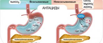
First, it is necessary to ensure adequate rehydration with physiological NaCl solution (0.9%). This will improve renal perfusion and increase the filtration of calcium ions by the renal glomeruli. Additionally, forced diuresis is performed using loop diuretics (furosemide). It is necessary to constantly monitor blood electrolyte levels. - Decreased absorption of Ca in the intestine.
Sodium or potassium phosphate salts are well suited for this purpose. Phosphates are strictly contraindicated for the treatment of secondary hyperparathyroidism caused by renal failure. Also, glucocorticosteroids (prednisolone, hydrocortisone) and synthetic antimalarial drugs (hydroxychloroquine, chloroquine) are used to suppress Ca absorption in the gastrointestinal tract. - Suppression of bone resorption.
An important stage in the treatment of hypercalcemia caused by hyperparathyroidism or cancer. The most effective drugs for preventing the progression of osteoporosis are bisphosphonates (pamidronic acid, zoledronic acid), which inhibit the activity of osteoclasts. The peptide hormone calcitonin and the cytotoxic antibiotic mithramycin have a similar mechanism of action, but a faster effect. - Suppression of the production of PTH and PTH-like protein.
For the pathogenetic treatment of primary and secondary hyperparathyroidism, calcimimetics (cinacalcet) are used, which increase the sensitivity of PTG cell receptors, thereby reducing PTH production. Gallium nitrate is used to treat hypercalcemia caused by a malignant tumor, which inhibits the secretion of PTH-like protein by tumor cells. - Intensive therapy.
For the treatment of severe life-threatening conditions (hypercalcemic crisis, calciphylaxis), as well as when other conservative methods of therapy are ineffective, hemodialysis using a low-calcium dialysate solution is an emergency measure to reduce Ca in the serum.
Surgery
Surgical removal of the parathyroid glands is the main treatment for primary hyperparathyroidism. The main indication for surgical intervention is a Ca level above 2.75 mmol/l. To prevent postoperative hypocalcemia (“hungry bone syndrome”), the patient is prescribed vitamin D and calcium supplements. Malignant tumors also need to be removed. To treat oncohematological pathologies, bone marrow transplantation is performed.
Experimental treatment
New drugs are currently being developed to treat this condition. The drug osteoprotegerin, which is a cytokine from the family of tumor necrosis factors, is at the stage of clinical trials. It inhibits the differentiation of osteoclasts and stimulates their apoptosis. In in vitro experiments, the calcitriol analogue EB 1089 suppressed the expression of the PTH peptide gene.
Use of calcium for hypocalcemia and other conditions[edit | edit code]
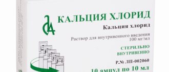
Calcium is used to compensate for the deficiency of this substance, and also as a dietary supplement. Calcium salts serve as a specific means of emergency treatment of hypocalcemic tetany, regardless of its etiology. For severe tetany, calcium salts are administered intravenously. For hypocalcemic tetany and laryngospasm, calcium chloride (CaCl2 x 2H20), containing 27% calcium, is usually used. It is administered intravenously, avoiding contact with tissue. Injections of calcium chloride are accompanied by dilation of peripheral blood vessels and a burning sensation of the skin. For intravenous administration, a 10% solution with a calcium concentration of 27 mg/ml (0.68 mmol/ml) is usually used. To avoid cardiac arrhythmias, the solution should be administered slowly (no faster than 1 ml/min). In this case, a slight decrease in blood pressure due to vasodilation is possible. Since calcium chloride acidifies urine, it is contraindicated in hypocalcemia caused by renal failure. Calcium glucoheptonate is administered intravenously for severe hypocalcemic tetany in a dose of 5-20 ml of a 22% solution with a calcium concentration of 18 mg/ml (0.45 mmol/ml). If administered too quickly, a temporary tingling sensation may occur. If intravenous administration is not possible, a solution of up to 5 ml is administered intramuscularly, which may cause a mild local reaction. A good source of calcium is calcium gluconate, a 10% solution of which contains 9.3 mg/ml (0.23 mmol/ml) calcium. IV administration of calcium gluconate is the mainstay of treatment for severe hypocalcemic tetany. For moderate and severe hypocalcemia, calcium gluconate is administered as an infusion at a rate of 10-15 mg/kg calcium over 4-6 hours. It is necessary to use many ampoules, since ordinary ampoules with a 10% solution have a volume of only 10 ml and contain 93 mg of calcium . Calcium gluconate cannot be administered intramuscularly, as there is a high risk of abscess formation at the injection site.
For mild symptoms of hypocalcemia, oral calcium is sufficient, usually in combination with vitamin D or one of its active metabolites. There are many calcium salts available for oral use. For hypocalcemia, the average dose is for calcium gluconate - 15 g / day (in fractions), for calcium lactate - 4 g / day (in fractions, with meals; lactose is taken simultaneously with calcium, 8 g / day), for calcium carbonate or phosphate - 1-2 g/day (in fractions, during meals).
In order to limit the absorption of phosphate in chronic renal failure and oxalate in chronic inflammatory bowel diseases, calcium carbonate or calcium acetate is prescribed orally. Rapid administration of calcium can be life-saving in severe hyperkalemia. Calcium gluconate (10-30 ml of a 10% solution) can temporarily reverse some of the cardiotoxic effects of hyperkalemia while efforts are made to reduce plasma K+ concentrations.
The use of calcium supplements for the prevention and treatment of osteoporosis is discussed: Osteoporosis.
Prognosis and prevention
Hypercalcemia is a severe and in some cases (especially in acute cases) a life-threatening pathological condition. In hypercalcemic crisis, the mortality rate is very high (60%). The frequency of deaths in chronic cases averages 20-25%. However, the prognosis is largely determined by the cause of the increase in Ca levels.
Prevention of this pathology consists in timely diagnosis and proper treatment of the diseases against which it develops. Before starting to take vitamin D or other medications that may increase Ca levels in the blood, a blood test should be performed to assess Ca levels.
Causes
The main causes of hypercalcemia are excess parathyroid hormone in the body (hyperparathyroidism), oncology and long-term use of calcium supplements. Hypocalcemia almost always develops against the background of parathyroid hormone deficiency, the production of which is responsible for the upper and lower parathyroid glands. By interacting with the hormone calcitonin (thyroid), the exchange of phosphorus and calcium in the body is regulated. Excess calcium, as well as deficiency, develops with deviations in the functioning of organs and systems.