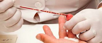
Blood tests
– the most accurate way to diagnose problems related to human health, as well as a necessary condition for the prevention of possible diseases. Under the influence of external and internal influences, blood is able to change its composition, which helps to see an objective picture of the patient’s health status, make a correct diagnosis and draw up the most correct plan for combating the disease.
There are many types of laboratory tests of blood, consisting of plasma and formed elements suspended in it. Pathological processes occurring in the body cause sudden changes in all indicators. Quite often, specialists resort to a series of studies to monitor the dynamics of the disease, as well as evaluate the effectiveness of the chosen therapy.
Clinical (general) analysis
Clinical (general) analysis
blood is prescribed both for preventive purposes and during illness. The required amount of biomaterial can be collected from a finger or a vein. Before the procedure, you must completely avoid eating and drinking 8 hours before the procedure. You can only drink drinking water without gas. The patient is advised to refrain from sports and strenuous physical activity.
A clinical blood test determines:
- The state of the body's immune system.
- Hemoglobin level.
- The presence and severity of the inflammatory process.
- The presence of allergic or parasitic diseases.
- Disorders of the blood clotting process.
Types of stool tests
Such studies are very informative and allow us to identify:
- disturbances in the functioning of the digestive system;
- helminthic infestation, bacterial infection;
- hidden blood
The following types of tests are common:
- coprogram. Analyzes the chemical and physical characteristics of feces, which allows you to evaluate the functioning of the stomach, liver, pancreas, intestines, and identify inflammation and colitis. The detailed analysis contains more than 20 indicators obtained as a result of chemical, macro- and microscopic examination;
- occult blood test. Prescribed for suspected bleeding in any part of the digestive tract. The coprogram does not show it, and such a study is needed for an accurate diagnosis;
- analysis for dysbacteriosis. Evaluates intestinal flora, identifies 15 main types of pathogenic bacteria, fungi, and microorganisms. Prescribed for disorders in the gastrointestinal tract, after long-term treatment, which can provoke them.
Blood chemistry
Blood chemistry
gives the most complete picture of the functioning of all human organs and systems, as well as the speed of metabolic processes occurring in his body. It is recommended to conduct a biochemical blood test annually. The biomaterial for research is taken from a vein in an amount of no more than 5 ml and distributed into several test tubes. Blood sampling is performed exclusively in the morning. Before analysis it is necessary to prepare:
Sign up for blood tests
Make an appointment
How is a general blood test performed and a hemogram drawn up?
How to donate a blood hemogram:
- It is determined from which vein the blood will be drawn.
- The elbow bend is treated with a swab moistened with an antiseptic.
- A not too tight tourniquet is applied to the arm just below the shoulder. You cannot tighten a limb for a long time.
- A vein is punctured.
- The required volume of blood is taken into a tube with EDTA.
- EDTA (ethylenediaminetetraacetic acid) is an anticoagulant needed to keep the blood fluid and not clot. The sample is then moved slowly around the tube several times.
- The material is sent to the laboratory.
The blood sample is examined in a special device called a hematology analyzer. The transcript of a blood test (hemogram) is a table containing the names of indicators, units of measurement, norms and actually detected deviations in the composition of the blood. They are identified by special markings or color changes.
The test results do not indicate diagnoses. It is transferred to the attending physician to make a final verdict (in conjunction with other studies, the patient’s complaints and his physical examination), since conclusions cannot be drawn only on the basis of the hemogram.
Blood grouping and determination of Rh factor
Blood grouping and determination of the Rh factor is one of the important studies that can save the life of a patient as a result of an accident. This laboratory analysis is also carried out:
Biomaterial is collected from a vein in the morning. Before the procedure, the patient should follow simple recommendations - do not consume food or alcoholic beverages for 4 hours, avoid smoking, and reduce the intensity of physical activity 12 hours before the test.
Test results and diagnosis
Normal values for each test may vary depending on the patient’s age, health status, past illnesses, and other factors.
Therefore, the laboratory test result itself cannot be regarded as a diagnosis. It definitely requires consulting a doctor. Thus, during examinations for elderly patients, the normal value changes for most laboratory parameters. Medical specialists evaluate them, taking into account the individual prognosis. The same approach is used in rehabilitation after injuries, illnesses, relapses and exacerbations, and alcohol addiction. This provides accurate information about the patient’s condition, allows you to prescribe safe and effective therapy, and achieve good treatment results. You have questions?
We will call you back within 30 seconds
or call the number
Clicking the "Submit"
, you automatically consent to the processing of your personal data and accept the terms of the User Agreement.
- Preventative medical examination for the elderly
- Types of instrumental examinations
- Laboratory diagnostics: main types of tests
- Examination by a neurologist and psychiatrist for the diagnosis of dementia and Alzheimer's disease
Coagulogram (hemostasiogram) or blood test
A coagulogram (hemostasiogram) or blood clotting test helps determine the quantity and quality of prothrombins, as well as their ability to form blood clots and prevent serious blood loss, as evidenced by the rate of formation of blood clots. If clotting rates are reduced, then there is a possibility of extensive bleeding even with minor injuries, which, in turn, can lead to serious health problems. Excessive blood clotting (increased thickness) can lead to the formation of blood clots that can block the most important vessels and arteries. Most often, a coagulogram is prescribed before surgery, for the diagnosis and treatment of thrombosis, during planning of pregnancy or its pathologies, for diseases of the hematopoietic organs, bleeding, diseases of the liver and cardiovascular system. To obtain reliable parameters for this type of study, the patient must follow some rules:
Blood is drawn from a vein in the morning. Before the procedure, you can drink a glass of plain water without carbon.
HEMOGRAM
HEMOGRAM
(Greek haima blood + gramma line, line, image; synonym
general clinical blood test
) - the results of a quantitative and qualitative study of blood composition. G. includes data on the number of red blood cells, their morphology, characteristics, the number of reticulocytes, the total content of hemoglobin in the blood, color index, the number of leukocytes, the ratio of their different types, the number of platelets, as well as some indicators of the blood coagulation system and physical-chemical. blood indicators. Depending on the patient population, the range of indicators can be expanded. In 1972, the All-Union Scientific Research Laboratory Center M3 of the USSR developed a new G. scheme.
The term “hemogram” was first proposed by V. Schilling in 1931 to characterize only the leukocyte composition of the blood, namely to determine the percentage of different types of leukocytes, including neutrophilic leukocytes with different nuclear shapes (see Leukocyte formula).
Rice. 1. A scarifier needle with a removable spear (1) for puncturing the skin when obtaining peripheral blood for research and a stand with removable scarifiers (2): in the middle of the stand there is a slot with fastenings for a scarifier needle. Rice. 2. A scarifier pen for puncturing the skin when obtaining peripheral blood for research.
It is recommended to take blood for testing in the morning on an empty stomach or an hour after a light breakfast. In laboratory practice, capillary blood is usually examined (blood can be taken from a vein). Blood is taken by pricking the flesh of a finger or earlobe with a needle, and in young children, the flesh of the heel. You should use scarifier needles with removable spears or scarifier feathers (Fig. 1 and 2), which after work are boiled in a sterilizer or placed in a drying cabinet for 2 hours at a temperature of 180°.
The skin at the injection site is wiped sequentially with two swabs: first moistened with alcohol, then with ether. With this treatment of the skin, the drop of blood protruding from the puncture does not blur. The injection should be done from the side, where the capillary network is thicker, to a depth of 2-3 mm, depending on the thickness of the skin, so that the blood flows freely. With strong pressure, tissue fluid will be mixed with the blood, which can affect the test results. It is important to follow the sequence of taking blood to study individual indicators: after removing (with a cotton swab) the first drop, blood is drawn to determine ROE, to determine the amount of hemoglobin, to count the total number of red blood cells, to count the total number of leukocytes, smears (usually two) are made to count the leukocyte formulas and studies of erythrocyte morphology; To count reticulocytes, smears are made on specially prepared slides.
To determine the clotting time and duration of bleeding, as well as to count the number of platelets, separate skin punctures are made.
Determination of hemoglobin amount
can be produced in various ways. In laboratory practice, a Sali type hemometer is often used for this purpose (see Hemoglobinometry).
Hemoglobin norms for men are 14.5 g% (range 13.0-16.0 g%) and for women 13.0 g% (range 12.0-14.0 g%).
A decrease in the concentration of hemoglobin in the blood is observed with anemia of various etiologies (with blood loss, deficiency of vitamin B12, iron, increased hemolysis of red blood cells, etc.). An increase in hemoglobin concentration occurs with erythremia, secondary, or symptomatic, erythrocytosis. When blood thickens, a relative increase in hemoglobin concentration may occur.
Total red blood cell count
counted in 1 µl of blood. Blood can be taken using the melanger method (in melangeurers or mixers) or the test tube method (in test tubes, according to N. M. Nikolaev), followed by counting red blood cells in a counting chamber under a microscope (see Counting chambers). Red blood cells are counted in a photoelectric erythrohemometer, as well as using a celloscope.
The normal number of erythrocytes in men is 4,000,000–5,000,000 per 1 μl of blood, in women 3,700,000–4,700,000. An increase in the number of erythrocytes is usually observed in diseases characterized by an increase in hemoglobin concentration (for example, polycythemia, secondary erythrocytosis and etc.). A decrease in the number of red blood cells is observed with a decrease in the erythroblastic function of the bone marrow (hypo- and aplastic processes), with pathologically altered bone marrow (leukemia, myeloma, metastases of malignant tumors, etc.), due to increased breakdown of red blood cells (hemolytic anemia), with a deficiency in the body iron, vitamin B12, for bleeding.
Color indicator
- an indicator expressing the relative content of hemoglobin in one red blood cell in Sali units. If the hemoglobin content is measured in gram percent, the color index is calculated by dividing three times the hemoglobin gram percent value by the first two digits of the red blood cell count (e.g., hemoglobin 14.0 g%, red blood cell count 4,200,000; color index = 1.0). If the hemoglobin content is expressed in Sali units, then this indicator is divided by double the first two digits of the erythrocyte content indicator (for example, hemoglobin 84 units, number of erythrocytes -4,200,000; color indicator 84/(2•42) = 1.0) . Normally, the color index ranges from 0.85 to 1.15. The value of the color index is important in determining the form of anemia: a color index below 0.85 is hypochromic, a color index of 0.85-1.15 is normochromic, a color index above 1.15 is hyperchromic.
Average hemoglobin content in one red blood cell
in abs. In calculus, it is usually denoted in picograms (pg). It is determined by dividing the hemoglobin content in 1 μl of blood by the number of red blood cells in the same volume. For example, the average value of red blood cells in 1 μl of blood is 5,000,000; the average hemoglobin content is 16.7 g%, which is 0.000167 g, or 167,000,000 pg in 1 μl of blood. Consequently, the hemoglobin content in one red blood cell will be 167000000/5000000 = 33.4 pg.
Practically, the average value of hemoglobin content in one red blood cell can be obtained by multiplying the amount of hemoglobin in gram percent by 10 and dividing the product of these numbers by the number of red blood cells in 1 ml of blood: 16.7•10/5 = 33.4 pg. The average hemoglobin content in an individual red blood cell in adults ranges from 27 to 33.4 pg.
Hyperchromasia (see Hyperchromasia, hypochromasia) depends on an increase in the volume of red blood cells (macrocytes, megalocytes), and not on the degree of their saturation with hemoglobin and is an indicator of impaired liver function, impaired metabolism of vitamin B12 or its deficiency in the body (Pernicious anemia). In these cases, the hemoglobin content in one red blood cell increases to 50 pg. Hypochromasia is observed with a decrease in the volume of red blood cells (microcytes) or a decrease in the hemoglobin content in a normal red blood cell. The average hemoglobin content in one red blood cell decreases to 20 pg.
Erythrocyte sedimentation rate
(ESR, see Erythrocyte sedimentation) is expressed in millimeters of plasma settling for 1 hour. Normally, in women it is up to 14-15 mm per hour, in men - up to 10 mm per hour.
Changes in erythrocyte sedimentation rate are not specific to any disease. However, acceleration of erythrocyte sedimentation always indicates the presence of a patol process.
Reticulocyte count
counted in blood smears stained intravitally with 1% brilliant cresyl blue solution in abs. alcohol Thin smears of blood are made on pre-prepared glass slides (preparation consists of applying paint in the form of a smear to a well-washed, degreased and heated glass) and the glass is immediately placed in a moist chamber (Petri dish, moistened gauze or cotton rolls are placed along the edges ). The camera is placed in a thermostat at t° 37° for 3-5 minutes. The smears are then air dried. Count the number of reticulocytes per 1000 red blood cells; The research results are expressed in ppm. Normally, their content in peripheral blood is 2-10‰.
Reticulocytes are young red blood cells in which a granular-mesh substance is revealed using supravital staining. The reticulocyte, upon exiting the bone marrow into the peripheral blood, turns into a mature erythrocyte. It is believed that their final ripening occurs within a few hours.
Platelet count
can be calculated using various methods.
1. In peripheral blood smears, the number of platelets is counted per 1000 red blood cells. Blood for smears is taken from a finger. A drop of 14% magnesium sulfate solution is first applied to the injection site; the released drop of blood is mixed with magnesium sulfate and a smear is made on the glass; smears are painted according to Romanovsky for 2-3 hours; Knowing the number of red blood cells in 1 μl of blood, calculate the number of platelets in 1 μl of blood. For example, when counting 1000 red blood cells, 60 platelets were found; the number of red blood cells in 1 μl of blood is 5,000,000, therefore, the number of platelets will be 60 × 5000, or 300,000.
2. In the counting chamber, platelets are counted after preliminary lysis of red blood cells using a phase contrast microscope.
3. The number of platelets can be counted using automatic counters, for example, a celloscope.
In adults and older children, the platelet count is 180,000–320,000 per 1 μl. With Werlhof's disease and symptomatic thrombocytopenia, the number of platelets can decrease sharply until completely disappearing.
White blood cell count
counted in the same way as the number of red blood cells. The average number of leukocytes in an adult ranges from 4000 to 9000 in 1 μl of blood. In children it is slightly larger, in newborns - up to 15,000-30,000. With a significant increase in the number of leukocytes, we can talk about leukocytosis (see), with a decrease - about leukopenia (see).
The leukocyte formula is examined in stained blood smears.
Hematocrit values
determined in hematocrit or, using the Van Slyke nomogram, by the concentration of hemoglobin in the blood (see Hematocrit number). The hematocrit number shows the volumetric ratio of the formed elements of blood and plasma. The results of the study are expressed either as a fractional number, in which the numerator is the volume of formed elements, and in the denominator - the volume of plasma, or as a percentage showing the ratio of the volume of formed elements to the volume of blood taken. Normal hematocrit values for men are 40/60-48/52 (or 40-48%), for women 36/64-42/58 (or 36-42%). An increase in the volume of erythrocytes is observed with erythrocytosis, a decrease - with anemia.
Morphology of red blood cells
(like leukocytes) is examined in stained blood smears. Before staining, blood smears are treated with fixing liquids (for example, methyl alcohol) in order to prevent the destruction of blood cells during the staining process. There are many chemical staining methods for blood smears. the affinity of cell elements for certain aniline dyes. When studying blood smears under a microscope, they get an idea of the size, shape and color of red blood cells, which can change in pathol conditions. The sizes of red blood cells, determined using an eyepiece micrometer (see), in healthy people vary within certain limits. This is the so-called fiziol, anisocytosis of erythrocytes. An accurate idea of the distribution of red blood cells by size is obtained by measuring their diameter and plotting an anisocytosis curve (see Erythrocytometry). A condition in which red blood cells of various sizes are found is called anisocytosis, which can happen, for example, with anemia. The vast majority of red blood cells have a diameter. 7-8 microns. Erythrocytes with a diameter of less than 6.5 microns are called microcytes, and the condition in which they predominate is called microcytosis (for example, with iron deficiency, with microspherocytic hemolytic anemia). Red blood cells with a diameter of more than 8 microns are called macrocytes. Their detection in newborns is considered a physiol phenomenon; macrocytes disappear by the age of two months. Macrocytosis is detected with increased blood regeneration, stomach cancer and polyps, decreased thyroid function, and myelomatosis. Red blood cells with a diameter of more than 12 microns are called megalocytes. They may have an oval shape. In addition to their large size, they are characterized by hyperchromia, lack of biconcavity (absence of a central lumen) and large thickness. Megalocytosis is detected when there is a lack of vitamin B12 in the body.
A change in the shape of red blood cells is called poikilocytosis. Red blood cells can become oval, pear-shaped, stellate, jagged, etc. Red blood cells with a pronounced oval shape are called ovalocytes, which are normally 5-10% (G. A. Alekseev).
Ovalocytosis (up to 80-90% of ovalocytes) can be a carrier state or a pathology, leading to the development of hemolytic anemia (see). Sickle-shaped red blood cells are found in sickle cell anemia (see).
Polychromatophils (see Polychromasia) are immature red blood cells containing, along with hemoglobin, remnants of a basophilic substance. Depending on the amount of hemoglobin, polychromatophilic red blood cells in ordinary smears (Romanovsky stained) have different shades, from blue to pinkish-gray. In ordinary smears stained with aniline dyes, reticulocytes are also polychromatophilic. Polychromatophils in a thick drop are counted. Smears with a thick drop are stained according to Romanovsky without fixation, while mature erythrocytes are hemolyzed, and young, immature ones appear as a basophilically stained mesh of bluish-violet color. Normally, in a thick drop, 1-2 erythrocytes with a basophilic mesh are found not in every field of view, and this is designated as P+, with 3-5 polychromatophils - P++, with 5-10 - P+++, with a more significant number polychromatophiles - P++++. This method is imprecise, but gives an idea of the increase or decrease in the number of young red blood cells. Polychromatophilia is an indicator of the regeneration of erythropoiesis and occurs with blood loss, increased hemolysis of red blood cells, etc. Normally, a large number of polychromatophils are found in the first days after birth and quickly decrease after two weeks.
With altered bone marrow regeneration, normoblasts emerge into the peripheral blood, and with pernicious anemia, megaloblasts can be found.
Nuclear forms of elements of the erythroid germ in the peripheral blood of adults are observed only in severe forms of anemia, their appearance is a sign of pathol. regeneration. In such cases, remnants of the nucleus can also be observed in the form of a thin ring, loop, figure eight of violet-blue color (Cabot rings), round formations of violet-red color 1-2 microns in size (Jolly bodies) or in the form of basophilic granulation in erythrocytes.
In case of malaria, Schüffner granularity is found in erythrocytes - small pink-red inclusions that fill almost the entire erythrocyte, or 10-15 inclusions of different sizes; the latter are sometimes called Maurer's spot.
In patients with severe intoxications (phenylhydrazine, nitrobenzene, aniline, berthollet salt, nitroglycerin, toluene diamine, etc.), treated with sulfonamides, as well as in persons working in chemical. industry, it is necessary to examine blood using the Dacey method in order to identify Heinz-Ehrlich bodies in red blood cells - round inclusions of purple-red color; sometimes they are found extracellularly.
Resistance of erythrocytes is the property of erythrocytes to resist destructive influences: osmotic, mechanical, thermal, etc. In wedges, in practice, the osmotic resistance of erythrocytes is usually studied: a series of test tubes with a solution of sodium chloride are prepared in increasing concentrations - from 0.28 to 0.56%. Normally, the minimum resistance of adult erythrocytes (when the first erythrocytes begin to be destroyed) ranges between 0.48 and 0.44% sodium chloride, the maximum (all erythrocytes are destroyed) is between 0.32-0.28%.
Leukocyte morphology
may change due to infectious diseases, exposure to chemicals. substances, diseases of the hematopoietic apparatus, the effects of ionizing radiation.
Anisocytosis (various sizes) of leukocytes is also found normally. However, pronounced anisocytosis is a manifestation of pathology.
Structural changes are found both in the nucleus and in the cytoplasm of the leukocyte. The following changes may occur in the nucleus: a) hypersegmentation of the nucleus (the term “hypersegmentation” is more often used to denote an increase in the number of segments of segmented leukocytes, the detection of which is of diagnostic value for pernicious anemia, radiation sickness); b) chromatinolysis - dissolution of nuclear chromatin while maintaining its contour; c) karyolysis - dissolution of part of the nucleus while maintaining the structure of the rest of the nucleus; d) fragmentosis - separation from the nucleus of 1-5 fragments, sometimes connected to the nucleus by filaments of basicchromatin; e) pyknosis - the nucleus becomes structureless due to compaction of the main chromatin; at the same time the size of the nucleus decreases; f) karyorrhexis - disintegration of the nucleus into pyknotic parts of a round shape and various sizes, not interconnected.
Toxigenic granularity in the cytoplasm of neutrophils in smears stained with mixtures of azure and eosin is not detected reliably. With special stains (Freifeld, Momsen, Shmelev) a bluish mesh with transitions into large lumps is found in the cytoplasm; sometimes the entire cell is strewn with small dust-like grains. Toxigenic granularity of neutrophils is observed in purulent processes, diseases associated with intoxication, and infections (diphtheria, measles, chicken pox, scarlet fever, rubella, pneumonia). The counting of neutrophils with toxigenic granularity is carried out in relation to the number of counted (and not per 100) neutrophils. For example, all neutrophils - 70%, including neutrophils with toxigenic granularity - 45%.
Cytolysis is the breakdown of a cell. Cytolysis is often characterized by the absence of cytoplasm, a nucleus with vague contours, the structure of the nucleus is preserved, and sometimes there is granularity near such a nucleus. These changes can also occur in normal blood, being signs of reverse cell development, but if detected in significant quantities, they are considered a pathology.
Platelet morphology
studied in thin blood smears taken without a stabilizer (magnesium sulfate), stained with a mixture of azure and eosin (according to Nocht).
The detection of aggregates of 5-6 platelets in a smear is noted as good agglutinability. In platelets, a central granular part (granulomere) and a peripheral homogeneous part (hyalomer) are visible. The granulomer consists of azurophilic grains with a lilac tint. It is also possible to distinguish finer details of the platelet structure - vacuoles, pseudopodia, thin antennae processes emanating from the granulomere. Based on the morphology and characteristics of platelets, a platelet formula is compiled, the edges can change with pathology (see Platelets).
Some blood clotting indicators
— bleeding time (see) and blood clotting time (see), included in G., are indicative for judging disorders of the blood coagulation system.
Rice.
3. Viscometer: 1 - capillary for distilled water; 2 — capillary switching valve; 3 - capillary for blood; 4 - a rubber tube with which water and blood are sucked. Blood viscosity
depends on the viscosity of the plasma, the number of red blood cells, hemoglobin, and the carbon dioxide content in the blood (see Viscosity, blood viscosity). Determined in a viscometer (Fig. 3). The determination principle is based on comparing the speed of movement of blood and dist, water in strictly identical capillaries and at the same vacuum in the viscometer system. The paths traveled by liquids in capillaries at the same time are inversely proportional to the viscosity of these liquids. Therefore, viscosity is expressed as the ratio of the length of the path traveled by water to the length of the path traveled by blood. Since blood is drawn up to the mark, the viscosity of the blood will be equal to the length of the path traveled by the water in the same time; the path length is measured on a scale. Blood viscosity in men ranges from 4.3 to 5.3, in women - from 3.9 to 4.9.
The causative agents of malaria can also be detected in ordinary blood smears, but in a targeted study for malaria plasmodia, it is also necessary to take the so-called. thick drop (see), the edges make it possible to detect parasites even if they are small in number. The following method of preparing a thick drop is recommended: prepare a regular blood smear on a glass slide and, while it is still wet, touch it to the drop of blood that has appeared again at the injection site. A drop of blood on a wet smear spreads evenly in a regular circle, eliminating the need to smear it. The thick drop is allowed to dry for about half an hour in air in a horizontal position. Unlike a regular smear, a thick drop does not need to be fixed; it is painted as usual with azure-eosin mixtures. When staining, hemoglobin is leached from red blood cells (due to lack of fixation), so only “shadows” of red blood cells can be distinguished. In the thick part of the smear, the probability of detecting a pathogen is greater, but sometimes they are less colored than parasites located along the edge of the drop.
See also Blood (research methods), Leukocytes, Thrombocytes, Red blood cells.
Bibliography:
Kassirsky I. A. and Alekseev G. A. Clinical hematology, M., 1970; Guide to Clinical Laboratory Research, ed. E. A. Kost and L. G. Smirnova, p. 44, M., 1964; S o k about l about in V.V. and Grib about in and I.A. Indicators of peripheral blood in healthy people, Laboratory, case, No. 5, p. 259, 1972; Handbook of Clinical Laboratory Research Methods, ed. E. A. Kost, M., 1975; Handbook of functional diagnostics, ed. I. A. Kassirsky, p. 304, M., 1970; T o d o r about in J. Clinical laboratory research in pediatrics, trans. from Bulgarian, p. 271, Sofia, 1968.
D. N. Ishmukhametova.
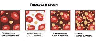
Blood sugar test
A blood sugar test determines the level of glucose in a person’s blood, the amount of which affects the well-being and functioning of all internal organs. Conducting such a study is indicated for people suffering from diabetes, as well as those who are at risk for this serious disease. Preparation for analysis is a set of simple activities:
Sign up for blood tests
Make an appointment
Basic blood diagnostic methods
Without a blood test, it is difficult to make a diagnosis for almost any disease. This is the most accessible and fastest diagnostic method. Modern laboratories equipped with advanced equipment produce research results by the evening of the same day on which the biological materials were submitted. You can sign up for a blood test in Moscow through the city doctor portal.
What types of blood tests are there?
With any ailment - mild or serious, the blood reflects this condition. The main indicators of the main body fluid change, making it possible to identify the disease and develop a treatment algorithm. After reviewing the results of the blood test, the attending physician will prescribe a number of additional tests.
The most commonly prescribed types of blood tests are:
- clinical or general;
- biochemical;
- immunological;
- analysis for hormonal group.
Clinical (general) blood test
Blood is taken from a vein or from a finger according to the doctor’s decision. Such a study reveals the slightest deviations in the body’s activity; it will tell about inflammatory processes. During the analysis, the following is determined:
- number of leukocytes (white blood cells). Many pathological conditions are diagnosed by changes in the leukocyte formula;
- erythrocytes (red blood cells) and the amount of hemoglobin;
- hematocrit - the ratio of formed elements to volume;
- platelet count . This indicator indicates the state of the coagulation system;
- number of eosinophils . It makes it possible to determine the degree of allergization of the body, as well as identify some infections.
Blood chemistry
Blood biochemistry is a popular and most informative type of research. It makes it possible to determine the following indicators in the subject:
- glucose level . An important factor in making a diagnosis of diabetes mellitus, as well as identifying other diseases associated with carbohydrate metabolism problems;
- triglycerides . A high rate indicates obesity or, on the contrary, starvation, anemia, large blood loss, poisoning, alcoholism, endocrine disorders;
- cholesterol _ The component is included in the composition of cell membranes and controls their permeability. If cholesterol is in excess, this indicates atherosclerosis, the risk of stroke and heart attack. A deficiency of the component is possible in some genetic disorders. During the study, lipoproteins - cholesterol transport complexes - are also determined;
- squirrels _ These are albumins, the level of which increases during inflammation, globulins, their amount increases with iron deficiency. Detection of immunoglobulins reports allergies. C-reactive protein indicates acute inflammation. Blood clotting proteins are also determined during the study. The detection of certain proteins or a violation of their ratio indicates a number of diseases;
- plasma enzymes . These are aminotransferases, the activity of which increases sharply in diseases of the liver and heart. Creatine kinase and lactate dehydrogenase report the stage of myocardial infarction;
- residual nitrogen . An increase in this indicator is observed with dehydration, tuberculosis, diabetes mellitus, pneumonia, kidney disease, cirrhosis of the liver;
- pigments . The main one is bilirubin. Based on its content, liver diseases are diagnosed;
- sodium _ The content of the element decreases with dehydration, insufficient dietary intake, intense sweating, vomiting, diarrhea, taking diuretics, renal failure;
- potassium _ Deficiency of the element is recorded with dehydration, metabolic disorders, long-term use of diuretics, and kidney disease. Excess potassium is observed in burns and serious injuries;
- acid-base state . This indicator reveals acidosis or alkalosis;
- phosphates, calcium . Abnormal content of these elements indicates many diseases - kidney failure, rickets, impaired absorption of nutrients in the gastrointestinal tract.
Immunological blood test
Blood is drawn from a vein. The study makes it possible to diagnose HIV, sexually transmitted diseases, various types of hepatitis, rubella, toxoplasmosis, and measles. This analysis is used in establishing oncological diagnoses and in allergological studies.
IMPORTANT! Antibodies, immunoglobulins, specific proteins, which are found in the membranes of lymphocytes, tissue fluid and blood serum, neutralize foreign viruses, bacteria and toxins.
For immunological studies:
- determine the Rh factor;
- analyze specific immune responses;
- detect C-reactive protein;
- carry out serological tests.
Hormonal blood tests
Research in the hormonal field identifies problems in the endocrine system. Based on blood taken from a vein, the amount of hormones of the thyroid gland, adrenal glands, pituitary gland and sex hormones.
Before taking any blood test, preparation is necessary, otherwise distorted results may be obtained. To do this, you should familiarize yourself with the rules for preparing for research.
The norms of blood parameters differ depending on the gender and age of the subject. In women, they even depend on the menstrual cycle. It is necessary to carefully study the obtained data and save it. The main thing is that only the attending physician can make a diagnosis based on test results.
As an advertisement
The most relevant in the category: Interesting
More interesting in the genre: News
Blood test for hormones
A blood test for hormones is prescribed by a specialist to determine the condition and correct functioning of many systems of the patient’s body. Hormones are biological active substances that regulate all important biochemical processes - growth, reproduction, metabolism and others. Changes in hormonal balance indicate pathological processes leading to the appearance of various diseases. As a rule, hormonal studies are recommended for suspected malfunctions of the internal secretion organs and associated abnormalities. The most common:
Blood test for hormones
taken in the morning, on an empty stomach. On the eve of the procedure, it is prohibited to drink alcoholic beverages or smoke. Eating should be stopped 8 hours before biomaterial collection. Taking hormonal medications must be stopped 7 days in advance, after consulting with a specialist. Women are advised to remember that hormonal levels change depending on the menstrual cycle. The most favorable days for taking hormonal tests are considered to be from 5 to 7 days from the first day of menstruation.
What do changes in blood parameters indicate?
Deviation of the blood hemogram from the norm in the table:
- A reduced level of hemoglobin is observed in anemia, leukemia, severe blood loss, pregnancy, congenital diseases of the circulatory system, and lack of iron and vitamins.
- Hemoglobin increases with congenital pathologies of the heart or lungs and their insufficiency, while at altitude, with dehydration caused by impaired renal function, diabetes mellitus and diabetes insipidus, vomiting or diarrhea, low fluid intake or heavy sweating.
- An increased level of leukocytes is observed in cases of cancer, purulent-inflammatory processes, burns or other injuries during which the soft tissues of the body are damaged.
- An increased number of lymphocytes indicates infection with any viruses, and neutrophils indicate bacterial infections. Eosinophils are considered to be markers of parasitic infestations and allergic reactions.
- An increased number of platelets occurs in chronic inflammatory diseases (tuberculosis, ulcerative colitis, cirrhosis of the liver), after operations, treatment with hormones, in some types of cancer, and iron deficiency anemia.
- A decrease in the number of platelets indicates heavy metal poisoning, blood diseases, kidney failure, pathologies of the liver, spleen, and hormonal disorders.
In addition, most drugs affect the results of laboratory tests of the above indicators.
We recommend
Therapeutic massage for the back: features, indications and contraindications Read more
Reasons that may affect the tests:
- The tourniquet was applied incorrectly when drawing blood or the application time was exceeded.
- Shaking the test tube.
- Insufficient amount of blood taken for testing.
- Mixed-up tubes with samples from different patients.
- The test tube filling standards were not met.
- Long-term storage of the collected blood sample.
- Biochemical parameters are disturbed.
- Blood is taken after the transfusion procedure.
- Technical failure of the equipment.
If the decoding of the hemogram does not correspond to the patient’s complaints and the results of the physical examination performed by the doctor, then an extended study is prescribed.
Immunological blood test and allergological studies
Immunological blood tests and allergological tests help assess the state of a person’s immune system, the presence of antibodies in his blood, and also determine the allergological status. This research method is recommended for use in cases where the patient is concerned about:
Blood is taken for examination from a vein in the morning, on an empty stomach. It is recommended to stop eating 12 hours before the procedure, and one day to exclude fatty and spicy foods, as well as alcohol, from the menu. Women should not get tested during their period. If the patient is prescribed special medications, then the doctor should be warned about this.
Blood test for PCR (polymerase chain reaction)
A PCR (polymerase chain reaction) blood test allows you to correctly diagnose any viral or bacterial infection at an early stage of the disease. Typically, this laboratory test is prescribed for serious pathological conditions:
You can undergo a similar study to determine the likelihood of infection after accidental unprotected sexual contact.
and during pregnancy in order to prevent diseases that can cause abnormalities in the development of the fetus. Preparation for a PCR blood test depends on the type of pathogen and the nature of the suspected disease. General recommendations are to exclude spicy and fatty foods and alcohol from the menu the day before the procedure. Blood is drawn from a vein in the morning, on an empty stomach. However, it is believed that the accuracy of the results does not depend on whether the patient had breakfast before the test or not.
Sign up for blood tests
Make an appointment
Blood hemogram - what kind of analysis is it?
It is carried out for the purpose of:
- Detection of anemia, leukemia, lymphoma.
- Diagnosis of bleeding and bleeding disorders.
- Detection of viral and bacterial infections.
- Detection of polycythemia vera (polycythemia vera).
- Studies of lymphadenopathy (enlarged lymph nodes).
- Detection of various allergies.
- Analysis of splenomegaly (enlarged spleen)
.
- Detection of thalassemia.
- Monitoring the treatment of liver diseases.
Currently, venous blood is an ideal material for this type of research. What is this - a blood hemogram? This is a quantitative and qualitative analysis of individual blood structures (erythrocytes, leukocytes, platelets, etc.).
What does a blood hemogram show:
- This examination method makes it possible to identify the nature of the occurrence of certain symptoms of pathology after a specialist has studied the patient’s medical history and physical examination.
- In order to monitor changes in blood counts during chemotherapy.
When to do this analysis:
- After injuries accompanied by internal and/or external bleeding.
- In case of malaise, fatigue, bleeding, bone pain, swelling of the lymph nodes, infection, etc.
- At the preparatory stage before surgery.
- To assess the quality of blood after transfusions.
Decoding a blood hemogram provides information not only about the number of cells. It can also indicate the physical characteristics of some of them, which helps the doctor make an accurate diagnosis.
There are cases when the analysis indicators do not change even in the presence of any disease.
Blood test for hCG
A blood test for hCG is the most popular test among women today. This method allows you to quickly and accurately determine whether a patient is pregnant at the earliest stages.
HCG is a human chorionic gonadotropin hormone that is produced by the chorion of the embryo. The level of this hormone in the blood indicates the onset of pregnancy or its absence. You can take the test starting from the first day of missed menstruation, or 14 days after sexual intercourse. You should refrain from eating 3 hours before the procedure. Blood is drawn from a vein. The results of the analysis can be known within a few hours.
Blood test for parasites
A blood test for parasites is the most accurate diagnostic method, allowing you to prevent serious inflammation of internal organs caused by various types of parasites entering the body. Studies are prescribed for adults and children. The material is taken from a vein. Preparing for analysis:
Study for the presence of tumor markers
Testing for the presence of tumor markers is one of the methods that determines the likelihood of a tumor process developing in the body. Using this analysis, it is possible to detect oncological diseases in the early stages, as well as prevent their relapses. A study for the presence of tumor markers is prescribed by a specialist. Preparation for the procedure includes excluding spicy and fat-rich foods from the diet, smoking and alcohol 3 days before the analysis. The patient is prescribed rest and minimal physical activity. You should not take medications other than vital ones prescribed by your doctor. Blood is drawn from a vein in the morning.
Other types of research
Histology
Biomaterial is a fragment of the epithelium of internal organs obtained through biopsy (tissue sample). It is studied under a microscope to identify malignant neoplasms, tumor cells, traces of hemorrhages, metastasis, and inflammation.
Cytology
Evaluates indicators of the cellular structure of sputum, urine obtained by puncture of fluids, scrapings, etc. Most often used in the diagnosis of cancer.
Bacterioscopy
Examination of a sample of fluid secreted by the body. Biomaterial is taken using a smear from the rectum and urethra. When using punctures, it is possible to analyze gland secretions, joint fluid, etc. In the laboratory, the flora is checked, protozoan microorganisms and fungi are identified (for this, bacteriological culture is performed).
Immunofluorescence assays
They evaluate the content of antigens in the collected biomaterial, allowing one to judge past or existing diseases. Can be used to detect malignant tumors at an early stage.
Polymerase chain reaction
PCR is a molecular diagnostic that shows traces or minimal concentrations of pathogens of infectious diseases and genetic diseases. Sensitivity against most viruses and infections is up to 100%.
Blood test for trace elements
A blood test for microelements allows you to most accurately determine the reserves of useful substances in the human body, identify disturbances in water-salt balance and various types of rheumatic conditions. Insufficient content of certain groups of vitamins and enzymes or their excess can cause the development of pathologies or deterioration of well-being. When testing blood for microelements, biomaterial is taken from a vein, exclusively in the morning and on an “empty” stomach. Before the procedure, eating and drinking alcohol is prohibited; it is recommended to limit physical activity 24 hours before. It is important to remember that a blood test for trace elements should be performed before starting a course of taking any medications or two weeks after stopping them.
Modern medicine offers a large selection of various blood tests that make it possible to identify various diseases even before the first clinical symptoms appear. With their help, the effectiveness of treatment and the correctness of selected medications for existing pathologies are assessed. For a correct diagnosis and accurate results, you need to know what blood tests exist, how and when they need to be taken.
Decoding the blood hemogram: table of normal indicators for an adult
- Erythrocytes are red blood cells that have a biconcave shape, small size and elasticity and ensure the passage of particles even in the narrowest capillaries. Their main function is to transport oxygen from the lungs to all organs and tissues of the body.
- Leukocytes are white blood cells that protect against infections, viruses and allergens. In addition, they “free” the body from cellular decay products.
- Platelets are the smallest cells that do not have a nucleus or color. This is one of the most important elements of blood, which is responsible for clotting. When the skin or organ tissue is damaged, platelets immediately “clog” the hole, forming a clot.
- Reticulocytes are “embryos” of red blood cells, capable of transforming into an adult cell under the influence of a special hormone.
- Eosinophils, basophils and neutrophils are types of white blood cells that perform a protective function against various types of bacteria and infections.
- Monocytes are large oval blood cells from the group of leukocytes with one nucleus. They do not contain granules (they are agranulocytes).
- Lymphocytes are a type of white blood cell. They are the main cells of the immune system, providing humoral (production of antibodies) and cellular (contact interaction with victim cells) immunity.
- Hemoglobin is a blood pigment, the main component of red blood cells. Its main function is to transport oxygen from the lungs to organs and tissues and remove carbon dioxide.
- Hematocrit is the ratio of plasma to formed elements, which allows us to determine the ability of blood to transport oxygen.
- ESR (erythrocyte sedimentation rate) allows you to estimate the ratio of plasma protein fractions.
- The color indicator of blood is the degree of saturation of red blood cells with hemoglobin.
Blood hemogram - interpretation in adults:
| Index | Norm |
| Red blood cells (RBC) | 4.3–6.2 x 10¹²/l – for men 3.8–5.5 x 10¹²/l – for women |
| Hemoglobin (HGB, Hb) | 120–140 g/l |
| Hematocrit (HCT) | 39–49% – for men 35–45% – for women |
| Red blood cell distribution width (RDWc) | 11,5–14,5 % |
| Mean erythrocyte volume (MCV) | 80–100 fl |
| Average hemoglobin content in erythrocytes (MCH) | 26–34 pg |
| Mean erythrocyte hemoglobin concentration (MCHC) | 30–370 g/l (g/l) |
| Platelets (PLT) | 180–320 x 109/l |
| White blood cells (WBC) | 4.0–9.0 x 10⁹/l |
| Lymphocytes (LYM), % | LY% 25–40% LYM# 1.2–3.0 x 10⁹/L (or 1.2–63.0 x 10³/µL) |
| Content of a mixture of monocytes, eosinophils, basophils and immature cells (MID, MXD) | MID# (MID, MXD#) 0.2–0.8 x 10⁹/l MID% (MXD%) 5–10% |
| Granulocytes (GRA, GRAN) | GRA# 1.2–6.8 x 10⁹/L (or 1.2–6.8 x 10³/µL) GRA% 47–72% |
| Monocytes (MON) | MON% 4–10% MON# 0.1-0.7 x 10⁹/l (or 0.1–0.7 x 10³/µl) |
| Erythrocyte sedimentation rate, ESR, ESR | Up to 10 mm/h for men Up to 15 mm/h for women |
Different laboratories may indicate different norms in the hemogram decoding forms. This is due to different methods for calculating parameters. Then the results should be interpreted based on the given standards.
Circumstances under which blood counts may change:
- Functional disorders in the human body that adversely affect the hematopoietic system.
- Bone marrow defects.
- Acute and chronic illnesses.
- Negative effects on blood cells outside the bone marrow.
A blood hemogram can be performed to prevent latent or indolent diseases, to confirm or refute a previously made diagnosis, to track the dynamics of the development of an already detected disease.
Recommended articles on the topic:
- How to massage the abdomen for weight loss: different techniques for health and beauty
- Stone massage: description, benefits, methods
- MRI of three parts of the spine: when is it necessary and what are the features of the procedure
Blood test cost
| Code | Name of the study | Biological material | Result | ****Execution period | Price | ***CIT | Note |
| 090001 | Total protein | blood (serum) | count | 1 k.d. | 230.00 rub. | 460.00 rub. | 0 |
| 090002 | Albumen | blood (serum) | count | 1 k.d. | 230.00 rub. | 460.00 rub. | 0 |
| 090003 | Protein fractions | blood (serum) | count | 1 k.d. | 345.00 rub. | 690.00 rub. | 0 |
| 090004 | Creatinine | blood (serum) | count | 1 k.d. | 230.00 rub. | 460.00 rub. | 0 |
| 090005 | Urea | blood (serum) | count | 1 k.d. | 230.00 rub. | 460.00 rub. | 0 |
| 090006 | Uric acid | blood (serum) | count | 1 k.d. | 230.00 rub. | 460.00 rub. | 0 |
| 090007 | Total bilirubin (TB) | blood (serum) | count | 1 k.d. | 230.00 rub. | 460.00 rub. | 0 |
| 090008 | Direct bilirubin (DB) | blood (serum) | count | 1 k.d. | 230.00 rub. | 460.00 rub. | 0 |
| 090009 | Total cholesterol | blood (serum) | count | 1 k.d. | 230.00 rub. | 460.00 rub. | 0 |
| 090010 | HDL cholesterol | blood (serum) | count | 1 k.d. | 230.00 rub. | 460.00 rub. | 0 |
| 090011 | LDL cholesterol | blood (serum) | count | 1 k.d. | 345.00 rub. | 690.00 rub. | 0 |
| 090012 | Triglycerides | blood (serum) | count | 1 k.d. | 230.00 rub. | 460.00 rub. | 0 |
| 090014 | Alanine aminotransferase (ALT, GPT) | blood (serum) | count | 1 k.d. | 230.00 rub. | 460.00 rub. | 0 |
| 090015 | Aspartate aminotransferase (AST, GOT) | blood (serum) | count | 1 k.d. | 230.00 rub. | 460.00 rub. | 0 |
| 090016 | Gamma glutamine transferase (GGT) | blood (serum) | count | 1 k.d. | 230.00 rub. | 460.00 rub. | 0 |
| 090017 | Alkaline phosphatase (ALCP) | blood (serum) | count | 1 k.d. | 230.00 rub. | 460.00 rub. | 0 |
| 090018 | Acid phosphatase* | blood (serum) | count | 1 k.d. | 345.00 rub. | 690.00 rub. | 0 |
| 090019 | Lactate dehydrogenase (LDH) | blood (serum) | count | 1 k.d. | 230.00 rub. | 460.00 rub. | 0 |
| 090020 | Alpha amylase | blood (serum) | count | 1 k.d. | 345.00 rub. | 690.00 rub. | 0 |
| 090021 | Creatine kinase | blood (serum) | count | 1 k.d. | 230.00 rub. | 460.00 rub. | 0 |
| 090022 | Creatine kinase-MB* | blood (serum) | count | 5-6 hours**** | 1725.00 rub. | CITO only | |
| 090023 | LDH 1st fraction (a-HBDH) | blood (serum) | count | 1 k.d. | 230.00 rub. | 460.00 rub. | 0 |
| 090024 | Myoglobin | blood (serum) | count | 5-6 hours**** | 2345.00rub | CITO only | |
| 090025 | Lipase | blood (serum) | count | 1 k.d. | 345.00 rub. | 690.00 rub. | 0 |
| 090026 | Cholinesterase* | blood (serum) | count | 1 k.d. | 230.00 rub. | 460.00 rub. | 0 |
| 090027 | Iron | blood (serum) | count | 1 k.d. | 230.00 rub. | 460.00 rub. | 0 |
| 090028 | Total iron binding capacity of serum (TIBC) | blood (serum) | count | 1 k.d. | 230.00 rub. | 460.00 rub. | 0 |
| 090029 | Vitamin B 12 (Cyanocobalamin)* | blood (serum) | count | 1 k.d. | 1035.00 rub. | 2070.00rub | 0 |
| 090030 | Folic acid* | blood (serum) | count | 1 k.d. | 1035.00 rub. | 2070.00rub | 0 |
| 090031 | Ferritin | blood (serum) | count | 1 k.d. | 920.00 rub. | 1840.00rub | 0 |
| 090032 | Transferrin* | blood (serum) | count | 1 k.d. | 690.00 rub. | 1380.00rub | 0 |
| 090033 | Calcium | blood (serum) | count | 1 k.d. | 230.00 rub. | 460.00 rub. | 0 |
| 090034 | Phosphorus | blood (serum) | count | 1 k.d. | 230.00 rub. | 460.00 rub. | 0 |
| 090035 | Magnesium | blood (serum) | count | 1 k.d. | 230.00 rub. | 460.00 rub. | 0 |
| 090036 | Ca2+/Na+/K+/Cl- | blood (serum) | count | 1 k.d. | 690.00 rub. | 1380.00rub | 0 |
| 090037 | Glucose | blood with sodium fluoride | count | 1 k.d. | 230.00 rub. | 460.00 rub. | 0 |
| 090040 | Rheumatoid factor RF | blood (serum) | count | 1 k.d. | 575.00 rub. | 1150.00rub | 0 |
| 090041 | Antistreptolysin-0 Asl-0 | blood (serum) | count | 1 k.d. | 575.00 rub. | 1150.00rub | 0 |
| 090042 | Glycosylated hemoglobin (HB A1C) | blood with EDTA | count | 1-2 k.d. | 690.00 rub. | 1380.00rub | 0 |
| 090043 | Zinc | blood (serum) | count | 1 k.d. | 345.00 rub. | 690.00 rub. | 0 |
| 090044 | Fructosamine | blood (serum) | count | 1 k.d. | 1380.00 rub. | 2760.00rub | |
| 090045 | Troponin I | blood (serum) | count | 5-6 hours**** | RUR 2530.00 | CITO only | |
| 090046 | Apolipoprotein AI (ApoAI) | blood (serum) | count | 3-5 k.d. | 690.00 rub. | 0 ₽ | 0 |
| 090047 | Apolipoprotein B (ApoB) | blood (serum) | count | 3-5 k.d. | 690.00 rub. | 0 ₽ | 0 |
| 090048 | Pancreatic amylase | blood (serum) | count | 1 k.d. | 460.00rub | 920.00 rub. | 0 |
| 090049 | Erythropoietin | blood (serum) | count | 1 k.d. | 1380.00 rub. | 2760.00rub | 0 |
| 090051 | Lactic acid (lactate)* | blood with sodium fluoride | count | 1 k.d. | 920.00 rub. | 1840.00rub | 0 |
| 090052 | Unsaturated iron binding capacity of serum (IBC) | blood (serum) | count | 1 k.d. | 460.00 rub. | 920.00 rub. | 0 |
| 090053 | Haptoglobin | blood (serum) | count | 1 k.d. | 1380.00 rub. | 2760.00rub | 0 |
| 090054 | Ceruloplasmin | blood (serum) | count | 1 k.d. | 1035.00 rub. | 2070.00rub | 0 |
| 090055 | Alpha-2 macroglobulin | blood (serum) | count | 1 k.d. | 1035.00 rub. | 2070.00rub | 0 |
| 090057 | Lipoprotein(a) | blood (serum) | count | 1 k.d. | 2300.00 rub. | 4600.00rub | 0 |
| 090059 | C-reactive protein (Highly sensitive method) | blood (serum) | count | 1 k.d. | 805.00 rub. | 1610.00rub | 0 |
| 090061 | VLDL - cholesterol | blood (serum) | count | 1 k.d. | 230.00 rub. | 460.00 rub. | 0 |
| 090068 | Ca2+ | blood (serum) | count | 1 k.d. | 460.00rub | 920.00 rub. | 0 |
| 090069 | Na+/K+/Cl- | blood (serum) | count | 1 k.d. | 460.00rub | 920.00 rub. | 0 |
| 090070 | Cystatin C | blood (serum) | count | 3-5 k.d. | 4600.00 rub. | 0 ₽ | 0 |
| 090078 | Vitamin B12, active (holotranscobalamin) | blood (serum) | count | 1-2 k.d. | 2300.00 rub. | 0 ₽ | 0 |
All our services and prices
Sign up for blood tests
Make an appointment