- Home >
- Clinic services >
- Neurology Clinic >
- Scheuermann-Mau disease
Scheuermann-Mau disease (dorsal juvenile kyphosis) is a progressive kyphotic deformity of the spine, mainly occurring during the period of intensive growth at the age of 10–16 years.
The disease occurs in 1% of adolescents, and is equally common among both boys and girls. The main clinical manifestation of Scheuermann-Mau is pathological kyphosis of the thoracic spine, resulting from a wedge-shaped deformation of several vertebrae of this section (most often 7, 8 and 9), in which the anterior part of the vertebral body decreases in height.
Such a change in the shape of adjacent vertebrae leads to curvature of the entire spinal column at the back and the formation of a hump. —>
At the MART clinic on Vasilyevsky Island
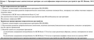
- Evidence-based medicine
- Experienced specialists
- Monitoring of patients for 6 months.
- Diagnostics (MRI, ultrasound, tests)
- Daily 8:00 – 22:00
Make an appointment
The human spine has four moderate natural curves, which in a healthy state allow you to evenly distribute vertical loads across the entire spinal column, help maintain balance and effectively absorb shocks and shocks that occur when walking, running, and jumping.
With Scheuermann-Mau disease, the angle of thoracic kyphosis, which is normally from 20 to 40 degrees, increases to 45–75 degrees. This leads to a significant redistribution of loads on all parts of the spine and entails the development of many complications.
Causes
Morphologically, the disease manifests itself in the fact that a wedge-shaped deformation of the thoracic vertebrae occurs, which generally leads to an increase in the posterior bending of the corresponding part of the ridge (thoracic kyphosis). The reliable mechanism for the development of deformation remains unknown today. There are several provoking factors, the impact of which significantly increases the development of the pathological process:
- Hereditary predisposition is one of the most common provoking factors, which is caused by changes in the genes responsible for the condition of the spine.
- Traumatization of the growth zones of the thoracic vertebrae, which occurred during the period of active skeletal growth.
- Congenital or acquired (the result of a metabolic disorder) osteoporosis, characterized by a decrease in the mineralization of bone tissue, a decrease in strength, which provokes a wedge-shaped deformation of the thoracic vertebrae.
- The prevalence of bone tissue growth activity in the posterior parts of the thoracic vertebrae, which causes their wedge-shaped curvature.
- Disruption of the anatomical structure and functional activity of the back muscles with a partial increase in their tone, which gradually leads to deformation of the vertebrae.
Knowledge of possible factors provoking the development of Scheuermann-Mau disease in certain cases makes it possible to carry out effective preventive measures.
Survey
- The main distinguishing feature of patients with Scheuermann's disease is thoracic kyphosis.
- Often, along with kyphosis, the patient is found to have cervical or lumbar hyperlordosis.
- Cervical lordosis can be increased with head protraction. The shoulders are mostly directed forward.
- These disorders may be accompanied by mild or moderate scoliosis.
Patients with Scheuermann's disease usually have well-developed muscles, in contrast to patients with postural kyphosis.
The examination consists of:
- Posture Assessment: Body position is assessed from the front, back and side.
- Neurological screening: in rare cases, compression of the spinal cord along the posterior surface of the vertebral bodies may occur at the site of greatest curvature. This may result in symptoms of impending paraplegia with clonus and hyperreflexia.
- Adam's forward-bend test: Scheuermann's kyphosis may be accompanied by scoliosis.
- Muscle Shortening Test: In Scheuermann's disease, the following muscles may be stiff: the pectoralis major, hamstrings, suboccipitals, and hip flexors. Contractures in the shoulder and hip joints are possible.
- Range of motion in the joints of the lower extremities and spine.
- Muscle Strength Test: The strength of the abdominal, core, trunk extensors, and gluteal muscles should be tested.
Symptoms
Sometimes, at the initial stages of the pathological process, even before the appearance of pronounced deformation of the vertebrae, pain may appear between the shoulder blades, which is usually a consequence of a change in the functional state of the back muscles. Against the background of pain, the subjective feeling of increased fatigue and discomfort during a long stay in a sitting position is also disturbing (this is often noted by children who feel tired after just a few lessons at school).
The most common objective manifestation of Scheuermann-Mau disease is poor posture and the development of stooping in a child or adolescent. Parents usually notice these symptoms and consult a doctor.
The pathological process progresses over a fairly long period of time. Without treatment, the deformation of the vertebrae of the thoracic spine increases. The range of motion worsens, it becomes difficult to bend forward and bend backward (development of rigidity). This leads to the appearance of a posterior bend (“hump”), as well as a violation of the shape of the chest, which is reflected in the functional state of the organs. Breathing and heartbeat may often be impaired, which is caused by compression of the lungs and displacement of the heart.
Consequences of the disease
In order to fully understand all the consequences of this disease, remember that the spine is a central organ: the most important blood vessels and nerve networks pass through it. It is through the spine that all organs of our body are controlled. Therefore, its deformation and disturbances in its work will lead to irreversible changes in all internal organs of our body and the musculoskeletal system. This in turn will lead to partial or complete loss of ability to work, and in advanced cases, death.
Kinds
Depending on the severity of the deformation of the thoracic vertebrae, as well as the intensity of subjective and objective clinical manifestations, 3 stages of the course of Scheuermann-Mau disease are distinguished:
- Latent (latent or orthopedic) course is the initial stage of the pathological process, which is characterized by minimal deformation of the vertebrae and a virtual absence of clinical manifestations. It is identified by a medical specialist after an objective diagnostic examination.
- Early neurological manifestations - more pronounced changes lead to the appearance of pain with a predominant localization in the back between the shoulder blades. They are inconsistent and have medium intensity. The mechanism for the development of sensations of discomfort is associated with compression of the sensitive fibers of the spinal roots in the area of their exit from the spinal canal. Typically, this stage develops between the ages of 16 and 20 years.
- Late neurological manifestations - against the background of severe deformation of the thoracic vertebrae, degenerative-dystrophic changes begin to develop in them, which include deforming spondylosis (changes affecting the processes of the vertebrae), the formation of herniated intervertebral discs, and osteochondrosis. This leads to constant pain in the back, impaired sensitivity of the skin in areas of the body (shoulder girdle, upper limb, chest). The shape of the chest changes significantly, which leads to complications in the lungs and heart.
Dividing Scheuermann-Mau disease into clinical stages allows the doctor to select the most optimal therapeutic measures for each of them.
Forms
The latent form develops in a child aged 8–14 years. Symptoms do not appear. If pain occurs, it is not very disturbing; the patient ignores it. It becomes uncomfortable after the patient does physical work. But deformation occurs at this stage every day. If a person is carefully examined, pathological changes will be revealed. Suspicion is raised by a very straight back with increased lumbar lordosis; kyphosis of the thoracic spinal segment may also increase.
Ankylosing spondylitis in men
The patient will not be able to reach his feet with his hands. An increased angle of the thoracic vertebral region is also present when there is a desire to straighten up. If a young person turns 15–20 years old, the following form of pathology will appear.
Pain syndrome of the lower back and lower thoracic spinal region is a frequent concern. Hernias form between the vertebrae. The severe course is aggravated by spinal compression. In the late form of the disease, osteochondrosis with hernias has already formed, spondylosis, spondyloarthrosis changes with ossifying ligamentosis appear.
This condition is observed in adults. The processes of dystrophy and degeneration lead to compression of the nerve roots. The mobility of the limbs will decrease, they will become less sensitive. The vertebral segments of the thoracic and lumbar regions can often be affected.
Diagnostics
Reliable identification of the development of Scheuermann-Mau disease, as well as determination of the severity of changes in the thoracic vertebrae, is carried out only by a medical specialist using several diagnostic measures:
- A clinical examination, during which a medical specialist finds out the patient’s complaints, the presence of a similar pathology in parents and close relatives, the presence of other pathological processes, including those not related to changes in the structures of the musculoskeletal system. Inspection and palpation (palpation of tissues) of the spine are also carried out.
- Visualization of the ridge using radiography is a common method of objective examination. The diagnostic criterion is to identify deformation of the intervertebral discs.
- Computed tomography or magnetic resonance imaging are modern techniques that have high resolution and make it possible to identify minimal changes in the components of the spine.
Based on the results of diagnostic studies, the doctor determines the stage of Scheuermann-Mau disease, which makes it possible to determine the appropriate treatment tactics.
Causes of development of Scheuermann-Mau disease
This disease occurs in adolescence, which is why it is also called “ juvenile kyphosis .” About 1% of people suffer from this problem. The exact reasons for the development of this disease have not yet been clarified. However, there are certain predisposing factors that may contribute to this problem:
- burdened heredity;
- spinal injuries;
- accelerated growth during puberty;
- endplate necrosis;
- muscle diseases;
- osteoporosis.
Treatment
Treatment of Scheuermann-Mau disease is complex. Therapeutic measures include:
- Conservative treatment with physiotherapeutic procedures - the prescription of drugs that strengthen bone tissue and improve metabolism (metabolic processes) in it. Exposure to physical factors (magnetic therapy, ozokerite, mud baths) has become widespread. While performing therapeutic measures, massage and special physical exercises are prescribed to strengthen the thoracic spine area.
- Surgical intervention is performed at the stage of early and late neurological manifestations, when conservative therapy does not provide the required effect. Includes implantation of special devices (hooks, knitting needles) that allow you to restore the shape of the thoracic spine.
- Rehabilitation, including long-term performance of special exercises, prevention of re-development of the pathological process through the use of orthopedic mattresses and special furniture.
The orthopedist selects the direction of treatment after diagnostic measures. He also monitors the effectiveness of treatment at every stage.
Shock wave therapy treatment
A modern effective and safe treatment method is shock wave therapy. The principle of the technique is exposure to high-energy ultrasound (shock wave), which causes microtrauma to tissues with subsequent activation of regeneration processes (recovery). With the correct choice of shock wave impact points, it is possible to gradually restore the shape of the vertebrae by destroying some areas and stimulating the growth of others. The treatment method allows achieving good therapeutic results without surgical intervention.
Early treatment of Scheuermann-Mau disease using modern techniques, including shock wave therapy, gives a favorable prognosis for the normal formation of the spine, and also prevents the development of various functional complications of the chest organs.
Patients turn to the doctor with various complaints: ⦁ cosmetic defect (Fig. 3): “crooked back”, asymmetry of the shoulders, shoulder blades, chest deformity, rib hump; incorrect position of the pelvis. ⦁ pain, which most often appears with a long course of the disease and its progression. Pain may appear with prolonged standing, with unusual loads, especially often in overweight patients and in the absence of regular physical activity; ⦁ the development of neurological symptoms also more often occurs with a long course of the disease and its progression, manifested by irradiation of pain to the lower extremities, impaired sensitivity, dysfunction of the pelvic organs; ⦁ in case of severe deformation of the chest and compression of the respiratory organs, respiratory dysfunction may occur.
Fig. 3 Cosmetic defect in scoliosis.
Diagnostics. 1. Inspection, physical examination (Fig. 4). 2. X-ray of the spine. X-ray images of the thoracic and lumbar spine are taken in a direct projection. For more accurate diagnosis and preoperative planning, it is necessary to take full-length images of the spine (spinal teleradiography), as well as functional images with lateral tilts. 3. Magnetic resonance imaging. MRI is necessary if there is a neurological deficit. Performed when planning surgical treatment to determine the level of implant fixation. 4. Computed tomography. In some cases, more detailed visualization of bone structures is necessary to determine the anatomical features and structural changes of the bones. 3D-CT reconstruction allows you to more accurately plan the operation and allows you to simulate the deformation on a 3D printer. Sometimes CT is performed with myelography to visualize the spinal canal, the contours of the dural sac and roots, determine compression of the dural sac and nerves, etc. In addition, it is recommended if there are contraindications to MRI.
Fig.4 Functional test with forward bend.
Conservative treatment is prescribed if the disease does not lead to disability and does not significantly affect the patient’s quality of life. In many ways, disability and deterioration in the quality of life are associated with the loss of the correct position of the spine in space (violation of the sagittal and frontal balance of the spine).
Recommendations for conservative treatment of pain syndrome in idiopathic scoliosis: ⦁ annual X-rays of the spine, dynamic observation by a doctor; ⦁ physical education: performing exercises aimed at strengthening the abdominal and back muscles, exercises to develop flexibility; ⦁ physiotherapeutic treatment; ⦁ corset therapy is often effectively used in growing patients with minor (up to 45°) deformities, most often lumbar or thoracolumbar, in order to prevent surgical treatment or delay surgery to a later time. The corset can be used for two to three hours a day or continuously. The method requires constant monitoring of the patient by a doctor and an engineer in corset technology for timely correction of the corset against the backdrop of a growing body. In adults, short-term use of corsets during physical activity is used to prevent stress loads on the vertebrae (long-term use is not recommended due to the risk of weakening one’s own muscle corset); ⦁ during an exacerbation of the disease, a course of drug blockades and infusions is prescribed, aimed at reducing swelling, improving blood supply to neural structures and transmission of nerve impulses, and relieving pain symptoms.
Indications for surgical treatment: ⦁ progression of deformity against the background of incomplete skeletal growth; ⦁ lack of effect from conservative therapy; ⦁ muscle weakness and impaired sensitivity in the lower extremities; ⦁ development of secondary degenerative manifestations with severe pain or neurological symptoms; ⦁ dysfunction of the lungs and heart (shortness of breath, heart failure) against the background of progression of the deformity; ⦁ pronounced restrictions on normal functional activity, which significantly reduces the quality of life in general; ⦁ cosmetic indications.
Goals of the operation: 1. Restoring the shape, balance and function of the spine, as well as the shape of the chest. 2. Correction of deformity. 3. Reducing pain. 4. Cosmetic effect.
Positive results of surgical treatment depend on the correct assessment of the examination results and their comparison with the existing symptoms. The patient’s psychological attitude and understanding from relatives (when treating children) are also very important.
The surgical operation is a complex process consisting of several stages: 1. Mobilization of the deformity. Mobility of the scoliotic arches is the success of any operation. It is checked by special x-ray tests during the preoperative examination. Small arches in children are most often quite mobile. In adult patients or with significant curvatures, rigidity increases. Mobilization can be achieved by performing special exercises before surgery, traction, or during surgery by excision of ligaments, intervertebral joints, discs or vertebrae. Such operations are called osteotomies. In addition to increasing mobility, they reduce the risk of neurological disorders during surgery, as they allow the deformity to be corrected without traction of the spine, thereby reducing the impact on the spinal cord. 2. Installation of implants. There are different types of implants: laminar systems (hook), screw, hybrid, ventral, etc. Their choice depends on the type of deformity, the purpose of the operation and the surgeon’s preferences. 3. Correction of deformity. There are various methods and techniques for correcting spinal deformities. It is carried out using special instruments to correct the arch and, what is important, eliminate rotation of the vertebrae. When correcting the deformity, physiological curves are restored (lordosis and kyphosis), and the costal hump is eliminated without resection. During correction, it is necessary to carefully monitor the function of the spinal cord to prevent neurological disorders (including paralysis). Therefore, the presence of neuromonitoring is a prerequisite for equipping the operating room in the treatment of this category of patients. 4. Spinal stabilization and spinal fusion are the final stages of the operation. The installed implants are securely fixed, after which autologous bone and (or) its substitute are placed on the bone structures in order to achieve fusion in the operated area in the future. This is a very important point, the quality of which determines the long-term result of the operation: whether the correction achieved during the operation will be lost in the future, or a fracture of the structure will occur, requiring repeated intervention. 5. Decompression of the spinal cord and spinal nerve roots. This intervention is used only if there are indications in the form of neurological disorders due to compression of the spinal cord. It is achieved by resection of ligaments, arches, articular processes or part of the vertebra.
Fig. 6 Full-length radiography of the spine before and after surgical treatment of a patient with idiopathic scoliosis.