Infectious disease specialist
Sinitsyn
Olga Valentinovna
33 years of experience
Highest qualification category of infectious disease doctor
Make an appointment
Toxoplasmosis, a common parasitic infection, develops in the human body as a result of infection with the protozoan microorganism Toxoplasma gondii. The disease spreads in different ways and occurs in acute, latent or chronic form. The pathogen is widespread on all continents of the planet, but the largest proportion of the infected population lives in developing countries, where Toxoplasma is present in the body of 9/10 of the population. The infection rate in developed countries varies from a quarter to half of the population; in Russia this figure is close to 30%.
Features and causes of the disease
The causative agent of toxoplasmosis is a single-celled parasite that lives in cells and intercellular space. In addition to the human body, it can penetrate into the bodies of many animals and birds, but to complete its full development path it needs the intestines of a domestic cat or other representatives of the cat family. Typically, a cat becomes infected by eating infected rodents or pigeons, after which Toxoplasma passes through its intestines and enters the human body through contact with cat feces.
Humans are an intermediate carrier for Toxoplasma. After entering the body, Toxoplasma penetrates the tissues, enters the blood through the lymph nodes and penetrates through the bloodstream into various organs, including the brain and organs of vision. Typically, infection occurs:
- when entering the digestive system after contact with soil contaminated with the feces of an infected animal, when eating unwashed fruits or vegetables, when meat is not properly cooked, etc.;
- transmissibly, through direct contact with the blood or saliva of an infected animal, in the presence of small wounds, scratches or abrasions on the skin;
- vertically, from the infected mother to the fetus, if the woman became infected during pregnancy.
The main measure to prevent toxoplasmosis is to eliminate the risk of the parasite entering the body. Good hygiene is most important in families with pets, especially cats. If the animal is free-ranging, precautions are especially important: wash your hands thoroughly after each contact, do not allow it to climb on the dining table or kitchen table, do not bring the animal to your face, and do not allow it to sleep in your bed. The risk of infection also remains if the cat does not go outside for walks: toxoplasma cysts can enter the house on the soles of outdoor shoes.
Clinic, diagnosis and treatment of toxoplasmosis
Toxoplasmosis is a widespread zoonotic parasitic infection, characterized by polymorphism of clinical manifestations and significant variability in the course of the process: from healthy, asymptomatic carriage to severe, lethal forms of the disease.
Etiology
The causative agent of toxoplasmosis, Toxoplasma gondii, belongs to the phylum Protozoa, subphylum Sporozoa, order Eucoccidia. T.gondii is an intracellular parasite, 4–7 µm in size.
In the human body, T.gondii can parasitize in the form of proliferative forms—endosites, in the form of pseudocysts, and in the form of true tissue cysts. In cats and other representatives of the feline family, toxoplasmosis can also be present in the intestines in the form of oocysts, which, when excreted with feces into the external environment, remain viable and invasive for 1.5–2 years. Toxoplasma cysts found in meat and meat products can remain viable at a temperature of 2–5°C for up to a month, but quickly die when heat treated or frozen at –20°C. The least resistant to environmental factors are endosites, which remain viable outside the body from 30 minutes to several hours.
Epidemiology
Toxoplasma or traces of its presence have been found in more than 200 species of mammals and 100 species of birds. The prevalence or infection rate of the population of the Russian Federation with Toxoplasma is on average about 20.0%. Infection rates are higher in regions with warm climates; among persons of a number of professional groups (epidemiological observations have proven an increased infection and incidence of toxoplasmosis in people who have professional contact with sources of toxoplasmosis infection (workers of meat processing plants and fur farms, livestock breeders, veterinarians, etc.). The infection rate in women is usually 2–3 times higher than men.
The incidence of toxoplasmosis is many times lower than infection rates, but diagnostic difficulties, despite mandatory registration, do not allow us to judge the true level of infection.
Susceptibility to toxoplasmosis is almost universal. The spread of infection is widespread in the form of carriage and sporadic diseases.
The main source of infection for toxoplasmosis is stray, wild and domestic cats, in whose bodies the pathogen goes through a full development cycle (tissue and intestinal) and is excreted in the form of oocysts with their feces. Cats shed the pathogen for an average of three weeks from the moment of infection. During this time, up to 1.5 billion toxoplasma enters the environment.
The main transmission factor is raw or insufficiently heat-treated meat, meat products with toxoplasma cysts in it. Additional factors for transmission of infection include poorly washed greens, vegetables, fruits (from the ground), dirty hands with pathogen oocysts on them.
The main and most common route of transmission of infection is oral; much less often, human infection can be carried out by transplacental (blood transfusion), percutaneous and transplantation routes.
A person with toxoplasmosis does not pose an epidemiological danger either to others or to medical personnel, which makes it possible to treat these patients both on an outpatient basis and in any somatic hospital.
Immunity. Immunity for toxoplasmosis is non-sterile and infectious. The immune state of the body is preserved simultaneously with the presence of the pathogen in the body, most often in the form of cysts. Antigenic metabolites produced by cysts maintain a certain level of humoral immunity and also cause the development of delayed-type hypersensitivity.
Pathogenesis
The pathogens released from cysts or oocysts invade the epithelial cells of the small intestine, where they multiply, forming a primary affect and then penetrating into the regional lymph nodes, and from them, with the lymph flow, into the blood. Dissemination of the pathogen leads to damage to a wide variety of organs and tissues.
Toxoplasma has a cytopathogenic effect on the cell, and inflammatory granulomas are formed at the sites of their penetration. Necrosis develops, in place of which lime salts fall out—calcifications characteristic of toxoplasmosis are formed. The degree of damage to a particular organ further determines the clinical symptoms of the disease.
The formation of immunity leads to the disappearance of the pathogen from the blood, and its reproduction in cells stops. True tissue cysts are formed, which can remain intact in the body for a long time, for decades (carriage of Toxoplasma). In humans, infection, as a rule, has a benign course, without the development of septic conditions.
Clinic
Most infected people have no clinical manifestations of the disease. In the vast majority of cases of toxoplasmosis, a healthy carriage of the parasite is observed, accompanied by a consistently low level of specific antibodies in the blood. Carriage does not require any therapeutic measures, and the carrier should be regarded as a practically healthy person.
At the same time, clinically pronounced variants of the course of the infection are possible, requiring careful diagnosis and specific treatment.
According to the nature of its course, acquired toxoplasmosis is divided into acute and chronic. In addition, depending on the duration of the disease and the severity of clinical symptoms, a subacute as well as inapparent (subclinical) course of the infection is possible, which is characterized by certain dynamics or a high level of specific antibodies in the blood, in the absence of clinical manifestations of the disease. Thus, the most convenient for practical healthcare, from our point of view, is the following classification of acquired toxoplasmosis: acute, subacute, chronic, inapparent and carriage.
The incubation period for toxoplasmosis lasts on average up to 2 weeks, although sometimes it can last up to several months. The disease, as a rule, begins gradually: general weakness, malaise, muscle pain, chilling appear, performance decreases, and the temperature rises to low-grade levels. The lymph nodes become enlarged: cervical, occipital, and less commonly, axillary and inguinal. Lymph nodes are soft, slightly painful on palpation. The size of the nodes is 1–1.5 cm, they are not fused with the surrounding tissues, do not form conglomerates, and the skin over them is not changed. Sometimes the mesenteric lymph nodes are significantly enlarged, which can simulate the picture of an acute abdomen.
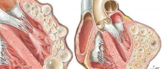
An acute onset, with a rise in temperature to 38°C and above, involving in the process, in addition to the lymphatic, nervous system, internal organs, muscle tissue, and organs of vision, is observed much less frequently. Patients may develop encephalitis, myocarditis, myositis, uveitis (chorioretinitis). In some cases, a short-term roseolous-papular rash and hepatolienal syndrome are observed. In patients with immunological disorders (especially in patients with AIDS), pneumonia, enterocolitis, severe central nervous system disorders of a cystic-necrotic nature, and a septic condition may develop.
Chronic toxoplasmosis is a long-term process with a general infectious syndrome and the presence of organ lesions of varying severity. The most characteristic signs of chronic toxoplasmosis are prolonged low-grade fever, intoxication and asthenia; fever can last for months, with slight temperature fluctuations, sometimes short periods of apyrexia, which is not amenable to conventional means of therapy. A common manifestation of toxoplasmosis is a generalized enlargement of the lymph nodes - occipital, cervical, inguinal and others.
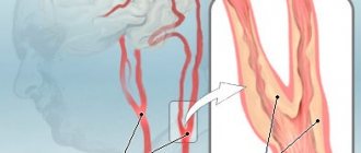
Damage to the central nervous system in chronic toxoplasmosis most often occurs in the form of cerebral, basal arachnoiditis; Hypertensive and diencephalic syndromes develop, vegetative-vascular disorders are detected, and episyndrome is noted. Sluggish myocarditis, myocardial dystrophy and myositis may be observed. Women may have specific inflammatory diseases of the genitals - toxoplasma salpingo-oophoritis (adnexitis); primary and secondary infertility develop.
Eye damage, both in acute and chronic acquired toxoplasmosis, occurs as posterior uveitis (focal chorioretinitis). Chorioretinitis is usually central, bilateral, and recurrent. The development of conjunctivitis, keratitis, iridocyclitis, central exudative retinitis, optic neuritis with outcome in dystrophy, complicated myopia is possible.
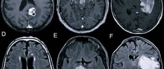
X-ray examination of a patient with toxoplasmosis allows in some cases to reveal the presence of calcifications in the soft tissues of the brain. Calcifications are usually small, multiple, round in shape, often with smooth contours.
In both acute and chronic toxoplasmosis, there are no special changes in the peripheral blood. Leukocytosis observed at the onset of the disease is replaced by normocytosis, relative lymphocytosis is detected, ESR is within normal limits.
It should be emphasized that with chronic toxoplasmosis there is no isolated damage to any one organ or system; we can talk about predominant organ damage against the background of the general process.
Diagnostics
Differential diagnosis. Toxoplasmosis should be differentiated from infectious mononucleosis, benign lymphoreticulosis, tuberculosis, brucellosis, listeriosis, mycoplasmosis, chlamydia, cytomegaly, herpes and a number of other bacterial, viral and parasitic infections. Oncological and systemic diseases should be excluded (for example: lymphogranulomatosis, rheumatism, etc.).
Laboratory diagnostics. For laboratory diagnosis of toxoplasmosis, serological methods are most often used: complement fixation test (CFR), indirect immunofluorescence reaction (IRIF), enzyme-linked immunosorbent assay (ELISA). The diagnosis is confirmed by a significantly increasing dynamics of the indicators of these tests, their high level or the presence of IgM class antibodies.
When giving a clinical assessment of the results of a serological examination of a patient, it is necessary to take into account that RNIF becomes positive from the first week of the disease and reaches its maximum values (1:1280–1:5000) usually by the second to fourth month of the disease; in low titers it can persist from a year to 15 years. RSC becomes positive from the second week of the disease, reaches maximum values (1:160–1:320) by the second to fourth month of the disease, but after 2–3 years it can disappear or decrease to values of 1:5–1:10. The interpretation of ELISA results is more objective, since it is focused on the WHO International Standard. Positive results may be indicated by indicators expressed in optical density units (OD ≥1.5); enzyme-linked immunosorbent units (EIU) ≥60; International units (IU) ≥125 and antibody titres (TA) ≥1:1600. The basic principle of serological diagnostics—the dynamics of the increase in indicators—is also applicable to this method. Of significant importance in the diagnosis of toxoplasmosis, especially in differentiating acute and chronic processes, is the determination of immunoglobulin classes, in particular IgM class antibodies. Toxoplasma infection can be reliably diagnosed only by comparing the results of serological reactions over time. Antibodies of all classes increase significantly by the end of the second - beginning of the third week from the moment of infection and reach a diagnostic level.
The diagnosis of toxoplasmosis, in the presence of an appropriate clinic, can be made with a positive serological conversion, when the second serum test becomes positive. When treating patients with already established positive reactions to toxoplasmosis, the question of diagnosis and activity of the infectious process can be resolved in the dynamics of serological studies. With fresh infection and disease, serological reactions are often positive with high titers of antibodies, and specific IgM is detected. When latent toxoplasmosis is reactivated, the IgM concentration may increase, but in this case the severity of the IgM response will be much less than during the primary infection. Positive RNIF in a low titer may indicate chronic toxoplasmosis or asymptomatic carriage of the pathogen. It should be noted that in ocular pathology, the presence of a fresh focus of inflammation, even with low antibody titers, indicates toxoplasmosis. In case of lymphadenopathy, even with high antibody titers, the diagnosis of toxoplasmosis is made only after histological examination of the lymph nodes and consultation with an oncologist. Based on the results of a single study, it is impossible to establish the duration of the infectious process and the exact correspondence to one or another of its stages, whereas this question is fundamental for assessing the risk of intrauterine infection of the fetus. Women who have had an infection before conception and women with chronic toxoplasmosis are practically immune from the risk of intrauterine infection of the fetus, while pregnant women infected in the first and early second trimester of pregnancy constitute the main risk group. However, it must be remembered that identifying and confirming the presence of IgM antibodies is not a clear indicator of termination of pregnancy. It is necessary to use additional methods to reduce the risk of inaccurate diagnosis.
Formation of diagnosis. When forming a detailed diagnosis of toxoplasmosis, the following should be indicated:
- form of toxoplasmosis (acquired, congenital);
- the nature of the process (acute, subacute, chronic, inapparent);
- organ or systemic pathology;
- severity of the current.
For example: acute acquired toxoplasmosis, lymphadenopathy, mild course; chronic acquired toxoplasmosis with primary damage to the eyes, chorioretinitis without exacerbation; pregnancy 24–26 weeks, inapparent toxoplasmosis.
When forming a diagnosis of toxoplasmosis, it is inappropriate to base the diagnosis of toxoplasmosis only on systemic or organ pathology (lymphadenopathic, cerebral, myocardial, ocular form, etc.), because toxoplasmosis must be considered as a general process involving many organs and systems.
When toxoplasmosis is excluded and a patient with positive reactions to toxoplasmosis is given another diagnosis, the medical history should also note the existing carriage of toxoplasma.
Treatment
The choice of treatment tactics depends on the form and nature of the disease, the severity of clinical symptoms, the severity of the course, the presence of complications and the predominant organ-systemic lesions.
The absolute indications for treatment are acute and subacute toxoplasmosis. Treatment of chronic toxoplasmosis is carried out depending on the severity of clinical symptoms and the nature of the predominant lesions. Inapparent toxoplasmosis detected in pregnant women also requires treatment.
The drugs Fansidar, Rovamycin and Biseptol are prescribed. Fansidar contains sulfadoxine 500 mg and pyrimethamine 25 mg. Etiotropic therapy consists of 2–3 cycles. Prescribed 1 tablet 1 time in 3 days (No. 8 tablets). Folic acid is prescribed between cycles. In case of intolerance to drugs of the pyrimethamine group, Rovamycin is prescribed, 1 tablet of which contains 3 million IU of spiromycin. Prescribe 3 million IU 3 times a day with a seven-day break. Rovamycin is well tolerated by patients, lack of drug interactions, and high efficiency allow it to be prescribed for the treatment of toxoplasmosis in all age groups.
It is possible to use combination drugs; Poteseptil (trimethoprim + sulfadimezine), Biseptol (trimethoprim + sulfamethoxazole), which are prescribed 1 tablet 2 times a day for 10 days (cycle), in the amount of 2-3 cycles (course). If these drugs are intolerant when taken orally, it is possible to prescribe Biseptol intravenously or drip: 10 ml per day, for 5 days (course). In the intervals between cycles (courses) of etiotropic therapy, folic acid is prescribed, on average up to 0.01 g per day.
If an immunodeficiency state is detected, immunotropic drugs are prescribed together with etiotropic therapy: Likopid, Cycloferon, Vitamedin-M, as well as natural calf thymus hormones and their synthetic analogues: Taktivin, Timamin, Thymogen, Dekaris.
Systemic enzyme therapy (SET) drugs, in particular Wobenzym and Phlogenzyme, are also used in complex therapy. To preserve the intestinal microbiocenosis, it is recommended to prescribe pro- and prebiotics.
Treatment and further follow-up of patients should be carried out by specialists depending on the nature of the prevailing pathology - infectious disease doctors, neurologists, ophthalmologists, obstetricians-gynecologists, etc. Hospitalization is carried out in a hospital of the appropriate profile (infectious diseases, neurological, ophthalmological, obstetric -gynecological, children's, etc.). This is due to the peculiarities of organ pathology, the specifics of the examination and the prescription of additional treatments.
Clinical examination
Dispensary observation is carried out by doctors according to the profile of the predominant pathology, in each individual case individually. After suffering from acute (inapparent) toxoplasmosis, patients are examined and examined every 3–4 months for a year, then 1–2 times a year. Patients with chronic toxoplasmosis are consulted 2 times a year.
Prevention
Prevention of acquired toxoplasmosis includes: eating only well-heat-treated meat products; eliminating the habit of tasting raw minced meat or raw meat; eating clean washed vegetables, herbs and fruits (from the ground); thorough hand washing after handling raw meat, raw meat products, after working in the garden, or for children - after playing on the playground, especially in the sandbox; fight against stray cats; treatment of patients with toxoplasmosis in domestic cats, prevention of infection of the latter. Specific prevention of toxoplasmosis has not been developed.
Diagnosis, clinical picture and treatment of toxoplasmosis in pregnant women
Diagnosis of toxoplasmosis in pregnant women includes the entire range of necessary clinical, paraclinical and special (immunobiological) studies that are used to diagnose toxoplasmosis in general.
A mandatory condition for examining a pregnant woman for toxoplasmosis should be a consultation with an infectious disease specialist to confirm or exclude a current infectious manifest or asymptomatic (inapparent) toxoplasma process. If the diagnosis is confirmed and the need for treatment is carried out, the latter is carried out by an obstetrician-gynecologist either on an outpatient basis (in a antenatal clinic) or in an obstetrics-gynecology hospital (maternity hospital). Considering the epidemiological safety of patients with toxoplasmosis for others, a pregnant woman with an uncomplicated obstetric history, but with a diagnosis of toxoplasmosis, can be hospitalized for examination, treatment and delivery in any (physiological) department of the maternity hospital. Pregnant women with a burdened obstetric history and diagnosed with toxoplasmosis are hospitalized in pregnancy pathology departments.
Screening of pregnant women for toxoplasmosis should be carried out by antenatal clinics when the pregnant woman first visits there. When conducting this examination, the obstetrician-gynecologist must remember that in our country, depending on the region, the percentage of infected women of childbearing age is on average 20–30%, i.e. every third of them can react positively to toxoplasmosis. As a rule, pregnant women with positive immunological reactions are healthy carriers of the pathogen and do not require any therapeutic, much less surgical measures. These women have virtually no complaints or objective manifestations of infection. Levels of specific antibodies remain stably at the same, usually low levels; there are no specific antibodies of the IgM class. 70–80% of women are free from infection and react negatively to toxoplasmosis. These women represent a “risk” group for congenital toxoplasmosis, since 0.5–1% of them become infected with toxoplasmosis during pregnancy. Of women primarily infected during pregnancy (high-risk group), 30–40% transmit the infection to the fetus. Consequently, non-immune (immunonegative) women are subject to dispensary observation for the prevention of congenital toxoplasmosis and dynamic examination (once every 1-2 months) during pregnancy in order to identify a fresh infection.
Clinical manifestations of toxoplasmosis in pregnant women do not have any significant differences. Acute toxoplasmosis is accompanied by an increase in temperature to febrile (usually subfebrile) levels, lymphadenitis is detected (usually posterior cervical and occipital), and possible disorders of the central nervous system, internal organs, eyes and muscles. When women become infected shortly before pregnancy or in the early stages of pregnancy, Toxoplasma may damage the fetal egg, usually leading to miscarriage. The obstetrician-gynecologist also needs to remember about the possible inapparent (asymptomatic) course of acute toxoplasmosis in women during pregnancy, when the development of the disease is recorded either by the significantly increasing dynamics of the level of specific antibodies, or by the detection of immunoglobulins of the IgM class in ELISA, which dictates the need for serological control (screening). ) for uninfected pregnant women throughout pregnancy.
Chronic toxoplasmosis in pregnant women is characterized by a general infectious syndrome (low-grade fever, generalized lymphadenopathy, chilling, decreased ability to work, etc.) with possible predominant organ damage from the internal organs, eyes, central nervous system and genitals.
The indication for prescribing etiotropic anti-Toxoplasma therapy to pregnant women is acute, subacute and inapparent toxoplasmosis. Treatment of chronic toxoplasmosis should be carried out strictly according to clinical indications either before or after pregnancy.
In the absence of complaints and clinical indications, women who have had toxoplasmosis before pregnancy do not need treatment. These women are regarded as practically healthy individuals who do not require special medical supervision.
Thus, the question of the time of infection of a pregnant woman is practically important: long before, immediately before or during pregnancy. The duration of infection is determined based on anamnesis and a comprehensive examination of the woman (cordocentesis and amniocentesis). Treatment of pregnant women should be carried out no earlier than 12–16 weeks of pregnancy (from the second trimester of pregnancy). Treatment is usually carried out with Rovamycin, Fansidar.
Rovamycin is prescribed in a daily dose of 3 million 2 times a day, the duration of administration is 7 days, the number of cycles is 2, the break between cycles is 1 month.
The concentration of Rovamycin in the placenta is 5 times higher than in the blood serum, which ensures cure. Rovamycin is the first macrolide used to treat toxoplasmosis in pregnant women. Rovamycin is well tolerated by patients, lack of drug interactions, and high efficiency allow it to be prescribed for the treatment of toxoplasmosis in all age groups.
Fansidar is prescribed 1 tablet 1 time every 3 days (No. 8 tablets). The break between cycles is one month. 3 such cycles are prescribed.
Considering the possible inhibition of hematopoiesis under the influence of etiotropic drugs, it is recommended to prescribe folic acid in medium therapeutic doses, as well as conduct general blood and urine tests.
In practical work, the question of indications for artificial termination of pregnancy during toxoplasma infection and recommendations for subsequent pregnancies is of particular importance.
Summarizing all of the above, it should be emphasized that only when infected in the first trimester of pregnancy and in the presence of clinical and immunological signs of acute acquired or inapparent toxoplasmosis, when the risk of giving birth to a child with gross organic lesions of the central nervous system and visual organs is greatest, the question of terminating the pregnancy for medical reasons can be raised ! Women infected during the second and third trimesters of pregnancy are subject to treatment. Termination of pregnancy for medical reasons is not indicated for women with chronic toxoplasmosis, and even more so with carriage of the pathogen, since in these cases there is no danger of infection of the fetus, because even an exacerbation of the process in pregnant women does not lead to repeated parasitemia and, therefore, to damage to the placenta, but through her and the fetus.
When giving recommendations for subsequent pregnancies, it is necessary to take into account that the same woman can have a child with congenital toxoplasmosis only once in her life (due to acute acquired or inapparent toxoplasmosis during pregnancy). During subsequent pregnancies, this woman may not be afraid of giving birth to a child with toxoplasmosis.
Prevention of congenital toxoplasmosis
Prevention of congenital toxoplasmosis should take into account the fact that only primary infection of a woman during pregnancy can lead to infection of the fetus. As mentioned above, out of the total number of non-immune women, up to 1% of pregnant women become infected during pregnancy. At the same time, infection of fetuses occurs in only 30–40% of them. According to the literature, the number of newborns with congenital toxoplasmosis is 1–8 per 1000 live newborns. Most often, the process in a child is asymptomatic, although the manifestation of the disease in the future cannot be ruled out. This can occur under the influence of immunosuppressive factors during the period of development of immunity, i.e. during the first 5–7–10 years of life.
Optimally, prevention of congenital toxoplasmosis should include screening women of childbearing age for toxoplasmosis before or, in extreme cases, during pregnancy. This examination is necessary to identify among them those who react negatively to this disease, i.e. non-immune women. The latter constitute the “risk” group for possible primary infection during pregnancy.
Non-immune pregnant women must be taken for clinical observation and examined for toxoplasmosis, but with the help of serological tests (RSK, RNIF, ELISA, etc.) throughout pregnancy, if possible, once every 1-2 months or at least 1 once every trimester.
If pregnant women at risk switch from negative serological reactions to positive ones and an increasing (3-4-fold) dynamics in the level of specific antibodies is detected, they need emergency preventive treatment. Treatment is carried out both in the manifest course of the infection and in the case of an in-apparatus course of the process. Children born to these women are subject to mandatory clinical and serological examination for toxoplasmosis and, if indicated, specific treatment. Children born to mothers with a clearly established primary infection during pregnancy are monitored until the age of 10, including regular clinical and immunological examinations in order to identify symptoms of congenital toxoplasmosis, which could be asymptomatic at birth.
Provided that at the last examination of pregnant women in the “risk” group, serological reactions remain negative, women (without clinical indications) do not need further special examination for toxoplasmosis and are dropped out of observation after childbirth. Children born to these women should be tested for toxoplasmosis only if clinically indicated. Pregnant women with negative immunological reactions are advised to strictly follow the basic rules for the prevention of toxoplasmosis.
Literature
- On the detection and prevention of toxoplasmosis in Moscow. Methodological recommendations (No. 25). M., 2007.
- Borisov B. A., Dzutseva F. K., Moroz B. V. Clinical and neurological manifestations of toxoplasma invasion. Tutorial. M., 2003.
- Bartlett J., Galant J. Clinical aspects of HIV infection. Johns Hopkins University School of Medicine. 2005–2006
F.K. Dzutseva , doctor of the highest category, head. City Center for Toxoplasmosis G. Yu. Nikitina , Candidate of Medical Sciences Yu. V. Borisenko , Candidate of Medical Sciences L. P. Ivanova *, Candidate of Medical Sciences, Associate Professor of the City Clinical Hospital named after. S. P. Botkina, *RMAPO, Moscow
Manifestations of parasitic infection
The duration of the incubation period from the moment of penetration of Toxoplasma into the body until the appearance of the first symptoms of toxoplasmosis varies from several days to three weeks. The disease can occur in acute, latent or chronic form.
- Acute toxoplasmosis begins with a sudden increase in temperature, fever, and manifestations of intoxication (chills, pain in muscles and joints, loss of appetite). The spleen and liver increase in size, the lymph nodes swell, and pink rashes may appear on the skin, distributed throughout the body. The disease may be accompanied by an inflammatory process in the myocardium, tissues and membranes of the brain. In severe cases of the disease, death cannot be ruled out.
- The chronic form of the disease is either asymptomatic or with inexpressive symptoms: the patient quickly gets tired, feels weak and generally weak, and may have a slight increase in temperature. If Toxoplasma penetrates the nervous tissue, brain or organs of vision, headaches, dizziness, blurred vision and memory are possible. If internal organs are damaged, myocarditis, hepatitis or pneumonia may develop. As a rule, the disease continues for many years with periodic exacerbations.
- The latent form is typical for the vast majority of people and occurs in 95-98% of cases of infection. Two to three weeks after the parasite enters the body, a person feels a slight malaise, which soon goes away on its own. The presence of toxoplasma is usually detected during the treatment of other diseases.
The disease can be acquired or congenital. Toxoplasmosis is most dangerous during pregnancy, since the parasite, penetrating the fetal tissues, disrupts their natural development. The result of infection can be a wide range of pathologies, from spontaneous abortion to various birth defects in the child, affecting the organs of vision, brain and spinal cord.
With intrauterine infection, the fetus develops irreversible changes that can appear immediately or after several years. Often children with a congenital disease lag behind their peers in development; they develop pathologies of the organs of vision and hearing, microcephaly, mental retardation and other disorders. However, negative manifestations are possible only when a woman becomes infected while carrying a child. If pregnancy occurs in a woman with a latent or chronic form of the disease, the child is protected by antibodies, which by that time have already been formed in the mother’s body.
Are you experiencing symptoms of toxoplasmosis?
Only a doctor can accurately diagnose the disease. Don't delay your consultation - call
Congenital toxoplasmosis occurring during pregnancy
Occurs when the parasite penetrates the placental barrier and infects the fetus. In most cases, it occurs during primary infection during pregnancy, less often - during relapse of toxoplasmosis associated with decreased immunity. The main risk group is women who were not infected with toxoplasma before pregnancy. If, as a result of contact with the pathogen, the disease manifests itself during pregnancy, the likelihood of developing congenital toxoplasmosis is quite high.
The survival rate of children with intrauterine infection depends on the period at which it occurred:
- If infected in the first trimester, the chance of fetal survival is 15%
- On the second – 30%.
- On the third – 60%
Even if pregnancy is successfully completed, an extremely high degree of development of congenital pathologies and congenital toxoplasmosis remains. The disease is severe, especially if infection occurs in the early stages. A characteristic tetrad of pathologies develops:
- Hydrocephalus.
- Bilateral retinochoroiditis.
- Delayed psychophysical development.
- Cerebral calcifications.
The prognosis for congenital toxoplasmosis is unfavorable; in most cases, the disease ends in the death of the newborn or severe disability. Even if, after intrauterine infection, an acute clinical picture does not arise, such children are at risk of developing:
- Mental deficiency.
- Epilepsy.
It is possible to develop many other pathologies that appear months and years after birth. For this reason, an acute form of toxoplasmosis that occurs during pregnancy is an indication for abortion even in the later stages. During pregnancy, it is necessary to undergo a lot of tests that help identify the presence of not only toxoplasmosis, but also CMV infection.
Diagnostic methods
Diagnosis of toxoplasmosis includes:
- blood sampling for a general analysis, the results of which indicate a reduced number of leukocytes and an increased number of lymphocytes and eosinophils;
- conducting a test with toxoplasmin, where a positive reaction means either a chronic infection or the presence of antibodies after an illness;
- spinal puncture performed according to indications;
- X-ray of the brain to identify an enhanced pattern of blood vessels, widened interosseous spaces, and the presence of calcifications;
- if necessary, ultrasound of internal organs.
Most often, an enzyme-linked immunosorbent assay or immunofluorescence reaction is performed to detect toxoplasma. It is repeated twice, at two to three week intervals, to detect the growth of IgM and IgG antibodies. Antibodies M appear in the blood two weeks after infection, their number begins to decrease no earlier than two months later. The presence of G antibodies indicates either the transition of the disease to a chronic form or a complete recovery.
To determine toxoplasmosis in newborns, their antibody titers are compared with those of the mother. A congenital disease is indicated by the presence in a child of IgM and IgG in quantities exceeding maternal values by at least four times. If the mother's antibody titer exceeds the child's, it means that immunoglobulins have been transferred, not the parasite.
Indications for examination
Analysis is most often prescribed in two cases:
- When planning pregnancy as part of a standard laboratory diagnostic package for TORCH infection.
- If you suspect toxoplasmosis and have certain symptoms.
Laboratory diagnostics are also used to identify symptoms of acute toxoplasmosis in adults or children.
In general medical practice, this test is prescribed if patients exhibit specific symptoms (visual impairment, seizures), as well as for HIV-infected patients.
How to get rid
The treatment method for toxoplasmosis is selected depending on the form and course of the disease, as well as the presence of damage to internal organs. As a rule, the patient is prescribed antiparasitic drugs - most often chloridine, which is combined with sulfonamide drugs that enhance its effect. During pregnancy, spiramycin is often prescribed.
Usually the drugs are taken in three courses lasting from 5 to 10 days, repeated at weekly intervals. To eliminate the cause of toxoplasmosis in the chronic form, these drugs are not enough; additional immunotherapy is necessary using intradermal injections of toxoplasmin. At the same time, anti-allergy medications, courses of vitamins and immune stimulants are prescribed.
If the brain, visual organs and internal organs are damaged, appropriate therapy is prescribed.
Differential diagnosis
It is carried out with diseases whose symptoms are similar to acute and chronic forms of toxoplasma infection. These include:
- Infectious mononuclease.
- Mycoplasmosis.
- Chlamydia.
- Cytomegaly.
- Tuberculosis.
It is also necessary to exclude oncological pathologies and systemic diseases (rheumatism, lymphogranulomatosis). The final diagnosis is established after receiving the results of specific serological tests and PCR. There are many laboratory methods for detecting specific antibodies to Toxoplasma. These include:
- ELISA. Linked immunosorbent assay.
- RNIF. Indirect immunofluorescence reaction. Becomes positive from the first week of the disease. High antibody titers can last up to 15 years.
- RSK. Complement fixation reaction. It becomes positive from the 10-14th day of disease development and persists for 2-3 years.
Examination in Medart
At the Medart Medical Center, a full range of serological tests are performed to detect toxoplasma in the patient’s body. An immunological test to identify specific immunoglobulins is part of comprehensive STD tests intended for couples planning to have a child. It is possible to take blood and other biological fluids to determine the presence of toxoplasma and clarify the clinical picture of the disease.
High-precision modern equipment allows you to obtain accurate research results in the shortest possible time.
Advantages of Medart Medical Center:
- Qualified specialists.
- The ability to quickly obtain accurate research results.
- Affordable price.
The medical center provides a full range of services, from preliminary appointments and consultations to establishing and clarifying the diagnosis and prescribing effective treatment regimens and prevention of toxoplasmosis and other diseases.
Screening for toxoplasmosis during pregnancy
If during pregnancy a pregnant woman develops symptoms that may indicate the development of acute toxoplasmosis, it is necessary to establish the level of specific immunoglobulins using serological methods.
The enzyme-linked immunosorbent assay (ELISA) is best suited to detect the acute stage of the disease. It most accurately shows the concentration of IgM, an increase in the level of which indicates ongoing or recently present acute toxoplasmosis.
Determining IgG levels is less informative since these antibodies persist long after infection and indicate carriage rather than recent infection or exacerbation. Women who have been infected with toxoplasma before pregnancy are insured against infection of the fetus and are not at risk.
It is also important to obtain an immunological picture over time, for which specific tests are carried out at least once every 2 weeks. Studying the dynamics of changes in antibody titers allows us to establish a diagnosis with greater accuracy.
It should be taken into account that the serological picture indicating infection with Toxoplasma is not a 100% indication for termination of pregnancy. In this case, additional tests will be required by taking fetal blood from the umbilical cord and samples of amniotic fluid by puncture.
Prevention measures
Prevention during pregnancy
It is especially important to prevent toxoplasmosis for pregnant women, for whom this disease poses an increased threat. To prevent toxoplasmosis, it is important to thoroughly wash your hands after handling raw meat, soil, or cat litter. Avoid eating food that is likely to be contaminated with cat feces. Meat should be cooked to a temperature of 73.9-76.7°C.
Pregnant women are advised to avoid contact with cats. If contact is unavoidable, it is recommended that you at least avoid cleaning litter boxes or wear gloves when doing so. Feeding your pets dry or canned meat is not dangerous for indoor cats that are fed dry or canned cat food.
Vaccinations for toxoplasmosis
Vaccinations against toxoplasmosis are given to domestic animals - cats. Such vaccination is the most effective means of preventing the disease in humans. In this case, a weak concentration of the pathogen is introduced into the body, to which the animal’s body begins to develop immunity. In newborn kittens, immunity from toxoplasmosis occurs due to mother's milk.
Very rarely, cats with toxoplasmosis die. More often they simply become seriously ill, which is also not very good, which is why vaccination for an animal may be extremely necessary. There are currently 2 types of cat vaccines known. The first vaccines are capable of protecting an animal from a specific disease, while the second are multicomponent and are immediately capable of forming immunity against a number of infections.
Even the high cost of constant vaccination of pets does not reduce the effectiveness of this procedure both for their population and for the people with whom they come into contact. Sometimes vaccinations may be offered to humans in addition to animals to increase protection.
Examination of newborns and young children
Aimed at early detection of the pathogen, before the onset of the acute phase of congenital toxoplasmosis and the development of severe complications. Prescribed when there is a suspicion of Toxoplasma infection, includes the following tests:
- Isolation of the parasite by inoculating material from the placenta and umbilical cord into living mice.
- Carrying out PCR analysis of amniotic and lumbar fluids.
- Computed tomography or MRI of the head. Allows you to identify specific changes in the brain, for example, hydrocephalus, at an early stage.
Serological techniques are also used, but they provide only additional information. The newborn’s immune system is not active enough and is often unable to produce a sufficiently high titer of specific antibodies.
What it is?
Toxoplasmosis is an infectious disease caused by Toxoplasma gondii. The disease can be asymptomatic, with manifestations of lymphadenopathy, mononucleosis-like symptoms, up to damage to the central nervous system in immunocompromised individuals.
Newborns may have chorioretinitis, seizures, and mental retardation. The diagnosis is confirmed serologically, HCC and histologically. Treatment is with perimethamine in combination with sulfadiazine or clindamycin. Glucocorticoids are used for chorioretinitis simultaneously with the main therapy.
The causative agent of toxoplasmosis
The causative agent of the disease is Toxoplasma gondii. This is the protozoan of the order Coccidia. It is mobile and has an arched shape. If you look at this living organism under a microscope, it resembles an orange slice.
The parasite can reproduce sexually and asexually. As a result of the sexual process occurring in the intestines, cysts are formed. These forms are very resistant to environmental factors: they are not afraid of low and high temperatures or drying. Together with feces, these cysts are released and lead to infection of other organisms. Asexual reproduction does not lead to the formation of persistent forms of the parasite.
Can you get toxoplasmosis from pets? Yes, because toxoplasmosis affects about 60 species of birds and 300 species of mammals (domesticated and wild). However, the sexual process occurs only in the intestines of felines. During 2 weeks of illness, a cat can secrete up to 2 billion cysts, which live in the external environment for up to 2 years.
Clinical symptoms
The acute phase of the disease affects almost all internal organs, infection is determined by:
- due to drowsiness, weakness;
- chills, elevated temperature;
- yellowness of the skin and whites of the eyes;
- enlarged liver and spleen;
- papular rashes, decreased muscle tone;
- strabismus.
With slow development of inflammation, less severity of symptoms is observed. Patients experience clouding of the lens, and dropsy gradually develops in the brain tissue.
The chronic course of toxoplasmosis is provoked by the occurrence of irreversible changes in the central nervous system, complete blindness, unreasonable irritability, and attacks of neurasthenia. Women experience irregularities in the menstrual cycle, men have problems with erection or complete impotence.
Modern methods of laboratory diagnostics
Today, the diagnosis of toxoplasmosis is based on the following studies:
- Serological method (ELISA);
- REEF;
- NRIF(IgM, IgG)
- PCR;
- Parasitological method;
- Allergological method.
Each method has its own specific indications for use. Let's take a closer look at them.
Serological method (ELISA)
This is an immunological study based on the “antigen-antibody” reaction. The method allows you to evaluate the qualitative and quantitative indicators of the pathogen in the blood. The reaction occurs thanks to specific reagents that are added to the patient's blood serum. If the antibodies in the blood react to the antigen that is in the reagent, then the diagnosis can be considered confirmed. The technique is widespread and is used to confirm clinical signs of toxoplasmosis.
REEF
The immunofluorescence reaction is based on the property of immune complexes to be illuminated by an immunofluorescent substance. If a reaction occurs, the medical worker sees a specific glow, evaluates its intensity and quantitative indicators. Surface and intracellular antigens can be assessed. The method is included in express diagnostics, does not take much time and is often used to make a diagnosis. A prerequisite is specific laboratory equipment and staff skills.
NRIF(IgM, IgG)
The indirect immunofluorescence method is not used so often. It allows you to determine the type of antibodies in different serum titers. Immunoglobulins of type M indicate an acute stage of the process, and class G antibodies are a sign of a chronic course or a previously suffered disease. Diagnosis should be carried out efficiently, because the use of low titers can easily be mistaken for a false positive result.
PCR
The polymerase chain reaction method is the most accurate and modern way of making a diagnosis. The study is quite expensive, as it requires specific laboratory equipment. The basis of diagnosis is the detection of nucleic acid sections of the genetic material of the pathogen, which makes the diagnosis as accurate as possible.
Parasitological method
The essence of the study is the direct detection of the pathogen in biological material. For this purpose, blood, saliva, cerebrospinal fluid, and biopsy specimens are examined. Microorganisms can be detected using special staining. The method has mixed reviews. On the one hand, it is possible to confirm the presence of a pathogen, but it is difficult to say how long the patient has been sick and at what stage of the process. In addition, the absence of parasites in the studied material does not always mean that the patient does not have toxoplasmosis.
Allergological method.
An allergy test is performed with intradermal injection of toxoplasmin. If the reaction is positive, this indicates that the patient is really sick and requires a more thorough examination. The test becomes positive starting from the fourth week of illness.
Examination of patients with HIV infection
Diagnosis consists of regular monitoring of immunoglobulin G titers using serological methods. Titration of immunoglobulin M is not informative since in most HIV-positive people the level of antibodies of this group is extremely variable.
Direct detection of the pathogen by microscopy of tissue samples or infection of laboratory animals is usually not necessary. The diagnosis can be established quite accurately through serological diagnosis and the presence of a specific clinical picture of toxoplasmosis. In HIV-positive patients it is more pronounced, which greatly simplifies the diagnosis.