Chronic sialadenitis is an inflammation of the salivary glands, in which periods of exacerbation alternate with periods of remission. This type of disease manifests itself differently in men, women and children. During reduced activity of the pathological process, symptoms of the inflammatory process are practically absent. During an exacerbation, there is a painful swelling in the cervical spine, an unpleasant taste in the mouth, and difficult salivation. Treatment is aimed at containing changes in the structure of the salivary gland tissue and eliminating painful symptoms.
Classification of forms of chronic sialadenitis
Depending on the area of localization and similarity of symptoms, three main forms of sialadenitis are distinguished:
- Parenchymal sialadenitis . Parenchyma is the collection of functional cells that make up the salivary gland. The inflammatory process is based on changes in cells of this type. Women are more often susceptible to this form of the disease. Parenchymal sialadenitis affects the parotid salivary glands.
- Interstitial sialadenitis. This type of inflammation occurs in the interlobular space of the gland. Most often it affects men. Interstitial sialadenitis affects the submandibular salivary glands.
- Ductal sialadenitis Occurs due to congenital pathology of the salivary ducts or as a result of their injury. Most often it occurs in older people.
Symptoms of chronic sialadenitis
At the initial stage of parenchymal sialadenitis , symptoms are usually absent. The main complaints are related to a feeling of dry mouth. In the future, there may be a painful swelling in the neck, an unpleasant taste in the mouth, and nagging pain. Without treatment, the surface of the salivary gland becomes lumpy, and saliva separation is difficult.
With interstitial sialadenitis, a separate area of the inflamed gland may bother you. Complaints of hearing loss are rarely observed. With increasing inflammation, the salivary gland increases in size; upon palpation, the unevenness of its surface is felt, and the secretion of saliva is impaired.
Ductal sialadenitis affects people of advanced age or with weakened immune systems. Inflammation is accompanied by an obsessive salty taste in the mouth. When pressing on the mouth of the duct, cloudy saliva is released. The mouth of the excretory duct itself is noticeably thickened.
Anatomy and pathology of the salivary glands. Sialadenitis, sialosis
In the article, the authors consider the normal and pathological anatomy of the salivary glands. Sialadenitis and sialosis are treated. They are trying to consider the macroscopic and microscopic picture of these diseases.
Key words: salivary glands, sialadenitis, sialosis, pathological anatomy, dental diseases, parotid gland, sublingual gland, submandibular gland.
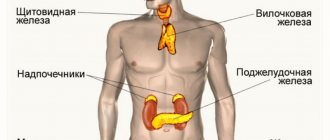
The salivary gland is an important part of the human digestive system. It performs exocrine, endocrine, filtration and excretory functions. The excretory ducts of the glands open into the oral cavity. There are large and small salivary glands.
The large salivary glands are located outside the oral cavity and include the parotid, sublingual and submandibular glands. The minor ones are located deep in the mucous membrane and are represented by the labial, buccal, palatine, molar and lingual glands.
Parotid gland, glandula parotidea
It is the largest gland. Located in the mandibular fossa, the excretory duct runs along the outer surface of the masticatory muscle and opens on the mucous membrane of the vestibule of the mouth in the area of the second upper molar [2].
In terms of its structure, it is complex alveolar, serous type, and has a lobular structure. The gland is covered with fascia, which encloses it in a capsule [4].
Submandibular gland, glandula submandibularis
Located in the submandibular space, the excretory duct opens on the sublingual papilla, like the duct of the sublingual gland. In structure it is a complex alveolar-tubular gland of a mixed nature, which also has a lobular structure. The submandibular gland secretes protein-mucosal saliva with a predominance of the protein component [4].
Sublingual gland, glandula sublingualis
It lies on the mylohyoid muscle, directly under the mucous membrane of the floor of the mouth, and has a thin connective tissue capsule. The excretory ducts of some lobules open independently into the oral cavity, and the main duct is located next to the duct of the submandibular gland [5].
The structure is a complex alveolar-tubular salivary gland, producing mixed saliva with a predominance of mucous secretion.
Diseases of the salivary glands can be either primary (independent) or secondary - a manifestation or complication of any systemic disease. Diseases can be caused by the development of infection, tumor, traumatic injury, as well as obstructive, tumor-like or autoimmune damage.
Sialadenitis
Inflammation of the salivary gland is called sialadenitis. Most often, the parotid gland is affected, less often - the submandibular gland, very rarely - the sublingual gland. Sialadenitis can be acute or chronic, and according to the etiology of the disease it can be viral, fungal or bacterial. The following routes of infection are possible: hematogenous, contact, lymphogenous and intraductal from the oral cavity of the body.
Acute sialadenitis
May occur due to exposure to local and general factors. Local factors - any surgical intervention, compression of the excretory ducts by a tumor, traumatic lesions. Common factors are diabetes mellitus, chronic gastrointestinal diseases, immunodeficiencies, cardiovascular diseases [1].
Acute sialadenitis is divided by morphology into gangrenous, serous and diffuse purulent. The salivary gland enlarges in this disease. Purulent sialadenitis is characterized by pronounced neutrophilic infiltration with an abundance of purulent bodies. With serous sialadenitis, swelling, neutrophilic infiltration, and plethora are observed.
Complications of acute sialadenitis include the occurrence of abscesses, sepsis and phlegmon of the neck tissue. Outcome: recovery or transition to chronic [3].
Chronic sialadenitis
Most often secondary, i.e. as a complication of traumatic injuries, infectious diseases, developmental defects. According to the degree of clinical manifestations, active and inactive sialadenitis are distinguished. Chronic sialadenitis often takes on a recurrent nature, so there are two stages: the remission stage and the exacerbation stage.
The salivary glands are enlarged, have a smooth surface and a densely elastic consistency. With exacerbation of chronic sialoadenitis, they acquire a doughy consistency
There are two morphological forms of chronic sialoadenitis: interstitial, ductal (Kussmaul sialodochitis).
Interstitial is characterized by focal or diffuse lymphohistiocytic infiltrates with macrophages, as well as the proliferation of connective tissue with atrophy of the epithelium of aciar structures. The preserved areas of the salivary gland in this case represent hypertrophied acini with hyperplastic cells of the intercalary and striated excretory ducts [3].
With ductal sialadenitis, between the acini and terminal ducts there are diffuse lymphohistiocytic infiltrates with an admixture of polynuclear leukocytes. The ducts are expanded and lined with multirow cubic epithelium. The cells of the epithelial layer of the interlobular excretory ducts are in a state of necrobiosis.
Outcome: with rare relapses, cirrhosis is possible, and with frequent relapses, the formation of large cysts with elements of purulent inflammation.
Full recovery usually does not occur. Preventive measures are aimed at preventing exacerbation of pathological processes and increasing the body's resistance.
Sialosis
It is characterized by hypertrophy of serous acinar cells, which contain mucoid substance, and is non-inflammatory in nature. It is based on dystrophic processes and changes. There is an enlargement of the glands, most often the parotid glands, painless or slightly painful swelling, and hyposalivation. Microscopically: narrowing of small and large ducts, as well as depletion of the gland parenchyma pattern due to hypertrophy and cell hyperplasia. There are three stages: the initial stage, characterized by hypersecretion, the clinically pronounced stage, which is characterized by depletion of secretion, dystrophic changes in the epithelium, and the late stage of lipomatosis and fibrosis [1]. Thus, the outcome is salivary gland lipomatosis.
Treatment is aimed mainly at eliminating the factor contributing to the development of sialosis, dystrophic changes in the salivary glands. In the late stage, there are indications for the use of surgical treatment methods: parotidectomy, extirpation of the excretory ducts of the salivary glands.
Pathology of the salivary glands is not so common, but still occurs, which is why it is important to know the normal and pathological anatomy of the salivary glands for the prevention, detection and treatment of pathological processes that occur in the oral cavity.
Literature:
- Afanasyev V.V., Abdusalamov M.R. Atlas of diseases and injuries of the salivary glands. - M.: VUNMC Roszdrav, 2008. - 192 p.
- Gaivoronsky I.V., Nichiporuk G.I. Anatomy of the digestive system: textbook. ELBI-SPb, 2006.-64 pp..
- Zavyalova M. V. Pathological anatomy of the head and neck: textbook / M. V. Zavyalova, S. V. Vtorushin, I. V. Stepanov; ed. V. M. Perelmuter; rec.: V. A. Shkurupiy, E. L. Kazachkov; Siberian State Medical University (Tomsk). - Electron. text data - Tomsk: Siberian State Medical University, 2013. - 167 p.
- Prives M. G., Lysenko N. K., Bushkovich V. I. Human anatomy. 9th ed. M.: Medicine, 1985. - 672 p. Textbook for students of medical institutes.
- Sapin, M. R. Atlas of human anatomy for dentists: atlas / M. R. Sapin, D. B. Nikityuk, L. M. Litvinenko. - Electron. text data - Moscow: GEOTAR-Media, 2013. - 600 p.
Diagnosis of sialadenitis
Diagnosis involves a thorough analysis of the patient’s complaints, as well as palpation of the salivary gland. To clarify the diagnosis, cytological studies of salivary secretions are possible. , radiography with a contrast agent may be prescribed . The purpose of the study is to evaluate the gland tissue and the patency of the salivary ducts. X-rays are performed only after acute inflammation has been eliminated. The goal of diagnosis is to differentiate chronic sialadenitis from other diseases with similar symptoms:
- acute bacterial or viral sialadenitis;
- benign neoplasms;
- Herzenberg's pseudomumps;
- sialadenosis.
Provoking factors
Sialodochitis can be provoked by diseases of the cardiovascular system and respiratory system, as well as gastrointestinal pathologies. Other factors that determine the development of specific changes are:
- injury to the ducts of paired glands;
- anorexia;
- rheumatoid arthritis;
- age-related changes;
- systemic lupus erythematosus;
- radiotherapy cycle (for the treatment of malignant tumors);
- hypercalcemia;
- compression of the excretory duct by a benign or malignant tumor or enlarged lymph node.
Of no small importance in the appearance of sialodochitis belongs to general somatic pathology.
Treatment of chronic sialadenitis
Treatment of chronic sialadenitis is aimed at preventing changes in gland tissue, normalizing saliva secretion and eliminating the inflammatory process.
During an exacerbation, the dentist prescribes a course of broad-spectrum antibiotics. In the absence of a bacterial infection, therapy may be limited to antiseptic rinses and the administration of a vitamin complex to support immunity. In complex therapy, it is possible to carry out physiotherapeutic procedures, such as electrophoresis or magnetic laser therapy. In rare cases, if conservative treatment is ineffective and the gland loses function, surgical intervention is possible.
Diagnostic criteria and measures
When clarifying the diagnosis, sialodochitis must be differentiated from parenchymal and interstitial sialadenitis, salivary stone disease, mumps, tuberculosis, syphilis, infectious mononucleosis, benign and malignant tumors, as well as cystic formations.
Additional examination methods help make a diagnosis:
- X-ray diagnostics;
- Ultrasound of the salivary glands;
- diagnostic puncture;
- bacteriological and cytological analysis of duct contents;
- biopsy;
- sialography;
- sialometry.
These procedures help to find out the true cause of the pathology and select the most effective treatment.
Therapy methods
Drug therapy is possible. Drug treatment of sialodochitis includes:
- the use of compresses with demixide;
- prescription of antibiotics.
Outside periods of exacerbation of the disease, which has become chronic, it is recommended to take vitamin and mineral complexes to increase immunity, and a special massage of the salivary glands.
If there is no effect of treatment, surgery is necessary. Depending on the need, perform:
- intraoral (along the duct) incision;
- external incision with washing of the salivary gland through the duct.
To speed up recovery after surgery, rinses with herbal infusions (chamomile, sage) and antibacterial compounds are prescribed, as well as a special diet that promotes salivation.
In later stages, as well as with regular exacerbations, resection of the salivary glands is recommended. If stones are found, they are removed. If treatment is started on time and completed until completion, recovery usually occurs within 2 weeks.
Complications of sialodochitis include the formation of stones, the appearance of localized, festering inflammations (abscesses) and phlegmon, as well as complete blockage of the ducts. In the most difficult cases, necrosis of the gland or its cicatricial change is observed.
Preventive measures
To prevent the development of pathological changes in the excretory ducts of the major salivary glands, it is necessary to strengthen the immune system, treat foci of chronic infection, and always monitor oral hygiene.
For complicated forms of somatic pathologies, it is recommended to rinse the mouth with antiseptic solutions (2% boric acid, 1% acetic acid, citric acid).
Sialodochitis is a disease that causes pain and discomfort. Diagnosis is usually not difficult. Timely treatment prescribed by a qualified specialist quickly eliminates the disorder. And taking preventive measures helps minimize the risk of relapse of the pathology in the future.
Possible complications
Most often, sialadenitis occurs without harm to health. In some forms of the disease (gangrenous), there is a risk of blood infection and sepsis. In the absence of timely treatment, a person may encounter other problems:
- duct stenosis;
- abscess;
- fistula;
- dysfunction of the salivary gland;
- salivary stone disease (sialolithiasis).
Dentists warn patients that inflammation of the submandibular salivary gland often leads to caries and benign formations in the oral cavity. In addition, gastrointestinal diseases develop due to constant infection.