Salivary glands
[
glandulae oris
(PNA, JNA, BNA);
synonym: glands of the mouth
, T.] - digestive glands that secrete into the oral cavity a specific secretion that is part of saliva.
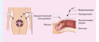
There are large - parotid, submandibular, sublingual and small salivary glands - buccal, molar, labial, lingual, hard and soft palate (Fig. 1). Rice. 1. Schematic representation of the localization of the main human salivary glands: 1 - molar glands; 2 - buccal glands: 3 - labial glands; 4 - anterior lingual gland; 5 - sublingual gland; 6 - submandibular gland; 7 - parotid gland; 8 - accessory parotid gland
Comparative anatomy and embryology
In animals that live in water, the glands of the mouth are poorly developed and are represented by simple glands that produce mucus. In terrestrial animals, due to the need to moisturize the oral mucosa and wet food, the salivary glands are more developed. Amphibians have mucous labial, palatine, lingual and premaxillary glands. In reptiles, in addition, sublingual glands appear; in birds, the sublingual and so-called glands are well developed. angular glands. In mammals (except cetaceans), in addition to numerous small salivary glands, large salivary glands appear, located outside the oral cavity.
In human embryogenesis, all glands of the mouth arise as a result of the ingrowth of cellular elements of the stratified squamous epithelium of the mucous membrane into the underlying mesenchyme. The minor salivary glands develop from the 3rd month of embryonic development; by the 5th month, excretory ducts are formed and the glands begin to function. Large S. develop from epithelial strands growing into the underlying mesenchyme, which during the growth process divide and form branching ducts and end sections. The formation of the parotid gland occurs at the 6th week, and the submandibular gland at the end of the 6th week. embryonic development. At 7-8 weeks. Several anlages of the sublingual glands appear, from which independent glands are formed; their terminal sections are united by a common capsule and open into the oral cavity with 10-12 separate openings.
Topography, anatomy
Depending on the location and place of confluence of the excretory ducts, the salivary glands are divided into glands of the vestibule of the oral cavity and glands of the oral cavity itself. The first group includes the molar (gll. molares), buccal (gll. buccales) and labial (gll. labiales) glands, as well as the parotid gland (see), the excretory duct opens into the vestibule of the oral cavity on the mucous membrane of the cheek on level of the upper second molar. The submandibular and sublingual glands, as well as the glands of the tongue (gll. linguales), hard and soft palate (gll. palatinae) belong to the glands of the oral cavity itself.
Large S. They are lobular formations that can be easily palpated from the oral mucosa (see Parotid gland, Submandibular gland, Sublingual gland).
Malye S. zh. have a diameter of 1 - 5 mm and are located in groups in the submucosa of the mouth (see Mouth, oral cavity). The largest number of small S. zh. located in the submucosa of the lips, hard and soft palate. Among the minor salivary glands of the tongue there are: Ebner's glands - branched tubular glands, the ducts of which open into the grooves of the circumvallate papillae and between the leaf-shaped papillae of the tongue; glands, the ducts of which open into the crypts of the lingual tonsil, as well as the anterior lingual gland (gl. lingualis ant.), which is a cluster of glands that open with 3-4 excretory ducts on the lower surface of the tongue and under it (nunova glands).
Operations
To remove the cyst together with P. g. An incision is made in the sublingual area. When isolating a cyst and gland, it is necessary to insert a probe or catheter into the duct of the submandibular gland to avoid injury to it. Isolation of pancreas. should start from the distal pole. In cases where part of the cyst is localized below the mylohyoid muscle, B. D. Kabakov proposed performing the operation in two stages. At the first stage, after dissecting the tissue in the submandibular or submental region, the cyst shell is isolated to its narrowed part at the mylohyoid muscle. This isthmus (the narrow part of the cyst) is ligated and divided. The part of the cyst separated from the surrounding tissue is removed. The wound is sutured in layers, leaving a small opening. At the second stage, the cyst is opened from the bottom of the mouth, widely excising the mucous membrane of the sublingual region covering the cyst, as well as the membrane of the cyst. After this, the cyst wall is sutured with knotted sutures to the edges of the mucous membrane of the sublingual region. The cyst cavity is packed.
See also Salivary glands.
Bibliography:
Kasatkin S.N. Anatomy of the salivary glands, Stalingrad, 1948; Guide to surgical dentistry, ed. A. I. Evdokimova, p. 226, M., 1972, bibliogr.; Sazama L. Diseases of the salivary glands, trans. from Czech., Prague, 1971, bibliogr.; Solntsev A. M. and Kolesov V. S. Surgery of the salivary glands, Kyiv, 1979, bibliogr.; Rauch S. Die Speicheldrusen des Menschen, Stuttgart, 1959.
I. F. Romacheva; V. S. Speransky (an., hist.).
Histology
The salivary glands are branched glands consisting of terminal, or secretory, sections and excretory ducts. Each gland is covered with a connective tissue capsule with layers of connective tissue extending from it into the organ, into which blood vessels and nerves pass. These layers divide the gland into lobes and segments, the basis of which is formed by the branches of the small excretory (intralobular) duct, passing into the terminal (secretory) sections. Terminal sections of the s. consist of glandular, secretory cells (glandulocytes) and myoepithelial cells (myoepithelial cells) located outside them. Secretion is formed in glandulocytes. According to the nature of the secretion, they distinguish between protein or serous (parotid gland and Ebner's glands), mucous (for example, palatine glands) and mixed (submandibular, sublingual, buccal, anterior lingual, labial) glands. According to the mechanism of secretion secretion, the salivary glands belong to the merocrine glands (see Glands).
Glandulocytes have a conical shape with a pointed apex and an expanded base. Electron microscopic studies (see Electron microscopy) have shown that on the lateral and basal surfaces of glandulocytes, the plasmalemma forms protrusions, folds and invaginations into the cytoplasm. The lateral surfaces have desmosomes (see) and end plates that provide communication between cells. Microvilli are detected at the apical edges, the number of which increases with increasing secretory activity of the gland. The cytoplasm contains a well-developed endoplasmic reticulum (see), ribosomes (see) and the Golgi complex (see Golgi complex).
The terminal sections of the protein (serous) veins. formed by conical or pyramidal-shaped glandulocytes with basophilic cytoplasm and rounded nuclei - the so-called. serocytes (serocytus). Between the serocytes there are thin intercellular secretory tubules that do not have their own walls, which are a continuation of the cavity of the terminal sections.
The terminal sections of the mucous membranes of the S. g. formed by glandulocytes that have a very light, poorly stained cytoplasm with numerous vacuoles and a dark nucleus - the so-called. mucocytes (mucocytus. The secretion in mucocytes is formed in the form of mucinogen granules, which merge into a large drop of mucus occupying the apical part of the cell, while the nuclei are shifted to the base of the cell and flattened.
Rice. 2. Schematic representation of the mixed salivary gland: 1 - protein end sections; 2 - mixed end section; 3 - crescent of protein secretory cells (serous crescent); 4 - mucous terminal section; 5 - intercalary duct; 6 - striated duct.
In mixed glands, along with purely protein end sections, there are mixed sections, which include both mucous and protein cells. In this case, the central part of the mixed section is occupied by large light mucocytes, and darker serocytes lie along the periphery of the terminal section in the form of a crescent - the so-called. serous crescent, or Januzzi's crescent - semilima serosa (Fig. 2).
Myoepithelial cells (myoepithelial cells) are located on the basement membrane of the stomach. outwards from the glandulocytes, enveloping them with their cytoplasmic processes, the contraction of which promotes the removal of secretions from the end sections and its movement along the ducts. The terminal sections pass into intercalary ducts (ductus intercalati), lined with low cubic or squamous epithelium. They are well developed in the parotid gland, shorter in the submandibular gland and almost completely absent in the sublingual gland. Intercalated ducts pass into striated ducts (ductus striati), or Pfluger tubes, lined with high cubic epithelium, the cytoplasm of which has a characteristic striation. Electron microscopic examination reveals two types of cells here: dark and light (more numerous). The striated ducts are credited with the functions of removing secretions and participating in the processes of its concentration. There is evidence that the cells of the striated ducts take part in the production of hormone-like substances, in particular insulin-like protein. There are no striated ducts in the mucous glands. Intralobular excretory ducts continue into interlobular ducts, lined with double-row epithelium, which, merging, form a common excretory duct, lined in the terminal section with stratified squamous epithelium.
Blood supply of the s. carry out the branches of the external carotid arteries (see), blood flows into the system of the external and internal jugular veins (see). A feature of the circulatory system of the stomach. is the presence of numerous arteriovenular and arteriovenous anastomoses, through which blood from the arteries and arterioles enters the veins and venules, bypassing the capillary bed, which contributes to the redistribution of blood in the gland.
Lymph flows into the chin, submandibular and deep cervical lymph. nodes.
Parasympathetic innervation is carried out by the upper salivary nucleus of the facial and lower salivary nucleus of the glossopharyngeal nerves, sympathetic innervation by the external carotid plexus, in the formation of which the branches of the upper cervical ganglion of the sympathetic trunk take part.
Pathological anatomy
Dystrophic changes in the salivary glands are often combined with a violation of their functions. Protein dystrophies (see Protein dystrophy) are characterized by cloudy swelling of glandular cells (granular dystrophy) and hyalinosis of interstitial tissue (see Hyalinosis). Granular degeneration of glandular cells is observed with sialadenitis (see), cachexia (see), as well as with poisoning with salts of heavy metals (mercury, lead, etc.), released with saliva and damaging glandular cells. Hyalinosis of the interstitial tissue leads to thickening of the interlobular septa; hyaline can be found in the walls of small vessels and in the basement membranes of the terminal (secretory) sections of the veins. With general amyloidosis (see), amyloid is occasionally deposited in the walls of blood vessels and basement membranes. Fatty degeneration of glandular cells (see Fatty degeneration) is observed in infectious diseases (diphtheria, tuberculosis) and chronic cardiovascular diseases. Lipomatosis S. is expressed in the growth between their lobules of adipose tissue (see Lipomatosis). Excessive development of adipose tissue in the thickness of the stomach. occurs in general obesity (see) and senile atrophy of the stomach.
Carbohydrate dystrophies (see) are observed in diabetes mellitus and are characterized by the appearance of inclusions of glycogen (see) in the cytoplasm of glandular cells.
Necrosis S. g. is rare and occurs. arr. with purulent denitis.
Disorders of blood and lymph circulation in the stomach. have no independent significance in the overall pathological picture. They are only local manifestations of general circulatory disorders, occurring in the form of venous hyperemia (see). Arterial hyperemia is observed, as a rule, during local inflammatory processes in the stomach. and in case of disruption of innervation. Hemorrhages in the S. g. are noted in blood diseases, injuries and certain inf. diseases (eg, typhoid and typhus).
An acute nonspecific inflammatory process in the gland—adenitis—occurs as a result of the introduction of pathogens of a viral or bacterial infection that penetrate the gland through the excretory duct, as well as by hematogenous or lymphogenous routes. There is a diffuse form of inflammation of the stomach. and limited (in the form of an abscess). Chronic nonspecific sleeping adenitis can occur primarily and less frequently, as an outcome of acute inflammation of the stomach. With chronic inflammation of the stomach decrease in size and acquire a dense consistency. Microscopically, infiltration of the stroma and parenchyma of the glands with lymphoid elements, atrophy of the terminal (secretory) sections and pronounced stromal sclerosis are noted.
Defeat S. zh. in tuberculosis it manifests itself in the form of a unilateral lesion and occurs secondarily as a result of lympho- and hematogenous dissemination. Development of tuberculosis S. begins with damage to regional lymph nodes of the gland, subsequently its parenchyma and stroma are involved in the process. In S. zh. detect miliary tubercles or small nodules undergoing caseous necrosis. Sometimes there is melting of caseous masses with the formation of a tuberculous abscess, which can cause fistulous tracts. With histol. The study reveals tuberculous tubercles of a normal structure (see Tuberculosis), and when the tissue melts, nonspecific inflammatory changes occur.
Defeat S. zh. It is rare in syphilis. Inflammatory changes are detected only in the secondary and tertiary periods of the disease and can be diffuse or limited in nature. With diffuse inflammatory infiltration (most often in the secondary period) S. It is affected throughout, and abscess formation and necrosis of individual areas of the gland often develop. Limited forms of damage are characterized by sclerosis and atrophy of the stomach. Histologically in the S. g. detect changes corresponding to the picture of specific inflammation in syphilis (see).
Actinomycosis of the salivary glands can develop primarily, as a result of the penetration of actinomycetes into the gland along the ducts, or secondarily, when the inflammatory process spreads from the surrounding salivary glands. soft tissues. Primary actinomycosis (see) proceeds slowly, with periodic exacerbations. In S. zh. a dense, limited infiltrate is revealed, which subsequently spreads to the entire gland with the formation of multiple abscesses. Secondary actinomycosis has less clear symptoms due to the development of a diffuse inflammatory process in the surrounding tissues. Specific inflammatory changes in actinomycosis of S. characterized by the development of granulation tissue with small pustules and pronounced signs of scarring. Druses of actinomycetes are often found in the inflammatory infiltrate, which, when localized in the excretory ducts, can be the basis for the formation of salivary stones.
Of the parasitic diseases of S. zh. Cases of echinococcosis are extremely rare. At the same time, S. are affected secondarily, and macro- and microscopic changes in them are similar to those with damage to other organs (see Echinococcosis).
Hypertrophy of the stomach. is a response to patol. processes occurring in the body. Increase in s. observed in endocrine diseases (eg, diffuse toxic goiter, hypothyroidism), cirrhosis of the liver and usually occurs as a result of reactive proliferation of interstitial tissue, which leads to interstitial sialadenitis. Hypertrophy of interstitial tissue is also observed in Mikulicz syndrome (see Mikulicz syndrome). In physiol. conditions of hypertrophy of the stomach. observed during pregnancy and the postpartum period. Sometimes, after removal of one of the paired glands, vicarious hypertrophy develops on the opposite side.
Atrophy of the s. characterized by a decrease in their size. Atrophic changes are observed when there is a violation of the innervation of the gland, age-related involution, as well as when the outflow of gland secretions is difficult, followed by atrophy of the parenchyma. Histologically, there is a proliferation of connective tissue with thickening of the interlobular septa, a decrease in the size of glandulocytes, and emphasized lobulation of the sternum.
Postmortem changes in S. zh. occur early (after 3-4 hours), which is associated with the self-digesting effect of salivary enzymes. Macroscopically, the glands acquire a reddish tint and soften. With pathohistol. The study reveals destructive changes in glandular cells, while interstitial tissue retains its structure much longer.
Examination methods include, in addition to general methods (questioning, examination, palpation, etc.), such special methods as probing the ducts, sialometry (see Salivation), cytol. examination of secretions, ultrasonic dowsing (see Ultrasound diagnostics), thermovisiography (see Thermography), scanning (see), sialography (see), pantomography (see), pneumosubmandibulography (see), computer tomography (see).
Research methods
Rice.
1. Pantomosialogram of unchanged submandibular glands: 1 - parenchyma of the gland, 2 - ducts of the gland, 3 - submandibular duct (the contours of the ducts are smooth, clear). To determine the nature of the pathological process, probing of the ducts, sialometry (measurement of the amount of secretion released from the duct per unit of time), cytological examination of the secretion (see Saliva), radiography, pantomography (see) with artificial contrast of the ducts, or pantomosialography (Fig. 1) are used ), thermal imaging (see Thermography), ultrasonic dowsing (see Ultrasound diagnostics) and scanning (see).