Osteoplastic operations are becoming more and more improved every year, as their demand increases. According to statistics, bone deficiency due to one reason or another occurs in every second dental visitor. In the vast majority of cases, insufficient jaw volume is detected before dental implantation. One of the most popular and reliable operations to restore the amount of bone in the upper jaw is considered to be a sinus lift.
Indications for its implementation:
- Long-term absence of anterior premolars and molars, which leads to resorption of bone tissue;
- Resolved inflammatory processes affecting bone tissue;
- Traumatic injuries to the jaw (most often surgical tooth extraction);
- Congenital structural features of the upper jaw and maxillary sinuses (physiological deficiency of bone tissue).
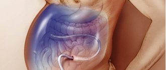
Depending on the severity of the bone tissue deficiency, open or closed volume restoration surgery is performed. The essence of the surgical intervention is to separate the mucous membrane lining the maxillary sinus, followed by filling the resulting cavity with bone grafts of various types. However, in the process of preparing for surgery, and sometimes during its implementation, obstacles to the procedure may be discovered. Most often, such unfavorable factors are cysts in the maxillary sinus.
General information
In the structure of diseases of the ENT organs, the pathology of the paranasal sinuses has occupied a leading place in recent years.
A paranasal sinus cyst is a pathological formation in the form of a benign formation in the shape of a sphere/ball, filled with liquid contents. The cyst shell is two-layered, in which the inner layer has an epithelial lining containing glands that produce fluid. Cysts gradually increase in size and never disappear on their own. The cyst is attached directly to the wall of the sinus, completely/partially filling it from the inside. Most often, the fluid that accumulates in a cyst is mucous in nature; purulent cysts are less likely to form, and serous cysts are extremely rare. Most often, cysts of the maxillary (maxillary) sinuses are formed (75-80%), somewhat less often - cysts of the ethmoidal labyrinth (about 15%) and extremely rarely - cysts of the frontal/sphenoid sinuses (about 5%). Most often, a cyst is discovered accidentally during an X-ray examination. There were no differences in the incidence of cysts in the left sinus and the right sinus. According to the classification of diseases (ICD-10), cystic pathologies of the paranasal sinuses belong to chronic forms of sinusitis.
You can suspect the presence of a cyst in the maxillary sinuses during periods of local hypothermia , acute respiratory viral infections , and exacerbations of chronic sinusitis . What can a maxillary sinus cyst lead to? An enlarged, untreated cyst is dangerous because it puts pressure on the nerve endings of the mucous membrane of the maxillary sinuses, provoking migraines . Also, an overgrown cyst in the maxillary sinuses often leads to blocking of the ducts between the nasal cavity and the maxillary sinuses, provoking the development of purulent sinusitis , which occurs in a severe form.
Diagnostic methods
If you suspect this pathology, you should consult a doctor - a dentist or otolaryngologist. The doctor will listen to your complaints, conduct an examination and prescribe additional tests. Based on the clinical picture, one can only suspect the presence of a pathological process in the projection of the upper quadrant. To clarify the diagnosis, additional examination is necessary.
| Research method | Explanation |
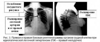
| Survey radiography | Plain radiography is the most accessible diagnostic method. It is carried out in two projections (direct and lateral). Plain radiography allows you to identify changes in the upper jaw, but this is not enough to determine the nature and localization of the pathology. |
| Contrast radiography | To clarify the location and size, contrast radiography is prescribed. A contrast agent is injected into the sinus through a natural opening, then an image is taken. |
| Probing | Probing makes it possible to penetrate the sinus cavity without opening it. |
| Computed tomography (CT), magnetic resonance imaging (MRI) of the brain | CT and MRI are the most informative research methods. They allow you to accurately determine the location of the cystic formation, its size and contours. |
Computed tomography and magnetic resonance imaging are the most accurate methods for diagnosing an intracranial cyst.
Pathogenesis
The pathogenesis of paranasal sinus cysts varies depending on the type of cyst. Thus, true cysts are formed when the lumen of the ducts of the glands of the sinus mucosa is obliterated due to squamous metaplasia of their epithelium and disruption of the process of outflow of their secretions. They develop against the background of inflammatory reactions caused by blockage of the lumen of the excretory duct with necrotic masses and stretching of the tissues of the gland and the proximal part of the duct. The leading mechanism of cystic transformation of the glands is a change in the composition of the secretory epithelium and hyperplasia of the glands. It is based on a sharp mucoidization of the secretory epithelium. At the same time, serous elements undergo pronounced changes, which, against the background of high mucoidization, are completely reduced. The corresponding transformation of the structure of the glands, together with goblet cell hyperplasia, leads to disruption of the transport function of the mucosa, excessive accumulation of secretions in the terminal sections of the glands, followed by cystic expansion, gradual enlargement and fusion into one common cavity. Also, the formation of cysts may be preceded by compression of the gland and duct by connective tissue.
False cystic formations develop primarily as a result of progressive disorganization of connective tissue (edema, degenerative changes in the mucosa and morphologically confirmed separation of connective tissue). At the same time, the leading role in the development of false cysts belongs to immunopathological mechanisms, as evidenced by the cellular composition of inflammatory infiltrates (eosinophils/mature plasma cells) and the predominant damage to blood vessels.
Odontogenic radicular cysts of the maxillary sinuses develop as a result of epithelial granulomas/necrotic changes in the apical part of a tooth affected by caries in combination with atrophic processes in the bone of the upper jaw.
The development of follicular cysts is based on inflammatory lesions of the impacted tooth germ of primary teeth. Congenital cysts develop against the background of various developmental anomalies of glandular tissue/ducts of the mucous glands.
Classification
The classification of maxillary sinus cysts is based on the factor of their origin and secondary changes in the sinus , which is due to the specific treatment of each form and the need for surgery. Based on the mechanism of occurrence and morphological characteristics, they distinguish:
- True (retention) cysts. They are formed as a result of complete/partial obstruction of the excretory ducts of mucus-producing glands. They have an epithelial lining with columnar ciliated epithelium. The reasons for their obstruction are: swelling , blockage , scarring or hyperplasia . The gland continues to function and produce secretions. Over time, the walls stretch, it overflows and closes the lumen of the sinus.
- False cysts (cyst-like formations). They do not have an epithelial lining and are formed in the thickness of the mucous membrane.
- Odontogenic cyst (radicular/follicular). Filled with pus. Radicular cysts form around the root of the inflamed upper tooth, penetrating into the sinus by growing through the atrophied bone tissue of the upper jaw. Follicular cysts form directly from the follicle of a baby tooth.
- Congenital cysts. Caused by developmental defects (anomalies of the mucous membranes of the maxillary sinuses/deformations of the upper jaw), contributing to the formation of cysts.
Rehabilitation period
Full recovery of the patient after surgery takes up to one week. Sometimes swelling and pain may occur. A visit to the clinic is not required during this period. To facilitate nasal breathing, the doctor prescribes nasal drops and painkillers.
Complications after surgery are extremely rare. In this case, rehabilitation is quite easy and quick. After just a few days, the patient feels well, nasal breathing is restored and the problem of growths in the nose no longer bothers him.
Causes
The most common cause of the development of cysts of the maxillary sinuses is local inflammatory processes of both infectious and allergic origin, causing blockage of the excretory duct of the glands, among which are:
- Chronic rhinitis.
- Sinusitis.
- Allergic rhinitis / polypous rhinosinusitis .
Also a common cause of polypous formations are congenital/acquired anatomical and topographical features of the structure of the nasal cavity (deviated septum, congenital deformities, polyp), which impede the movement of air through the nasal canals.
Another reason is inflammatory diseases of the teeth ( pulpitis , periodontitis ) with the spread of infection to the root of the tooth and root canals. Quite often, a foreign body, mainly filling material, gets into the maxillary sinuses. As a rule, filling material enters the maxillary sinuses after filling teeth (“sixes” of the upper jaw) or with an open wound.
The consequences of filling material getting into the maxillary sinuses can be severe and cause severe purulent inflammation and subsequent formation of cysts. Another reason may be long-term use of vasoconstrictor nasal drops.
Removal of sinus cyst without surgery
If a sinus cyst is detected, treatment without surgery is carried out using decongestant sprays, antibiotics, painkillers, mucolytics, steroids and antihistamines. They treat concomitant diseases - allergies, sinusitis, inflammatory processes of the gums, teeth, nasal mucosa. In combination with these drugs, various means for rinsing the nasal cavity, regenerating and restorative sprays are used. Treatment is prescribed by the doctor based on the results of the patient’s examination.
Symptoms
Symptoms of maxillary sinus cysts, the size of which does not exceed 1 cm, often do not manifest themselves clinically. And only as its size increases, patients begin to complain of discomfort in the area of the maxillary sinus, chronic nasal congestion localized on the affected side, a feeling of “fullness” in the cheek area, headaches poorly controlled by analgesics , periodic mucous discharge from the nose, pain when pressing /biting on the causative tooth, redness/ swelling of the gums, less often - asymmetry of the face with pain in the area of the anterior wall of the sinus. A maxillary sinus cyst can also manifest itself with non-specific symptoms such as irritability , dizziness and sleep disturbances .
Cyst in the sinus: surgery, reviews
Patient reviews give preference to the endoscopic method of cyst removal. When performing the classical method and laser vaporization, an incision is required under the upper lip to gain access to the cyst. Classic removal of a sinus cyst involves dissection of the soft tissue under the upper lip from the frenulum to the first molar (Caldwell-Luc method) or using the Denker method, which is also carried out through the facial part. Classic operations are more traumatic, with long postoperative recovery, but these techniques allow you to gain access to a cyst located in a hard-to-reach place. Endoscopic removal is carried out using an endoscope, the cyst is removed through a small burr hole, the operation is performed under local anesthesia.
At the Yusupov Hospital, patients will be able to undergo a full examination using modern diagnostic equipment. The hospital hosts doctors from various fields, there is a clinical laboratory, and a rehabilitation center. In the hospital you can recover from surgery or illness, and patients have a 24-hour inpatient facility.
List of sources
- Zakharova, G.P. Clinic, pathogenesis and surgical rehabilitation of patients with cyst-like formations of the maxillary sinuses / G.P. Zakharova // Sat. scientific tr. XV Congress of Otorhinolaryngologists of Russia. - St. Petersburg, 1995.-T. 1.-S. 96-102.
- Daynyak, L.B. Nose and paranasal sinuses / L.B. Daynyak // Guide to otorhinolaryngology. M.: Medicine, 1994. - P. 222.
- Guryev I.S., Dolzhikov A.A. Features of patho- and morphogenesis of cysts of the paranasal sinuses. Russian rhinology. 2002; 2:53-54.
- Allahverdiev S.A., Lopatin A.S. Choosing the optimal surgical approach for maxillary sinus cysts. Russian rhinology. 2010;(1):34-35.
- Kulakov A. A. Surgical dentistry and maxillofacial surgery. National leadership / ed. A. A. Kulakova, T. G. Robustova, A.I. Nerobeeva. - M.: GEOTAR-Media, 2010. - 928 p.