Causes
Erythema can be both physiological and non-physiological in nature.
In the first option, erythema may occur as a result of various psycho-emotional states or short-term abnormal phenomena. This kind of redness goes away very quickly on its own and is not the cause of certain disorders in the body. If erythema is of non-physiological origin, then it can be considered a separate disease, which is accompanied by prolonged redness of the skin and the presence of an inflammation process. Erythema can have many causes. These are viruses, infections, various diseases of the skin and connective tissues, as well as physiotherapeutic procedures that involve the use of sources of thermal or chemical action. Also, causes of erythema may include poor circulation, allergic reactions, regular rubbing of the skin, exposure to cold or chemicals.
Often reddened areas of the skin are a consequence of Crohn's disease, ulcerative colitis and pregnancy. Erythema can also be caused by medications such as penicillin, hormonal contraceptives (pills), non-steroidal anti-inflammatory drugs, anticonvulsants and sulfa drugs.
Diagnostics
Diagnosis of erythema is carried out by a dermatologist, dermatovenerologist or allergist. Diagnosis of erythema includes taking a patient’s medical history, external examination, taking a scraping from the rash, general urine and blood tests, and a smear for streptococcal infection. If the erythema is rheumatological in nature, a consultation with a rheumatologist, radiography, and MRI are required.
Symptoms
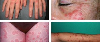
Erythema is usually divided into several types. Each of them has its own specific symptoms, as well as symptoms common to erythema - rash and redness.
Erythema is divided into two groups - infectious and non-infectious. The first occurs against the background of various infectious diseases, the causes of which are viruses and other microorganisms.
Erythema infectiosum of Rosenberg
It mainly affects older schoolchildren and people under 25 years of age. The signs are quite acute: the patient may experience a high fever, severe headache, suffer from insomnia and joint pain. After a few days, the skin may become covered with an asymmetrical, patchy, red rash. Erythema also affects the oral mucosa. The rash goes away within 5–6 days after its appearance and leaves behind lamellar peeling of the skin. The disease lasts for 7–13 days.
Erythema infectiosum of Chamera
The causative agent of this type is considered to be parvovirus. Children are usually susceptible to it, and it often occurs without any symptoms. If they do exist, they appear from the first day. This is a small rash on the face that eventually merges into one large spot. Rashes can also appear on the skin of the arms, legs, and torso and become pale over time. Relapses of the disease often occur. In general, this erythema goes away easily and lasts no more than 2 weeks.
Erythema nodosum
Its main symptom is the occurrence of nodular subcutaneous inflammations, which are mainly located on the anterior parts of the lower extremities, on the forearms and on the thighs. They are distinguished by their round shape and noticeable swelling, their structure is dense, and their sizes can range from 1–10 cm in diameter. These inflamed areas can be painful, especially in the case of mechanical impact (pressure, etc.). Other symptoms include joint pain and a feeling of fatigue.
Erythema nodosum can be a symptom of a more serious disease, such as rheumatism or tuberculosis, or be independent. In the latter case, the origin of the disease often remains unclear, although the cause is often streptococcal infection, mononuculosis, or the use of contraceptives and sulfa drugs. The disease can last for two weeks or one and a half months. Over time, redness and swelling will subside, and bruising may remain on the affected areas, which will go away on its own.
Multiforme or polyform exudative erythema
This form of the disease is manifested by fever, headache, joint pain and discomfort in the muscles. After a few days, the skin of the body and limbs may become covered with a severe rash, which can cause itching and burning. Erythema multiforme may be accompanied by the formation of blisters filled with exudate. After their rupture, painful erosion may remain.
Even more severe forms of the disease can manifest as the Stevens-Johnson symptom, which is characterized by the appearance of blisters on the mucous membranes of the mouth, eyes, throat, and genitals. Lyell's syndrome may also occur, which is accompanied by a skin rash that turns into large blisters with serous-hemorrhagic content. In half of the patients, the exact cause of the disease cannot be identified. In other situations, manifestations of this type of disease such as erythema exudative multiforme are provoked by infectious viral microorganisms - measles, herpes, scarlet fever, etc. or taking certain medications.
Sudden erythema
As the name suggests, this type of erythema develops quite quickly. Symptoms include a sharp increase in body temperature with enlarged lymph nodes under the jaw, headache, aching joints and general weakness. Within four days the temperature returns to normal, and after that the skin of the face, limbs and torso becomes covered with a spotty rash. Sometimes the spots can merge and turn into erythematous fields.
Erythema migrans
May be a sign of Lyme disease, which is predominantly transmitted by a tick bite, although the cause is often unclear. The incubation period lasts 1–3 weeks. A ring-shaped spot appears at the site of the bite. It grows quickly and reaches 30 cm in diameter. Then this ring may turn pale and disappear altogether. The illness can last from 2 weeks to a couple of months. Erythema annulare cannot be treated and goes away on its own over time. This species poses a particular danger to a pregnant woman and her baby.
If we talk about the second, non-infectious form of erythema, then this includes diseases that appear as a reaction to a certain irritant, or as a manifestation of an allergy. These include the following types of pathologies:
X-ray erythema
It is manifested by inflammation of the skin and is characterized by a rash in response to prolonged or repeated exposure to X-ray waves. This type of erythema manifests itself on the irradiated area of the skin as a pronounced red spot that appears approximately 7 days after irradiation. This stain lasts 10 days, after which it darkens and acquires a brownish tint. Peeling may also appear at the site of the lesion.
Infrared or heat erythema
It appears due to prolonged or regular exposure to heat, which is not enough for a full-fledged burn. It appears as a mesh or pigmented rash appearing on the skin.
Persistent elevated erythema
It is a sign of allergic vasculitis. The disease has two forms: symptomatic, which manifests itself as a side effect, that is, as an allergy to taking certain medications, or against the background of polyarthritis, and idiomatic, which is associated with hereditary etiology and manifests itself as a nodular rash of a purple tone.
In the structure of the group of figured erythemas, one of the significant places is occupied by Afzelius-Lipschütz migratory erythema (EA), which is a pathognomonic sign of Lyme borreliosis, a widespread vector-borne disease on almost all continents. In the Russian Federation, the annual number of people sick with tick-borne borreliosis ranges from 7 to 9 thousand [7, 8]. In 2011, the incidence rate of ixodid tick-borne borreliosis was 7.02 per 100 thousand population [4]. In recent decades, there has been an increase in the frequency of severe, chronic, torpid forms of Lyme disease (LD) [7, 8, 24, 45].
ME refers to the early skin manifestation of BL, a manifestation of an inflammatory-allergic skin reaction to tick suction, observed in 50-80% of patients, and if it persists for more than 4 weeks, it can be classified as chronic. Ixodid tick-borne borreliosis (TB) is a naturally occurring, focal, vector-borne infectious disease that was first described in Europe, including Russia, at the end of the 19th century [5, 27, 48]. At the beginning of the twentieth century (1908), the Swedish dermatologist Arvid Afzelius [10] established a relationship between the appearance of ring-shaped ME on the skin (“Erythema of Afzelius”, “migratory dermatitis”) and mite suction. The modern name was given by researcher Lipschutz in 1913 [37]. In 1975, in the USA, in the town of Lyme, cases of tick-borne borreliosis in a group of children in combination with ring-shaped ME after tick ingestion were first described [39, 41]. Until the early 80s of the 20th century, BL and chronic ME were considered as two independent diseases with an unclear etiology. In 1982, American microbiologist Willy Burgdorfer [19] first isolated and identified previously unknown spirochete-like microorganisms from Ixodes dammini
(modern name -
Ixodes scapularis
).
Later, these microorganisms were isolated from the blood, skin biopsies, and cerebrospinal fluid of sick people [13, 16, 40]. Pathogens B.L. belong to the order Spirochaetales
, family
Spirochaetaceae
, genus
Borrelia
.
For Eurasia, the most important epidemiological significance in the development of BL is the ticks Ixodes ricinus
and
Ixodes persulcatus
, in the USA
- I. scapularis
.
Borrelia enters the human body with tick saliva. A ring-shaped ME develops on the skin at the site of its suction. With the flow of lymph and blood, the pathogen enters the internal organs. Under the influence of nonspecific defense factors of the macroorganism, some borrelia die, releasing endotoxin, which triggers a cascade of immunopathological reactions [34]. Currently, the pathogenicity of 3 species of B. . burgdorferi sensu lato : B. _ burgdorferi sensu stricto , B. _ garinii
and
B. _ afz elii
.
The species of borrelia of the B. burgdorferi sensu lato
, have adapted in the process of evolution to survive in various biological objects of the environment: ticks and their “feeders” - vertebrates, which is achieved through the functioning of various regulatory genes that change the level of expression of a number of proteins to throughout the life cycle [12, 29, 48]. Borrelia have groups of antigens: surface (OspA, OspB, OspD, OspE and OspF), flagellar and cytoplasmic. Surface antigens are characterized by variability. The outer shell proteins determine the species of the pathogen and are the main immunogens.
The incubation period is 3-30 days (average 7 days). The ability of Borrelia to undergo independent forward movements in tissues is reflected in the characteristics of local inflammation. In the center of the erythema (at the site of initial accumulation of the pathogen), borrelia are actively exposed to inflammatory factors, they lose their mobility and their number decreases, resulting in the formation of a clearing zone in the center of the erythema. The appearance of new rings of hyperemia is associated with new generations of Borrelia and usually leads to the development of characteristic ME.
The pathohistological picture in the early stages of ME development (the first 15 days) is characterized by signs of exudative-productive inflammation and is represented by subepidermal and subthreshold blisters, characteristic of the central areas of erythema. In the epidermis, hyperkeratosis, hydropic degeneration of epithelial cells of the basal layer, and rarely koilocytosis are observed. In the papillary layer of the dermis, pronounced edema, pericapillaritis and interstitial band-like cellular infiltrates are observed, consisting of lymphoid and a small number of plasma cells, histiocytes, eosinophils, and rarely neutrophils. Stasis is detected in the capillaries, and rarely - erythrocyte thrombi. In the peripheral areas of erythema, hydropic degeneration of the epithelium of the spinous layer and pericapillaritis in the papillary layer of the dermis are found. Diffuse or focal cellular infiltrates of varying degrees of severity are also detected there. In the central areas of erythema, at a later date (15-40 days), the presence of pericapillaritis and fibrosis of the vessel walls is revealed, and rarely, hydropic degeneration of epithelial cells [9, 23].
Differential diagnosis of ME must be carried out primarily with other figured erythema: annular centrifugal erythema of Darier, rheumatic erythema of Lehndorff-Leiner, figured persistent erythema, neutrophilic erythema, eosinophilic annular erythema, tortuous creeping erythema of Hammel. The problem of differential diagnosis of ME is also due to the variety of nonspecific exanthematous rashes with figured outlines, most often caused by viral infections. The differential diagnosis of ME in the form of a homogeneous focus should be carried out with the erythemal form of erysipelas, the spotted form of toxicerma, including fixed erythema, with a reaction to insect bites, granuloma annulare, microbial eczema and other dermatoses manifested by ring-shaped lesions.
The prognosis for ME is usually favorable. ME regresses spontaneously (usually within 1–2 months), but can persist for a long time from 6 to 12 months [30]. Therapy B.L. is most effective in the early stages of infection; a favorable outcome of the disease and recovery depend on the timeliness of the initiated etiotropic treatment. Therefore M.E. requires etiotropic therapy. Therapy for M.E., as well as other manifestations of BL, includes the prescription of antibiotics. First-line drugs are doxycycline and cefuroxime; amoxicillin and erythromycin can be used. Amoxicillin may become the drug of choice in the treatment of children and pregnant women. The duration of antibacterial therapy is usually 14-21 days. However, the study showed the same high effectiveness of 10- and 20-day courses of doxycycline M.E. therapy. The effectiveness of antimicrobial prophylaxis using doxycycline at a dose of 200 mg once in the first 72 hours after tick bite is controversial [33]. A good effect was obtained as a result of the use of azithromycin in the treatment of ME according to the regimen of 1000 mg on the 1st day and 500 mg daily for the next 4 days [15]. Parenteral administration of ceftriaxone at a daily dose of 2 g or penicillin G IV at 18-24 million IU daily for 10-14 days can be used in cases of multiple ME and in patients with immunodeficiency [31]. Antibacterial therapy at stage I of infection significantly reduces the likelihood of developing neurological, cardiac and arthralgic complications, promotes the disappearance of erythema in a shorter period of time, and most importantly, prevents the development of late stages of BL [20, 47].
To prevent BL, vaccines containing antibodies against the surface lipoprotein A spirochete OspA (Lymerix) are used, the effectiveness of which is 70-80% [42]. However, evidence has accumulated on the adverse effects of vaccination and the presence of post-vaccination complications caused by the arthritogenic potential of the OspA lipoprotein. Currently, researchers [44] are focusing on potential antigens for the second generation of vaccines (OspC, DbpA, OspB, p35, p37, p66).
Despite powerful antibiotic therapy for ME with a long course and relatively large single doses, the formation of chronic forms of infection occurs in 3.5-30% of patients. When treating chronic forms of BL, the effectiveness of etiotropic therapy decreases and amounts to 43.2–48.7% [6].
When studying the pathogenesis of the disease in patients with Lyme borreliosis, evidence was obtained that borrelia lipoproteins, in particular OspA, are potential activators of the inflammatory response due to binding to CD14 and TLR2 on macrophages [11]. Activation of receptors TLR2, TLR6, TLR½, TLR5, TLR9 leads to the secretion of proinflammatory cytokines [46]. Receptor dimers TLR2/TLR6, TLR2/TLR1, through the recognition of triacylated lipoproteins, such as Borrelia
burgdorferi
OspA, flagellin, peptidoglycans and zymosan, are involved in the activation of the nuclear transcription factor NfkB [25, 28, 35].
J. Salazar et al. [36] in a study of peripheral blood and aspirates from patients with ME also found high expression of TLR2, TLR1 and TLR4 on the surface of monocytes, macrophages, and dendritic cells. During the development of ME, flagellin production promotes pronounced expression of TLR5 and increased functional activity of TLR5 on human keratinocytes. It was revealed that peripheral blood mononuclear leukocytes, by the action of Borrelia
, reduce the levels of TLR2 and TLR4 mRNA, while the expression of the corresponding proteins on monocytes does not decrease [22]. As a result of experimental work, it was revealed that mice deficient in TLR2 or TLR1 had low levels of antibodies to OspA after vaccination, which indicates a violation of the cooperation and functioning of adaptive immune effectors [17]. During experimental work on a mouse model of ixodic tick-borne borreliosis, it was revealed that from the 14th day of infection, stimulation with the DbpA antigen increased the number of T-lymphocytes carrying TLR2 receptors: the number of CD4+ increased by 2.3-7 times, CD8+ by 2, 2-5.6 times. In the 1st week after infection, the proportion of CD4+ cells synthesizing IFN-γ increased 2.5 times, but after 14 days it decreased to normal levels. The subpopulation of CD4+ producing TNF-α increased on the 7th day, but their level normalized by the 30th day. During the development of the disease, there was an increase in the Th2 subpopulation that synthesizes IL-4, and a decrease in the Th1 subpopulation that synthesizes IFN-γ [3].
Data were obtained on high spontaneous and antigen-stimulated production of IL-2, an increase in spontaneous and suppression of induced production of IFN-γ in patients with chronic BL, with a high level of spontaneous production of IL-4 and induced production of TGF-β, which indicates involvement in immune regulation mechanisms limiting the functional activity of Thl cells. According to the authors [4, 34], the data obtained may indicate the formation of chronic specific inflammation of the type of delayed-type hypersensitivity (DTH), characteristic of a chronic infectious process with intracellular localization. Under B.L. a Th1-mediated immune response develops with high levels of IFN-γ production. When studying phagocytosis in patients with BL, an increase in the phagocytic index was revealed in the acute form of the disease compared to the chronic form [1]. A number of studies have shown that in the chronic course of BL there is a decrease in the functional activity of neutrophils compared to a group of healthy individuals, a decrease in the number of CD3+, CD4+, CD22+ cells, CD16+ NK cells, against the background of an increase in CD8+, serum immunoglobulins of the classes IgG, IgM, IgA, circulating immune complexes (CIC). The identified disturbances in the immune system in patients with chronic BL indicate, according to the authors, a decrease in the activity of the body’s nonspecific resistance mechanisms, which can create conditions for long-term persistence of the pathogen. Insufficiency of CD22+ cells and an increase in the level of CEC creates conditions for the development of autoimmune pathology [2, 4]. Blood cells from patients with persistent BL release low levels of the proinflammatory cytokines TNF-α and IFN-γ. However, the authors' opinions on this issue are mixed. There are studies that show opposite results. Borrelia lipoproteins, particularly OspA, are potential activators of the inflammatory response by binding to CD14+ and Toll-like receptor-2 (TLR2) on macrophages. As a result, the secretion of pro-inflammatory cytokines (IL-1b, IL-6, IL-10, IL-12) and TNF-α increases [22, 36, 46].
When studying the influence of B. burgdorferi
human monocytes have been shown to induce the expression of TLRs and produce pro- and anti-inflammatory cytokines (TNF-α, IL-6, IL-10 and IL-1β), epithelial IL-8, and IFN-β.
In turn, TGF-β in combination with IL-6 induces the synthesis of proinflammatory cytokines IL-1β and TNF-α [18, 43]. It is suggested that OspA of B. burgdorferi
may be involved in the triggering of autoimmune inflammation and provoke the development of Lyme arthritis through the differentiation of Th cells that express IL-17.
An experiment on B. burgdorferi
-vaccinated mice also showed that IL-17 plays a key role in the development of arthritis.
Moreover, IL-17 can stimulate fibroblasts and synoviocytes to produce proinflammatory cytokines, including IL-6 [21]. Induction of IL-17 in the ME area leads to a decrease in the number of positive epidermal Langerhans cells (CD1a), which ensure the “capture” of Borrelia in the early stages of infection, thereby contributing to the chronicity of the infectious process [14]. In the development of Lyme arthritis, the leading role was traditionally previously attributed to Th1 cytokines, such as IFN-γ, but recent studies have revealed that OspA B. burgdorferi
may be involved in the triggering of autoimmune inflammation and provoke the development of arthritis through the differentiation of Th cells, expressing IL-17 [26, 38]. In addition, the study showed that IFN-γ not only does not induce the synthesis of anti-OspA antibodies, but also plays an important role in suppressing the production of anti-borrelia antibodies, which may justify the development of highly effective vaccines [32].
There were no differences in the subpopulation composition of peripheral blood lymphocytes in acute and chronic forms of BL [1]. In Lyme borreliosis, humoral immunity is characterized by the predominant synthesis of IgG1 and IgG3 subclasses of antibodies involved in complement activation and the process of borrelia opsonization. The production of immunoglobulins of the IgG2 subclass is insignificant, and IgG4 is absent [8].
Thus, at the present stage, ME, being the most common nosological form in the group of figurative erythema and a pathognomonic sign of the early stage of a common transmissible disease - BL, is characterized by a torpid chronic course and is accompanied by disturbances in the innate and adaptive immune system. Development of M.E. in BL occurs against the background of insufficiency of cellular and/or humoral factors of nonspecific resistance of the body, which determines the need for timely etiotropic therapy and the direction of pathogenetic therapy of the early stage of BL in order to prevent the development of severe torpid forms.
There is no conflict of interest.
Treatment
The course of treatment for erythema depends on the degree of skin damage and the individual characteristics of the patient’s skin. First of all, it is necessary to sanitize foci of infections, avoid physical influences that can affect the skin (massage, sunbathing, etc.), eliminate contacts between the skin and chemical irritants.
For erythema disease, treatment should include antibiotics, corticosteroid drugs, iodide alkalis, agnioprotectors that improve the rheological properties and microcirculation of the blood, as well as disaggregants, hemokinators, adaptogens and special drugs that strengthen the walls of blood vessels. For local therapy, occlusive dressings with corticosteroid and butadiene ointment and dimexide applications are used.
A patient with erythema needs to remain in bed, especially when the erythematous lesions are concentrated on the legs. Gymnastics is also very useful, as it helps improve blood circulation. Experts advise sticking to a healthy diet that does not include fried, salted and smoked foods, preservatives, alcohol, strong coffee and tea, and chocolate. It is also better to exclude factors that may cause a relapse: prolonged walking or standing, smoking, exposure to low temperatures, lifting heavy objects.