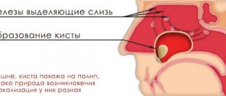
A sinus cyst is a fluid-filled formation with elastic walls; the location of the cyst affects the symptoms of the disease. Most often, cysts form in the maxillary sinuses. The reasons for the development of cysts in the nasal sinuses are varied; there are a number of reasons for the appearance of a neoplasm:
- Blockage of the ducts of the mucus-secreting glands.
- Dental diseases, tooth roots protruding into the paranasal sinuses.
- Allergic diseases of the nose.
- Frequent inflammatory processes.
- Anatomical features of the structure of the nose.
- Mechanical injuries of the nose.
In the oncology department of the Yusupov Hospital, you can undergo examination using MRI, CT, ultrasound, diagnosis of diseases of the paranasal sinuses if a nasal tumor is suspected. The patient will be able to undergo a full examination at the diagnostic center, take tests in the hospital laboratory, and receive advice from an otolaryngologist, oncologist and other specialists.
The maxillary cyst is most often located in the nose area
The maxillary sinus cyst consists of epithelial cells of the mucous membrane. Inside it can be filled with mucus, serous contents or pus. Most often it is localized in the area of the sinuses, that is, the paranasal sinuses, which is why the pathology is also called a maxillary sinus cyst.
A maxillary cyst is often called a maxillary sinus cyst.
Possible complications
After surgery to remove a cyst from the maxillary sinus, swelling of the mucous membrane, a feeling of dryness or profuse nasal discharge, and pain in the area through which the sinus was accessed may appear. These symptoms are not complications of surgery and usually resolve within a few days. If they persist and intensify, you should consult a doctor.
The nature of complications after surgery depends on the technique used:
- if access to the sinus was carried out through the alveolus of the tooth, an incision or puncture in the upper jaw, a fistulous tract may appear;
- with micromaxillary and radical maxillary sinus there is a risk of damage to the infraorbital or trigeminal nerve with the development of neuritis. In this case, areas of the skin, nasal mucosa and mouth become numb, which requires additional treatment;
- inflammation and/or suppuration of the wound develops if it becomes infected (with poor quality oral care, in cases where the patient does not follow the doctor’s orders).
There are several types of pathology
Cysts of the maxillary sinuses are different:
- odontogenic: inflammation begins to form at the root of a poorly treated or completely untreated tooth located in the upper jaw. As the tumor increases in size, it destroys bone tissue and gradually grows into the sinuses. Such neoplasms are most often filled with pus,
- retention: formed due to disruption and obstruction of the glands that produce mucus.
Doctors also classify false cysts as a separate category - such formations do not contain epithelial cells and appear due to pathologies and structural disorders of the maxillofacial apparatus.
One of the most common causes of tumor is a bad tooth.
A maxillary sinus cyst can form due to a reason such as dental disease. More precisely, when treatment was not carried out, or it was performed poorly. Most often, the development of pathology is provoked by advanced caries, pulpitis and periodontitis on molars and premolars (these teeth are separated from the paranasal sinuses by a thin septum).
Teeth with cysts and granulomas were previously more often removed than treated. Today, in some clinics, high-precision tooth-preserving operations are carried out, where all stages of treatment are accompanied by work on a dental microscope. A competent endodontist carefully removes the cyst under a microscope using special instruments, then the canals are sterilized to their entire length and filled with filling material. Treatment under a microscope is accurate, fast, without risks or complications, and, most importantly, the natural tooth remains intact.
The prerequisite for the appearance and growth of a tumor may be the individual anatomical characteristics of the patient, for example, a deviated nasal septum or jaw abnormalities. Another reason is the systematic blockage of the ducts of the glands located in the nose, against the background of rhinitis, runny nose, and allergic reactions.
Diagnostics
Figure 1. Cyst in the maxillary sinus.
Source: Contemporary clinical dentistry / Open-i (Attribution-NonCommercial-ShareAlike 3.0 Unported) Before surgery to remove a sinus cyst, you need to take tests (for HIV, syphilis, hepatitis B and C, general blood test).
Usually, to make a diagnosis, the doctor only needs to collect an anamnesis and a general physical examination, including the nose.
Other diagnostic tests include:
Endoscopy of the nose. The doctor performs a detailed examination of the nose and sinuses using a narrow tube with a lighted magnifying lens or small camera.
Visualization methods. Images obtained from a computed tomography (CT) scan can help the doctor accurately determine the size and location of cysts in the deeper areas of the sinuses, and assess the degree of swelling and inflammation.
Test for cystic fibrosis. If a child needs diagnosis, the doctor may suggest testing for cystic fibrosis. This is an inherited disease that affects the glands that produce nasal mucus, tears, sweat, saliva and digestive juices. This is usually a non-invasive sweat test that can determine whether your sweat is more or less salty than most people.
A small cyst can only be detected by x-ray
When the neoplasm is small, less than 10–15 mm, it is almost impossible to find out about its existence, since no alarming signs are observed. At this stage, the only option to identify pathology is to do a computed tomography scan. A dentist may also suspect its presence during a routine examination or other doctors if a person needs to visit them.
At the initial stage, the problem can only be detected on an x-ray
Signs of a large cyst are similar to signs of acute sinusitis
When the cyst begins to grow, a person develops alarming symptoms, which, based on clinical manifestations, can be confused with acute purulent sinusitis. Let's list them:
- severe headache: unpleasant sensations spread to the cheekbones, temples and the back of the head. The pain intensifies when the head is tilted forward, pressure surges, or diving under water. Discomfort may persist even after taking analgesics,
- difficulty breathing through the nose, constant nasal congestion,
- nasal discharge: clear, less often yellow,
- a feeling of heaviness and fullness in the areas under the eyes,
- the presence of a viscous mucous lump in the throat immediately after sleep and in the morning.
The signs of a cyst are similar to the signs of sinusitis.
However, the signs become more pronounced when the lumen in the nasal sinus is practically blocked. Facial asymmetry and swelling of the cheek may appear. If there are associated problems, for example, a bad tooth, swelling of the gums, pain when chewing food and any mechanical impact are disturbing.
What is a sinus cyst?
This is a large formation in which mucus or pus accumulates. The cyst is not dangerous, it does not degenerate into cancer, but over time it can grow and put pressure on the walls of the sinus. Because of this, breathing problems appear, the nasal mucosa swells, a runny nose almost never goes away, and recurrent bronchitis and pneumonia may occur. The cyst is sometimes located in such a way that there are no symptoms, and it is discovered by chance during another examination - for example, before dental prosthetics.
What causes cysts to form in the sinuses?
To ensure that the mucous membrane of the respiratory organs does not dry out, is not overcooled and is protected from dust, it contains glands that constantly secrete moisturizing mucus. This mucus flows to the surface through the ducts of the glands. If at least one of them is blocked, mucus continues to be produced, but will accumulate inside the gland, filling it and stretching the walls. As a result, a cyst appears.
The formation of cysts can occur due to:
- frequent infections;
- severe allergies;
- structural features of the nasopharynx;
- injuries and curvature of the nasal septum;
- inflammatory processes in the upper jaw (usually dental diseases).
The most common symptoms of a cyst are headaches, difficulty in nasal breathing due to swelling of the mucous membrane, discomfort or pain over the upper jaw.
If left untreated, vision and more can seriously deteriorate.
Cysts of the maxillary sinuses are dangerous because if they are not treated, they can become a source of very serious health problems.
The infection can spread to the nearby teeth of the upper jaw and along the chain take over the entire row, as a result of which you can lose all your teeth. Pathology can lead to deformation of the skull bones and jaw fracture, the development of purulent processes - abscess, phlegmon, osteomyelitis, meningitis.
The tumor provokes the development of sinusitis and chronic sinusitis. Its presence is always accompanied by a runny nose and inflammation of the sinuses.
If the problem is not addressed, it can affect vision
The growth of the tumor often leads to blurred vision, “double vision.” If the cyst is purulent, it can lead to brain damage and encephalitis. When a tumor grows, there is a risk that it will completely block oxygen and complicate breathing, which is especially dangerous during sleep, since a person may not wake up.
Symptoms of a sinus cyst
The cysts and polyps themselves are soft and lack sensitivity, so if they are small, many people may not notice them at all. However, multiple growths or a large cyst can block the nasal passages and sinuses.
Common signs and symptoms of nasal cysts include:
- runny nose
- reduced or absent sense of smell
- loss of sense of taste
- facial or headache
- pain in upper teeth
- feeling of pressure on the forehead and face
- snore
- frequent nosebleeds
The main method of treating pathology is surgery
To treat a tumor, doctors combine therapeutic and surgical methods. First, surgery is used to remove the tumor, then drug therapy is used to eliminate signs of the inflammatory process and avoid complications, speed up rehabilitation and tissue healing.
If the maxillary sinus cyst is small (less than 1 cm), then drug therapy is used for treatment and dynamic observation is carried out, and there is no need for surgical removal of the tumor. But there are two important points. The first is that rarely do any patients go to the clinic until the cyst has grown and began to cause significant problems. Secondly, therapy can take a long time (4-6 months) and the result will not always be positive.
When treatment is carried out in dentistry
Most often, patients go to clinics when the cyst reaches 1 cm or more in diameter. In this case, its removal can be carried out in different ways. If the cause of the problem is a diseased tooth, then treatment is carried out by a dental surgeon. The doctor removes the tumor by performing a cystectomy if its size is no more than 1–2 cm. Or by cystotomy1, when the size is more than a few centimeters and the tumor has grown into the sinus.
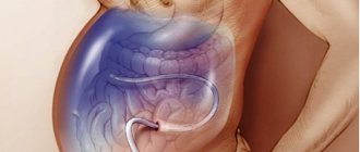
The photo shows an operation to remove a cyst.
The main tasks of the dentist in this case:
- eliminate the source of inflammation in the oral cavity and stop the inflammatory process,
- ensure the outflow of purulent contents, if any, using drainage,
- save the diseased tooth, if possible: in cases where the root is slightly immersed in the tumor, its resection (removal of the apex) is carried out, and the tooth itself is preserved. When the root is immersed in the cyst by more than 1/3, its complete removal is recommended to avoid relapses,
- fill the cavity left after tumor removal with bone material,
- close the communication between the oral cavity and the sinus.
When treatment is carried out at the direction of an ENT doctor in a hospital setting
Maxillary sinus cysts are not always associated with diseased teeth. But regardless of the cause of their appearance, advanced tumors almost always lead to inflammatory processes in the nasal mucosa, congestion, sinusitis, and mucus discharge from the nasal passages. Therefore, many patients with such symptoms turn to an ENT doctor.
In this case, the tumors are removed through surgery. For example, using a classic operation, when a deep incision is made in the upper jaw, through which the formation is removed. This method is the most affordable, but it is quite traumatic, and the patient then requires a long postoperative recovery.
“Two years ago I had a maxillary cyst removed. My background was like this: snot was either pouring out of my nose, or there was constant congestion, and for a long time I could not smell or breathe normally, so I went to an otolaryngologist. The doctor sent me first for an x-ray, and then for tests and surgery. The operation was performed in a hospital and under anesthesia, I was very afraid of how everything would go, and especially how I would recover from the anesthesia, but everything was tolerable. Then I was in the hospital for about 4 days, but some people stay longer. I felt discomfort for another ten days after that.”
[email protected] , fragment of a review from the site woman.ru
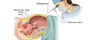
However, today the most common surgical method is endoscopy. The operation is performed through an opening in the facial wall of the maxillary sinus, or through the nasal passages. The entire treatment process is monitored on the monitor screen, so the doctor can control his actions. The technology is considered the most gentle, atraumatic; after its use, the patient has no stitches, incisions or scars on his face. The procedure is quick (duration is about 40 minutes), tissue restoration takes about 7 days.
An endoscope is an optical device used in all areas of medicine for high-quality examination and treatment of hollow internal organs. Endoscopes can be inserted through natural body openings (in this case, the nose) or through surgical incisions.
The operation can be performed using an endoscope through the nose
The operation can also be performed using a laser. This approach almost completely eliminates the likelihood of developing postoperative complications. But due to the use of laser equipment, the cost of treatment increases significantly.
Cyst of the maxillary sinus
Maxillary sinus cyst
is a benign neoplasm that appears on the mucous membrane inside the paranasal sinuses.
A maxillary sinus cyst is a rounded sac, with a membrane in the form of a thin film, filled with fluid. It happens that the cyst shell spontaneously ruptures, and this is manifested by copious clear discharge from the nose. Retention cysts
in the sinuses are formed due to prolonged disruption of the normal movement of air in the nasal cavity against the background of sluggish chronic inflammation (for example, allergies or chronic sinusitis). Odontogenic cysts occur in the sinus in response to inflammation in the area of the roots of the teeth of the upper jaw or as a reaction to the installation of dental implants.
WHAT IS THE DANGEROUS OF A PARONAL SINUS CYST?
Cysts of the paranasal sinuses are not tumors
, do not become malignant, and do not pose a threat to life.
A small cyst may be located in the sinus and not manifest itself in any way for many years. If the cyst does not grow and does not bother you, no treatment is required. On the other hand, if the cyst occupies more than half the volume of the sinus
, this leads to impaired ventilation and stagnation of mucus. A large cyst completely clogs the sinus anastomosis, preventing the normal movement of air and the outflow of secretions. There are often cases when, against the background of an acute respiratory infection, a maxillary sinus cyst suppurates - acute purulent sinusitis occurs, which is already fraught with complications. For this reason, large cysts are recommended to be removed.
TREATMENT OF MAXILLARY SINUS CYST
Cysts of the paranasal sinuses can be cured only in one way - surgically remove them.
. Once formed, the cyst will no longer be able to resolve on its own or with the help of medications. If the cyst bursts during a sinus puncture or is “cauterized” with a laser, it will grow again in the same place. To avoid recurrence of the cyst, it is important to carefully remove all its membranes. This is only possible under visual control, during endoscopic surgery.
ENDOSCOPIC MAXILLARITY IN THE ENT CENTER
At the ENT Clinic, treatment of paranasal sinus cysts is carried out only by the endoscopic method.
according to the principles of FESS - functional endoscopic surgery of the paranasal sinuses.
This method involves the most gentle approach through the natural anastomosis of the sinuses, small incisions, minimal damage to the mucous membrane and intranasal structures. Endoscopic surgery to remove a maxillary sinus cyst is called endoscopic maxillary sinusotomy
.
METHODS OF TREATMENT OF MAXILLARY SINUS CYST
Cysts of the paranasal sinuses can be cured only in one way - surgically remove them. Once formed, the cyst will no longer be able to resolve on its own or with the help of medications. Traditional methods of treatment (rinsing the nose and sinuses with solutions of salt or medicinal plants), nasal sprays and antibiotics are ineffective against cysts. Non-surgical treatments can temporarily shrink the cyst by draining the fluid. If the cyst is burst with a needle during sinus puncture or cauterized with a laser, it will grow in the same place again in 2-3 months. To avoid recurrence of the cyst, it is important to very carefully remove all its membranes. This is only possible under visual control, during endoscopic surgery.
MODERN OPERATIONS FOR THE TREATMENT OF MAXILLARY SINUS CYST
At the ENT Clinic Center, treatment of paranasal sinus cysts is carried out only by the endoscopic method according to the principles of FESS - functional endoscopic surgery of the paranasal sinuses. Endoscopic surgery to remove a maxillary sinus cyst is called endoscopic maxillary sinusotomy. This method involves the most gentle approach through the natural anastomosis of the sinuses, minimal damage to the mucous membrane and intranasal structures. The advantages of endoscopic surgery are that everything is done inside the nose - there are no swelling, incisions, stitches or scars on the face or under the lip after it. The endoscope gives the surgeon a good overview, a wide field of view and bright light, which allows the entire cyst to be removed completely without leaving any membranes.
HOW THE OPERATION WORKS:
- The anesthesiologist and operating nurse prepare the patient for surgery.
- The surgeon examines the nasal cavity with an endoscope and identifies the natural anastomosis of the maxillary sinus on the lateral wall of the nasal cavity.
- With the help of special instruments, the sinus anastomosis expands to approximately 9-10 mm.
- The internal contents of the sinus are examined with an angled endoscope.
- Special micro-forceps are inserted through the enlarged anastomosis, the cyst is captured and removed entirely along with the membranes.
- The sinuses are washed and examined.
- After the operation, special breathable sponge tampons are installed in the nose.
The result of endoscopic maxillary sinus surgery will be the restoration of the normal volume of the sinus, the natural outflow of mucus and the movement of air in it.
Dear patients! Please note that the stated cost of the operation does not include the cost of laboratory blood tests and other examinations. The cost of treatment depends on the chosen method of anesthesia (local or general) and the length of stay in the hospital room. Only a surgeon after a face-to-face consultation can tell you the exact amount in your specific case.
Endoscopic surgery is used in the ENT Center clinic to treat not only maxillary sinus cysts, but also chronic sinusitis of any nature, polyps and other sinus tumors.
ENDOSCOPIC SURGERY IS INDICATED FOR DISEASES:
- Cysts of the maxillary sinuses
- Polyps of the nasal cavity and polypous rhinosinusitis (from a single polyp to widespread polyposis).
- Chronic and recurrent purulent sinusitis (sinusitis, ethmoiditis, sphenoiditis, frontal sinusitis, or their combinations - polysinusitis, pansinusitis).
- Odontogenic sinusitis and foreign bodies of the maxillary sinuses (after treatment by a dentist and cementing material entering the sinuses).
INDICATIONS FOR ENDOSCOPIC SINUS SURGERY:
A maxillary sinus cyst accidentally discovered on a CT scan does not always require urgent surgery. If it occupies less than 1/3 of the sinus, does not complicate breathing and does not cause pain, observation by an ENT doctor is sufficient. You should think about endoscopic cyst removal if:
- One or both halves of the nose are always stuffy, so you have to breathe through your mouth.
- There is an excess accumulation of mucus in the nose and nasopharynx.
- The constant feeling of heaviness, pressure, distension in the sinus area or headache is disturbing.
- Acute purulent sinusitis occurs on the side of this sinus.
WHAT YOU NEED TO PREPARATE FOR ENDOSCOPIC OPERATION:
1. First, you need to undergo an examination by an ENT surgeon to make an accurate diagnosis and determine clear indications for surgery. 2. Be sure to do a CT scan of the nose and paranasal sinuses for individual planning of the scope of the intervention. CT scans also help the surgeon navigate the procedure. 3. After the ENT doctor has confirmed the need for surgery, you need to undergo a general preoperative examination on an outpatient basis and obtain a physician’s opinion on the absence of contraindications. You can undergo the examination both at the ENT Center clinic and at your local clinic. You can download the list of required tests in PDF format from this link.
REHABILITATION AFTER ENDOSCOPIC SURGERY IN THE NASAL CAVITY
- After endoscopic surgery, special sponge breathable tampons are placed in the nose, which the surgeon removes after 1-2 days. Tampons sometimes cause a feeling of pressure in the nose and a headache, these symptoms quickly disappear after removal.
- The patient spends a day in the hospital and goes home the next day after the operation.
- At the appointed time, the patient visits the surgeon on an outpatient basis: to remove tampons, rinse the nose and monitor healing.
- In the first week after surgery, the patient is not recommended to drink hot and alcoholic drinks, take a hot shower, or perform physical exercise (so as not to provoke dilation of blood vessels in the nose and nosebleeds). It is best to follow a home regime and avoid colds.
- As it heals over the course of 1-2 weeks, clots of mucus and crusts constantly form in the nose - this is normal. The doctor will prescribe saline solutions to moisten the nose and other medications to reduce swelling and speed up healing. Nasal breathing is completely restored gradually, usually by the 3rd week after surgery.
After removal of the cyst, you need to adhere to restrictions
Treatment of tumors in most cases involves their surgical removal. And naturally, after surgery the body needs time to recover. Usually 7–14 days. During this period, swelling, soreness, and difficulty breathing are normal. To make unpleasant symptoms go away faster, you must follow these tips:
- use antibiotics, antiseptics and vasoconstrictors prescribed by your doctor,
- avoid actions that provoke active use of facial muscles: long conversations, laughing, yawning, blowing your nose,
- do not visit saunas, steam rooms, do not take hot baths,
- follow a gentle diet with a predominance of soft and not too hot food,
- reduce the intensity of physical activity as much as possible; it is better to avoid visiting gyms altogether for a while,
- buy a high pillow: when sleeping, your head should be higher than your body level.
Sleeping on a high pillow promotes a speedy recovery after treatment.
Doctors consider the main measure to prevent maxillary sinus cysts to be timely treatment of dental problems and ENT diseases, sanitation of the oral and nasal cavity, as well as correction of jaw and nasal abnormalities (for example, the nasal septum), if they occur. be.
Notice
: Undefined variable: post_id in
/home/c/ch75405/public_html/wp-content/themes/UltraSmile/single-item.php
on line
45 Notice
: Undefined variable: full in
/home/c/ch75405/public_html/wp-content /themes/UltraSmile/single-item.php
on line
46
Rate this article:
( 4 ratings, average: 3.25 out of 5)
flux
- Sirak S.V. [and others] Surgical treatment of odontogenic cysts of the upper jaw penetrating into the maxillary sinus, based on clinical and morphological research data // Medical Bulletin of the North Caucasus. – 2014.
Expert “In advanced dental diseases, such as pulpitis and periodontitis, there may be several mechanisms for the development of a maxillary cyst. The first is that the formation appears at the root of the diseased tooth, and then grows into the sinuses. The second is that the infection spreads upward through the root canals, and a cyst begins to form in the sinus. In this case, it is possible to prevent the development of pathology if “dental problems” are solved in a timely manner. Dentist-therapist Elena Vladimirovna Orlova
Consulting specialist
Dzhutova Aida Vladimirovna
Doctor rating: 8.8 out of 10 (5) Specialization: Implant surgeon, periodontist Experience: 9 years
Comments
The other day I visited LORA, because... I'm worried about constant nasal congestion. HE examined and diagnosed “odontogenic sinusitis” and prescribed symptomatic treatment. He said that in order for everything to return to normal, you need to go to the dentist and look for the cause. No cyst was found. Explain what could be the reason and why this is dangerous?
Tanyusha (06/03/2020 at 17:53) Reply to comment
- Dear Tanyusha, the diagnosis of odontogenic sinusitis indicates that the problem should be looked for in the oral cavity. The cause of its development is often periodontitis, perforation of the sinuses during dental treatment, impacted and dystopic teeth. If the exact cause is not identified and eliminated, then the formation of a cyst will not take long to occur.
Editorial staff of the portal UltraSmile.ru (06/08/2020 at 09:16) Reply to comment
Hello. Today I had an MRI about pain on the right side of my face. A cyst was discovered in the maxillary sinus on the right. Tell me where to run and what to do? Thank you.
Victoria (08/17/2021 at 04:57 pm) Reply to comment