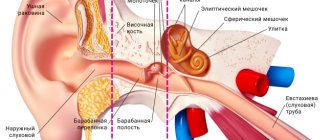
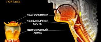
AUDITORY TUBE
[
tuba auditiva
(PNA),
tuba pharyngotympanica
(JNA),
tuba auditiva (Eustachii)
(BNA);
synonym: Eustachian tube, pharyngeal-tympanic tube, salpinx auditiva
] - a canal connecting the nasopharynx with the tympanic cavity; in humans it was first described in detail in 1564 by B. Eustachius. Since the term “Eustachian tube” (E.t.) has become widespread and traditionally established in clinical practice, it is given below.
In phylogenesis, the Eustachian tube (Eustachian tube) and the tympanic cavity (see Middle ear) develop in terrestrial vertebrates as a result of transformation of the cavity of the first visceral fissure. In ontogenesis, the Eustachian tube and tympanic cavity develop from the first pharyngeal pouch simultaneously with the development of the apparatus that perceives sound stimulation. During development, the first pharyngeal pouches grow laterally until the endoderm lining them is in contact with the ectoderm of the first branchial groove on each side of the body. The tympanic cavity develops from the distal part of the pouch, and the proximal part narrows and forms the Eustachian tube, which maintains communication with the pharyngeal cavity.
Anatomy
Eustachian tube - a paired canal 30-40 mm long; the diameter of its lumen is 1-2 mm. The axis of the Eustachian tube is inclined downward and inward, forming an angle of approx. 45° sagittal and approx. 30° with horizontal plane. The walls of the Eustachian tube are formed partly by bone, partly by cartilage and connective tissue, and therefore there are bone and cartilaginous parts in it.
The bony part of the Eustachian tube (pars ossea tubae auditivae) is approx. 1/3 of its length; It opens on the anterior (carotid) wall of the tympanic cavity with the tympanic opening of the Eustachian tube (ostium tympanicum tubae auditivae). It represents the lower part of the muscular-tubal canal of the temporal bone - the semicanal of the Eustachian tube (semicanalis tubae auditivae), located at the junction of the stony and squamous parts of the temporal bone. Medial to it is the carotid canal with the internal carotid artery passing through it (see Carotid arteries).
The cartilaginous part of E. t. (pars cartilaginea tubae auditivae) is approx. 2/3 of its length; it lies on the outer base of the skull in the groove at the posterior edge of the greater wing of the sphenoid bone, with its medial end approaching the base of the medial plate of the pterygoid process of the sphenoid bone. This part of the E. t. is formed by cartilage (cartilago tubae auditivae) and a connective tissue membranous plate (lamina membranacea). The cartilage has the appearance of a groove, facing downwards and laterally, and consists of medial and lateral plates (laminae med. et lat.). The edges of the groove are closed by a membranous plate. The cartilaginous part of the E. t. ends with the pharyngeal opening (ostium pha-ryngeum tubae auditivae), located on the side wall of the nasopharynx (see Pharynx). In the area of the opening, the medial plate forms a thickening in the form of a roller (torus tubarius). The transition point between the bony part and the cartilaginous part is called the isthmus of the Eustachian tube (isthmus tubae auditivae). The lumen of E. t. in the isthmus region is narrowed.
Three muscles of the soft palate originate from the walls of the E. t.: the muscle that strains the velum palatine (m. tensor veli palatini), the muscle that lifts the velum palatine (m. levator veli palatini), and the tubopharyngeus muscle (m. salpingopharyngeus). The muscle that strains the velum palatine lies anteriorly and laterally and separates the E. t. from the ear ganglion, the mandibular nerve and its branches, the chorda tympani and the middle meningeal artery. Part of the fibers of this muscle, starting from the lateral plate of the E. t. cartilage and the membranous plate of its wall, is often called the dilator of the Eustachian tube. From the lower part of the cartilage of the E. t., near its pharyngeal opening, the tubopharyngeal muscle begins, and from the medial plate of the cartilage and the membranous plate originates the muscle that lifts the velum palatine. The contraction of these muscles, which causes displacement of the cartilaginous and membranous plates of the walls of the E. t., affects the size of its lumen. During swallowing movements, the muscles open the E. t., and air freely penetrates into the tympanic cavity.
In a newborn (see) E. t. is shorter and wider than in an adult, and has the shape of a cylinder. The tympanic opening of E. t. in newborns is projected in the upper segment of the tympanic membrane, and in adults - in the inferoanterior segment. This projection and shape of E. t. in newborns contribute to easier penetration of infectious agents into the supratympanic cavity of the tympanic cavity.
The arteries of E. t. are branches of the ascending pharyngeal, middle meningeal arteries and the artery of the pterygoid canal. The veins accompany the arteries and flow into the venous pterygoid plexus. Lymphatic vessels are directed to the lateral retropharyngeal and deep cervical lymph. nodes. Innervation of E. t. is carried out due to the branches of the tympanic plexus and the pterygopalatine ganglion.
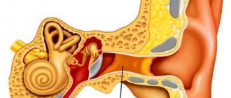
Where is the Eustachian tube located?
The Eustachian tube (auditory tube) connects the eardrum and the nasopharynx. It was first described by the scientist Bartolomeo Eustachio, which is why the canal was named in his honor. The auditory tube is S-shaped, length – 3-4 cm, diameter – 2 mm. As a person grows older, it increases.
The cavity of the auditory tube is covered with mucous epithelium, which becomes inflamed when pathogenic microorganisms penetrate from the nasopharynx into the eustachian opening. Air movement is disrupted even if it is slightly narrowed.
Histology
The mucous membrane of the wall of E. t. is a continuation of the mucous membrane of the tympanic cavity on one side and the nasopharynx on the other. The epithelium is lined with multirow ciliated epithelium containing goblet mucous cells. The movement of the cilia of the epithelium is directed towards the nasopharynx. On the surface of the epithelium, ducts of the mucous glands (gll. tubariae) open.
Near the pharyngeal opening of E. t. in the mucous membrane of the nasopharynx, an accumulation of lymphoid tissue is concentrated, forming the tubal tonsil - tonsilla tubaria (see Tonsils).
Treatment of acute eustachitis
When the Eustachian tube becomes inflamed, it requires complex therapy. The treatment regimen depends on the results obtained after the diagnosis.
The otolaryngologist takes into account the patient’s condition, the degree of development of the inflammatory process, where the pathological focus is located and selects the most effective means. Treatment is carried out using traditional means; in the absence of serious contraindications, folk remedies can be used.
Drug therapy helps relieve tissue swelling, eliminate allergic reactions and inflammation, and also restore the patency of the auditory tube.
Patients are prescribed the following medications:
| Group of drugs | Name | Application |
| Vasoconstrictor nasal agents | Sanorin, Tizin | Medicines help to quickly relieve swelling. Adult patients are prescribed 1-3 drops in each nasal passage 3-4 times a day. The course of treatment depends on the severity of symptoms, on average it lasts 3-7 days. |
| Antihistamines | Fenistil, Loratadine | The products reduce the allergic reaction. It is recommended to take medications internally. The adult dosage is 2 drops per 1 kg of body weight per day, it should be divided into 3 doses. |
| Nasal saline solutions | Humer, AquaMaris | Medicines clear the mucous membrane of the eardrum of accumulated mucus. Prevent infection from entering the ear cavity. Reduce swelling of the mucous membrane. Sprays are recommended to be used 4-8 times a day. Inject the product 2-3 times into each nostril. The course of treatment lasts 2-4 weeks. |
| Antiviral agents | Derinat, Nazoferon | The drugs increase the body's immunity and slow down the proliferation of pathogenic flora. They also reduce inflammation, swelling and the risk of complications. The medicine is used 3-6 times a day. It is recommended to drop 3-5 drops into each nasal passage. The duration of therapy is 1-2 weeks. |
| Antibacterial drugs | Azithromycin, Ampicillin | Medicines are prescribed to patients in case of infection of the Eustachian tube. The drug should be taken before meals or after meals 1 time per day. The recommended dosage for an adult patient is 0.5 g for 3 days. |
| Hormonal agents | Nasonex, Avamis | The therapeutic dosage is 2 inhalations into each nasal passage once a day. |
Treatment of acute eustachitis lasts on average 3-7 days. The chronic form requires more time for therapy. It is important to strictly adhere to the recommendations and prescriptions of your doctor to prevent serious complications.
Physiology
The Eustachian tube performs ventilation, drainage and protective functions. The ventilation function, or barofunction (see), is to maintain equal pressure on both sides of the eardrum (see). Under this condition, the vibrations of the chain of auditory ossicles reach their greatest strength, which ensures the best sound conduction. Changes in pressure in the tympanic cavity occur due to the constant resorption of gases from the middle ear into the tissue and due to pressure changes in the environment. When atmospheric pressure increases, air from the nasopharynx penetrates through the E. t. into the tympanic cavity, and when it decreases, on the contrary, from the middle ear into the nasopharynx. The main physiological mechanism, thanks to Krom, opens the lumen of the E. t. and air exchange takes place in the middle ear, is the act of swallowing (see). At the moment of swallowing, the muscle that strains the velum palatine contracts, while the membranous plate of the cartilaginous part of the E. t. is pulled back and its lumen opens. The opening and closing of E. t. is influenced by the pressure of surrounding tissues. Ventilation of the middle ear is ensured by reflex regulation of the lumen of the E. t., while the receptive field of reflexes is the tympanic plexus. If this reflex mechanism is disrupted, the E. t. can be completely impenetrable for air and does not provide ventilation of the tympanic cavity. If the barofunction of E. t. is impaired, there is a decrease in hearing and a decrease in the stability of the eardrum in relation to barotrauma (see). The drainage function of E. t. is aimed at removing transudate or exudate from the tympanic cavity. Fluid resorption by the middle ear mucosa plays a lesser role. The drainage function is ensured by the opening of the E. t. during the act of swallowing, the equalization of pressure in the middle ear, the flickering of the cilia of the epithelium of the mucous membrane of the E. t. towards the nasopharynx and the force of gravity of the fluid accumulated in the tympanic cavity. The protective function of E. t. is ensured by the bactericidal properties of its mucus secreted by the mucous glands.
Diagnostics
Diagnosis of the disease is not difficult for an experienced ENT doctor. The complaints that the patient describes are quite typical.
An appointment with a doctor begins with collecting an anamnesis of the patient’s life and health. The otolaryngologist asks the patient about the symptoms that are bothering him and the time of their onset.
This is followed by a direct examination of the ear cavity (otoscopy) and nose (rhinoscopy).
To determine the degree of hearing loss, an audiometric study is performed. To study the functions of the components of the middle ear, a tympanometric study is performed.
To determine the type of inflammatory agent and its sensitivity to antibiotics, secretions from the ear canal are sent for laboratory analysis. If the allergic nature of the disease is suspected, the patient is referred for allergy testing.
Research methods
Research methods are reduced mainly to determining the permeability of the E. t. for air. For this purpose, increased air pressure is created in the area of the pharyngeal opening of the E. t. and its passage into the tympanic cavity is recorded. The pressure is changed by the patient himself when performing a swallowing test (see Ear manometry) or in the Valsalva experience (see Valsalva experience) or is created artificially by blowing the ear with a Politzer balloon (see Blowing the ear). The research results are recorded by auscultation using an otoscope (see Otoscopy) or an ear manometer (see Ear manometry). In recent years, converting and recording devices have been used that allow for objective graphic recording of the ventilation function of the Eustachian tube.
Folk remedies
The Eustachian tube is located in a place where pathogenic microorganisms easily enter. Recipes from witch doctors and healers are used at home in addition to traditional medications, but after consultation with the attending physician.
It is necessary to exclude the possibility of an allergic reaction or individual sensitivity. Folk remedies include decoctions that should be taken orally or instilled into the ear, and compresses should also be made.
Effective non-traditional methods for damage to the Eustachian tube:
| Name | Recipe | Application |
| Herbal infusions | Mix chamomile, yarrow, lavender, St. John's wort and mint in equal parts. Pour 2 tbsp. herbal mixture with boiling water (1 tbsp), leave for 40-60 minutes and strain. | It is recommended to take the finished product orally, 1 tbsp. every day. |
| Aloe juice | Grind the plant and squeeze the juice through cheesecloth. Before use, it is recommended to dilute in equal parts with warm boiled water. | Aloe juice should be instilled into the nose, 5 drops every 4 hours. |
| Potato | Potatoes must be boiled without adding salt. | It is recommended to inhale the vapors of the evaporating liquid over the pan with your head covered with a towel. 10-15 minutes is enough. The procedures should be repeated every day. |
| Onion | Garlic and onion juice must be mixed in equal proportions. The resulting solution is used for instillation into the ears. | Patients are recommended to drop 3-4 drops into each ear 3-4 times a day, and close the ear canal with a cotton swab. |
| Garlic | Chop 3-4 cloves of garlic. Mix the resulting pulp with vegetable oil at room temperature (25 g). The resulting mass must be covered and left for 30 minutes. | The finished product should be used to lubricate the nose with a cotton swab 3 times a day. |
Boric alcohol has a bactericidal effect. It is recommended to put 3 drops into the ear canal every day and keep your head tilted to the side for 10 minutes.
Breath
- Standing, inhale deeply through your nose (nostrils flared and tense), the stomach protrudes, exhale slowly through the mouth, the stomach retracts.
- Standing, inhale deeply through your nose (nostrils flared and tense), stomach protrudes, hold your breath, then bend forward, arms relaxed down, exhale.
- Sitting, inhale deeply through your nose (nostrils flared and tense), exhale through your nose.
- Open your mouth wide - yawn. Then swallowing, i.e. closing the upper palate with the back of the tongue.
- Open your mouth wide, take a deep breath, close your mouth, swallow.
- Open your mouth wide, breathe deeply through your mouth.