Vinogradov D.D., Khromova M.R.
General characteristics of roundworms
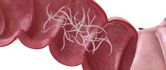
Nematodes, or roundworms themselves (Nematoda), are a type of protostomes, protocavity, bilaterally symmetrical moulting animals.
Spreading. Nematodes are one of the most widespread types of animals that have been able to colonize a variety of habitats - from the interstitium (the space between grains of sand) and moss communities to arctic ice (such as Theristis melnikovi and Cryonema crissum, found in the thickness of multi-year ice in central part of the Arctic Ocean). Parasitic nematodes are of particular interest to researchers, including due to the wide variety of their hosts.
Building plan. Thin fusiform body, tapering towards the ends, round in cross section. The mouth is located at the front end, and the powder (anus) is at the rear. The outside of the body is covered with a multilayer elastic cuticle - a non-cellular formation secreted by the hypodermis. The hypodermis, or epidermis, is located under the cuticle. The muscles are represented by a layer of longitudinal obliquely striated muscle fibers. The primary body cavity (schizocoel), devoid of its own epithelial lining, is filled with fluid.
Digestive system. The oral opening at the anterior end of the body is surrounded by protrusions - lips (usually three) and leads into a muscular ectodermal pharynx with a triangular lumen. The pharynx leads into the endodermal midgut from a single layer of columnar epithelial cells. Next comes a short ectodermal hindgut, which opens into the anus.
Excretory system. Excretory organs are unicellular glands that replaced protonephridia. There is usually one cervical gland in the anterior part of the body, from which a short excretory duct arises. There are also “storage kidneys” - phagocytic organs that accumulate insoluble metabolic products that are not removed from the body.
Circulatory and respiratory systems. These systems are missing. Breathing occurs through the skin. Anaerobic metabolism is also possible (anaerobic breakdown of glycogen to butyric and valeric acids in parasites).
Nervous system. The nervous system is of the scalariform type. Represented by a nerve ring and six longitudinal trunks. The two nerve trunks running along the ventral and dorsal lines are more powerful and are connected by semicircular nerve bridges (commissures).
Sense organs. There are papillae and setae - organs of touch located around the mouth. Some marine representatives have primitive eyes - pigment spots. Chemical sense organs, amphids, usually have the shape of a pocket, spiral or slit. They are located on the sides of the head end and are especially well developed in males, as they help in finding females.
Reproduction and development. Nematodes are dioecious animals. The internal genital organs are paired and have a tubular structure. Reproduction is only sexual. Sexual dimorphism is pronounced: females are larger, in males the posterior end of the body is curved. Fertilization is internal and viviparity occurs. In development, nematodes go through four larval stages, separated by molting, which are accompanied by shedding of the cuticle. The third stage in some species (including the famous Caenorhabditis elegans) under unfavorable conditions is modified into the so-called dauer stage - a resting larva.
Parasitism. Currently, of the more than 24,000 described species of nematodes, about half are parasitic. They can affect almost all tissues and organs: connective tissues, muscles, blood and lymphatic vessels, gonads, sensory organs, as well as the body cavity, etc. Among them there are both ecto- and endoparasites of plants, vertebrate and invertebrate animals, including other nematodes, and even protozoa.
The following are descriptions of the most significant representatives of roundworms from the point of view of medical parasitology.
Characteristics of pinworms
Today, all helminths can be divided into 3 classes. There are flatworms, tapeworms and roundworms. Pinworms belong to the class of nematodes (roundworms).
The disease in humans is caused by pinworms of the genus Enterobius vermicularis. Pinworms have a small, elongated body. The length of females reaches 10-12 mm, and males - 2-5 mm.
These organisms live in humans for 3-4 weeks, after which they die.
Pinworms live in the lower part of the small intestine, cecum and ileum. A special feature of these roundworms is the ability to actively move and exit the anus onto the human skin. In this case, the female lays several thousand eggs in the perianal area.
This happens at night when the anal sphincter relaxes. After laying, the pinworm dies. The second distinctive feature of these helminths is the high contagiousness of enterobiasis. Eggs can be transmitted from one person to another through close contact (a simple handshake).
This explains the outbreak of enterobiasis in children's institutions and adult groups.
The development cycle of pinworms is quite simple. Human infection occurs through contact, food, household or through self-infection. Eggs from dirty hands or food enter the human intestines.
Soon the eggs hatch into larvae that attach to the intestinal wall. After a month they can lay eggs.
After laying eggs, a person experiences itching, scratches the skin, and the eggs are brought back into the oral cavity with their hands.
Enterobiasis ranks 1st among all helminthiases in terms of prevalence. The main source of infection is a sick person. He is also the definitive host of pinworms.
Factors in the transmission of eggs can be food, various toys and household items, and contaminated hands. Eggs can be inhaled along with dust, but this mechanism of transmission is of minor importance.
With the development of enterobiasis, the following symptoms may be observed:
- itching in the anal area;
- nausea;
- vomit;
- loss of appetite;
- bowel dysfunction;
- pain in the abdominal area;
- rash;
- difficulty urinating (infantile enuresis);
- development of inflammatory diseases of the genital organs (in girls);
- fatigue;
- irritability.
The incubation period from the moment of infection is 3-6 weeks. Complications of this disease include purulent inflammation of the skin, paraproctitis, vulvovaginitis, enterocolitis, acute appendicitis, peritonitis, salpingitis.
Trichinella (Trichinella spiralis)
Description. Small nematode 2-4 mm long (Fig. 5). Parasitizes the mucous membrane of the small intestine. Distributed in Eurasia and North America.
Rice. 5. Trichinella
Life cycle. For the development of Trichinella, a change of hosts is necessary. Usually these are wild animals (foxes, wolves, bears, wild boars), as well as people and livestock. Females are anchored by the anterior end of the body into the intestinal epithelium and give birth to 1-2 thousand larvae. Ovoviviparity is typical: the hatching of larvae from eggs occurs in the female genital tract. The larvae are carried throughout the body through the blood and lymphatic vessels and settle in the striated muscles. At this stage, they have a stylet, they use it to destroy muscle tissue, causing the host to form a capsule in which, curled up, they reside in the future. After a few months, the capsule is soaked in lime. Such muscle trichina can exist for several years and survive even after the death of the owner and the decomposition of his corpse.
Once in the stomach of the new host (after it has eaten the corpse of the previous one), the larvae are freed from the capsule (Fig. 6), penetrate the mucous membrane and within a couple of days, having undergone four molts, turn into adult worms.
Rice. 6. Development of Trichinella in the human body
Clinical picture of trichinosis. Increased temperature, puffiness of the face, muscle pain, allergic reactions.
Prevention. Trichinosis is transmitted by food through contaminated meat. Therefore, to prevent the disease, meat must undergo a veterinary examination and be properly prepared - boiled for 2-3 hours. Cooking methods such as smoking and salting do not destroy Trichinella.
Human roundworm
Another prominent representative of roundworms is the roundworm. These helminths are large in size. Females can reach a length of 40 cm, males - up to 25 cm. Most often, parasites live in the small intestine. Roundworms are classified as geohelminths.
This means that their development cycle does not require intermediate hosts. A sick person excretes ascaris eggs along with feces, which then fall into the soil. This is where the larvae develop. The soil has optimal temperature and humidity for this.
It takes about 2 weeks for the eggs to become infective.
Human infection occurs through the fecal-oral mechanism (through food, water and dirty hands). In the stomach, the shells of the eggs are destroyed and the larvae emerge.
They live in the intestines, often leading to injury and obstruction. Sometimes the larvae are carried through the bloodstream to various organs (heart, lungs, brain, sinuses).
It is important that the development of larvae does not necessarily have to take place in the soil. Autoinvasion (self-infection) often occurs.
Bancroft's string (Wuchereria bancrofti)
Appearance . White thread nematode, females 10 cm long, males 4 cm long (Fig. 12).
Rice. 12. Bancroft's filaria
Spreading. Tropics, subtropics of Asia, Africa, Central and South America.
Life cycle. Adults usually occur in the lymph glands and vessels, obstructing the drainage of lymph and causing persistent swelling. Females produce larvae - nocturnal microfilariae, which appear in the peripheral blood at night, and during the day go deep into the body (into the pulmonary vessels and kidneys). This is due to the fact that the intermediate host is mosquitoes, which usually suck blood in the evening and at night. The larvae enter the stomach of the mosquito, then into the body cavity, where they grow, after which they accumulate near the proboscis, from which they are transmitted to humans by sucking blood. Bancroft's filaments cause elephantiasis, or elephantiasis, or elephantiasis. It is worth noting that this disease can also be caused by other nematodes.
Clinical picture and treatment of elephantiasis. An enlargement of any part of the body occurs (Fig. 13) due to hyperplasia (painful growth) of the skin and subcutaneous tissue, which is caused by inflammatory thickening of the walls of the lymphatic vessels and stagnation of lymph, which occurs due to clogging of the lymphatic vessels by adult Bancroft's filamentosa. The skin on the diseased part of the body becomes covered with ulcers.
Treatment of elephantiasis is aimed at improving fluid outflow. The use of anthelmintic drugs such as avermectin is effective. In later stages, surgery may be required.
Rice. 13. A patient suffering from elephantiasis (according to Brunt)
Manifestations of ascariasis
The development cycle of these worms is about 3 months. This period is the incubation period of the disease. Clinical symptoms of ascariasis are varied. They include:
- the appearance of a rash;
- skin itching;
- slight increase in body temperature;
- decreased appetite;
- nausea;
- flatulence;
- abdominal pain;
- hypersalivation;
- drowsiness;
- weakness;
- irritability.
In severe cases of ascariasis, signs of intestinal obstruction are possible: pain, bloating, constipation. In some cases, adult helminths can be excreted in the feces. Less commonly, ascariasis affects other organs. If roundworms are localized in the eye, oculomotor disorders and hemorrhages are formed. With pulmonary localization, suffocation may occur.
Clinic.
The incubation period is 2-4 weeks.
The symptomatology of enterobiasis is varied and is determined by the intensity of the invasion and the individual reaction of the host. The main symptoms are: perianal itching, burning (sometimes in the perineum) (due to irritation by parasites) usually at night, local skin irritation. Possible sleep disturbances, weakness, headaches, decreased ability to work, and fatigue. Loss of appetite and weight loss (more common in children). Nausea, abdominal pain (sometimes acute - pinworms can penetrate deeply into tissues, penetrating into the abdominal cavity and internal organs), diarrhea with copious mucus and sometimes blood.
- Based on the nature of the flow, the following forms can be distinguished:
- Asymptomatic - in most cases.
- The clinically pronounced course, in turn, is distinguished:
- subclinical course (with low intensity of invasion) - usually itching in the rectum, in the anus, less often the vulva in the evening and at the beginning of the night, sometimes causing short-term insomnia. Itching is determined by the characteristic frequency of pinworms: 1-2 or more days in a row, then completely disappears for 2-3 weeks.
- intestinal form (with more intense infestation) - crawling out of parasites (with corresponding symptoms) is more often observed not only in the evening, but also during the day, and free spaces - in short, sometimes completely disappear. Abdominal pain appears - it can be vague, diffuse, or localized in some parts of the intestine (usually in the area of the cecum and in the appendix), often with palpation pain in these areas. Copious laxative stools with mucus, less often with blood. These symptoms accompany or precede the abundant release of parasites. Associated symptoms may include nausea, loss of appetite, headaches, and dizziness.
- nervous form - the disease occurs with pronounced damage to the nervous system (intestinal phenomena can be completely obscured) - painful, sometimes unbearable itching (can last around the clock, intensifying towards night) leads to severe insomnia, decrease and loss of memory, and a sharp decline in working capacity. Children often become capricious, absent-minded, disobedient, and school performance decreases. Depletion of the nervous system leads to neurasthenia, sometimes even to obsessive thoughts of suicide (with a long course of the disease). This form often occurs in adults, sometimes in elderly people, more often in men.
- enterobiasis vulvovaginitis - is often the main and only manifestation; they can creep into the vulva in large quantities, causing disruption of the mucous membrane, toxic irritation, introducing intestinal microflora from the rectum. Scratching caused by itching increases irritation and inflammation; hymen rupture is occasionally observed; irritation from pinworms can lead to masturbation. Often, with severe vulvovaginitis, the complaint of perianal itching fades into the background and is identified only with appropriate questioning from the doctor; such patients can be observed for a long time by venereologists. In the literature there are indications of the detection of pinworms (or their eggs) in women at all stages of migration (up to and including the abdominal cavity).
- skin form - as a result of constant scratching, cracks (sometimes deep and painful) may appear; the introduction of microflora and constant mechanical and toxic irritation of these cracks by pinworms contribute to the appearance of rashes, abscesses, and eczema (often weeping). Skin lesions, starting in the anus and perineum, gradually spread to the genitals, thighs and even the lower abdomen.
- enuresis - more often in children.
Treatment and prevention of ascariasis and enterobiasis
Treatment of ascariasis involves taking anthelmintic drugs. These include tablets Albendazole, Vermox, Levamisole. Therapy for ascariasis includes following a diet (table No. 13 is prescribed), taking enzyme preparations, probiotics and prebiotics, vitamins, and antihistamines.
Compliance with the rules of personal hygiene (regular washing of hands and body, washing, washing and ironing clothes, cutting nails, wearing thick underwear, changing bed and underwear) and wet treatment of household items is of great importance. Preventive measures against ascariasis and enterobiasis include thoroughly washing fruits and vegetables, limiting contact with infected people, and boiling water.
Thus, pinworms and roundworms are the most significant helminths from an epidemiological perspective.
List of sources
- Medical parasitology: textbook / G.I. Myandina, E.V. Tarasenko. M: Practical medicine. 2013. 256 p.
- Avdyukhina T.I., Konstantinova T.N. et al. Enterobiasis. Clinic, diagnosis, treatment, epidemiology, prevention: Textbook. manual for doctors. M., 2003. 56 p.
- Epidemiological characteristics and basics of prevention of contact helminthiases: educational and methodological. allowance / I.N. Valchuk, T.E. Doroshenkov, G.N. Chistenko. – Minsk: BSMU, 2017. –40 s.
- Shreiner E.V. Helminth infections in pediatric clinical practice: issues of diagnosis, therapy, prevention. Rus. honey. magazine 2013.
- Bronshtein A.M., Malyshev N.A., Luchshev V.I., Davydova I.V. Social and epidemiological problems and basic issues of pathology and chemotherapy of helminth infections of the digestive organs. Ross. honey. magazine 2007; 2:33–6.
Differences between roundworms and pinworms
Most often, these parasites have the same method of infecting the human body. If you do not wash your hands after using the toilet, after going outside, or before eating food, then you are likely to introduce parasites into your body. But there are some differences in infection with worms, since roundworms are quite tenacious and can be found on different objects and in different places. For example, in raw, untreated water or on objects in an unsanitary environment. They remain on fruits and vegetables if they are not washed properly. It is also worth remembering that worm larvae can be introduced through food if it is poorly processed or has not undergone heat treatment at all.
It is not always possible to become infected with helminths from another person, since the larvae can remain on underwear or bedding. Therefore, we must not forget that simply taking a course against worms is not enough. It is definitely recommended to change your underwear and bed linen. In addition, eggs can be carried by insects that come into contact with food, such as flies or cockroaches. Moreover, eggs can be inhaled into the lungs after inhaling dust.
Ways of infection with ascariasis
The main difference in infection mechanisms is that the eggs mature in different environments. For pinworms, these are the folds of the perianal zone of an infected person. From here they enter the mouth and then the intestines of the same person. Or into another person's mouth and intestines.
The method of infection by roundworms is determined by the fact that in order for their eggs to mature, they need to lie in the soil. Leaving the human intestines along with feces, the embryos of the parasite end up in the ground. After lying there at a certain temperature (1336 C) and humidity (not lower than 4%), they become ready for invasion. The optimal temperature for eggs is 24 C, at which maturation occurs in two weeks.
This method of infection is called fecal-oral. Eggs are capable of infecting humans only after they have remained and matured in the ground.
The principles of infection with roundworms in humans can be different.
- When eating thermally unprocessed food.
- When eating poorly washed fruits and vegetables.
- Drinking raw, unboiled water from open reservoirs and water pipes.
- When swimming in open waters.
- From pets that have parasite germs on their fur.
- From flies and other insects. Crawling on uncovered food, they infect it.
- Upon contact with things and interior elements that could somehow get worm eggs.
Contributes to parasite infection:
- working with soil in the garden and vegetable garden, especially if feces are used to fertilize them;
- improperly constructed cesspools in private farmsteads.
Similarities
The main similarity between these parasites lies in their structure. Like pinworms, roundworms are also roundworms and nematodes. They have the same light color. There are also other similarities:
- Both types of parasites are localized in the intestines and affect other organs to a much lesser extent;
- Both parasites have the same transmission pattern - exclusively fecal-oral, that is, for the development of invasion, you need to ingest a certain number of parasite eggs;
- The transmission routes are the same. These are unwashed vegetables and fruits, raw meat, contaminated water, contact with pets or contaminated surfaces;
- They have some similarities in symptoms. Pinworms and roundworms cause mainly intestinal symptoms, intoxication and an allergic reaction;
- There are similarities in the parasite's life cycle. In particular, the peculiarity is that the invasion develops with constant repeated self-infection;
- In both species, males are much smaller than females;
- Both species are present in the intestines in entire colonies.
It is important to take into account that mixed invasions are currently widespread, when several types of parasites are present in the intestines at the same time. Roundworms and pinworms may well exist together in the body, causing the characteristic symptoms of both invasions.
Diagnostics.
Diagnosis of enterobiasis presents certain difficulties. Requires persistence and an integrated approach:
To monitor the effectiveness of treatment, scrapings are carried out 3 times every 2 weeks at weekly intervals.