Causes of stones in the ureters
The main and most common cause of stones in the ureter is considered to be stones that have descended into this organ from the kidneys. More often it is only one stone stuck in the excretory duct - a very narrow tube up to 30 cm long. But there are cases when there are several stones. For a stone to become stuck, it must be larger than 2mm. Causes of stones in the ureter that have entered it from the kidneys:
- disorders of salt and purine metabolism;
- pyelonephritis;
- urethritis;
- prostatitis;
- cystitis;
- chronic gastrointestinal diseases;
- hereditary predisposition;
- drinking hard drinking water.
If stones descend into the ureter from the kidneys, they are called secondary. Such stones are more common than primary ones, which form in the duct itself due to the following problems:
- ectopia or tumor of the ureter;
- strictures;
- foreign bodies;
- ureterocele.
The causes of stones in the ureter in men are the same as in women. The difference may be prostatitis. This condition occurs only in men and can cause stones. In some cases, the formation of a calculus in the duct is associated with a grain of sand that moved and caught on the mucous membrane of the organ. Salts build up on it and over time a stone forms.
Symptoms of stones in the ureter
The danger of ureterolithiasis is that stones in the ureters, compared to other locations (in the bladder, urethra, kidneys), cause more severe and serious complications. They are also accompanied by pronounced symptoms. The main symptoms of stones in the ureters:
- Pain in the lower abdomen - when the stone is located in the lower part of the ureter.
- Dull pain covering the entire abdomen - when a stone forms in the upper part of the duct.
- Pain radiating to the pubic area - when a stone forms in a distant part of the ureter, which is located in the bladder.
Additionally, the following may appear:
- elevated temperature;
- general malaise;
- problems with stool;
- increased tone of the abdominal wall;
- increased gas formation.
The stage of self-removal of stones from the ureter is the most difficult, since the duct is very narrow. Only loose formations can pass through it. But even they cause severe pain.
A characteristic symptom if a stone is stuck in the ureter is very sharp pain in the lower abdomen. It is especially strong when the urge to urinate appears. If a headache, chills and weakness appear, this may indicate urinary retention and intoxication of the body. In this case, you should immediately consult a doctor.
Ureteral and bladder stones - find and destroy
Kryshko Denis Konstantinovich
Urologist
April 26, 2021
Urolithiasis (UCD) is a striking example of how urology has made a powerful leap in its development over the past decades. Today, classical operations for urinary tract stones are becoming a thing of the past. Patients no longer have to make incisions to remove stones. In most cases, operations are performed using the endourological method, when through natural openings it is possible to reach the stone, see it and, depending on its size, structure, location, either remove it or crush it into the smallest particles, including using a laser. This became possible thanks to the widespread introduction into practice of modern high-tech methods for the treatment of urolithiasis.
Denis Konstantinovich Kryshko, Candidate of Medical Sciences, Assistant of the Department of Urology of St. Petersburg State Pediatric Medical University, talks about the features of this disease, methods of diagnosis and treatment.
— How common is urolithiasis, how often do you make this diagnosis at appointments?
Urolithiasis (UCD) is a disease that occurs quite often. Stones (or, as it is correct to say, calculi) of the urinary tract are the second most common urological problem after infectious and inflammatory diseases. In Russia, ICD is diagnosed in 32-40% of cases of all urological diseases. Stones are found in at least 1-3% of people of active age (20-50 years). Urinary tract stones can be detected at any age, but more often this happens in working age. In childhood and old age, cases of primary detection are quite rare. Men get sick 3 times more often than women.
— What types of ICD are there?
Typically, stones form in the kidneys, from where they descend into the bladder and ureters; these are the so-called primary stones. The least difficult problems are caused by ureteral stones; in almost any part of it, a stone can be seen using ureteroscopy and, using a special tool, pulled out or crushed (evaporated) with a laser.
Secondary stones formed in the bladder are much less common, mainly as a result of adenoma and stagnation of urine. There are also ligature stones, which are formed as a result of the deposition of salts on ligatures (threads) after previous operations on the urinary tract. But this is a topic for another discussion.
In most cases, stones form in one of the kidneys, and in 9-17% of cases, urolithiasis is bilateral. Kidney stones can be single or multiple (up to 5000 stones). The size of the stones is very different - from 1 mm, to giant ones - more than 10 cm and weighing up to 1000 g, but such extreme figures, of course, are very rare.
— What are the manifestations of urolithiasis?
This is classic renal colic: severe pain, nausea, difficulty urinating, urinary retention or, conversely, frequent urination, pain, false urges. Pain can be in the lower back and lower abdomen and radiate to the leg and groin area. Taking painkiller pills can reduce symptoms for a while, but, firstly, they blur the symptoms and complicate diagnosis, and secondly, they lengthen the time before seeing a doctor. You should contact a urologist immediately if these symptoms occur, since delay can lead to unwanted infectious (purulent) complications.
— What contributes to the formation of stones?
Unfortunately, there is no clear answer to this question, and therefore it is almost impossible to guarantee the occurrence of stones. Urolithiasis occurs from a combination of several factors. According to modern ideas, this disease is most associated with enzymopathies - disturbances in the functioning of the enzyme system that controls the exchange of salts from which stones are formed.
But heredity, characteristics of traditional cuisine, lifestyle, and environment also play a role. For example, it has been statistically proven that residents of the Caucasus, Central Asia, the Volga region, and northern regions suffer from this problem more often. This is due to the characteristics of the diet, water hardness, intensity of solar insolation, etc. The onset of the process can be triggered by a sharp change in the usual diet, accompanied by a disturbance in the outflow of urine, when salts begin to precipitate and stones form.
— Does urolithiasis always end with surgery to remove the stone, or is conservative treatment possible?
You can get rid of some stones conservatively, prescribe drug therapy and wait to see if the stone comes out on its own, but this is associated with prolonged and severe pain, limited ability to work and activity. I have had patients who compared the intensity of pain from a stone passing naturally with labor pains. In addition, during this period the kidney, urinary tract, and nerve endings suffer.
Therefore, it is better not to allow such a state to occur; if a stone has discovered itself, there is no need to wait and “grow” it. It is easier and safer to remove it while it is small, with the help of a urologist and modern equipment - gently, less traumatic, with anesthesia. Some stones, the so-called urate stones, can be dissolved, but the best effect in this case is also achieved by combined therapy - crushing and dissolving.
Once a stone has formed, it cannot dissolve on its own. Stories in which stones were first discovered and then disappeared are mainly due to inaccuracy of diagnosis.
— What diagnostics are carried out for urolithiasis?
It all starts with an ultrasound; this is a quick, inexpensive and painless way to determine whether there is a stone or not and make a preliminary diagnosis. But ultrasound is not a 100% method, even with a qualified specialist using good equipment, not always what looks like a stone turns out to be really a stone. Therefore, a diagnosis previously made by ultrasound requires intravenous urography (x-ray with contrast) or computed tomography for confirmation. Having confirmed the presence of a stone, determining its position and size, a decision is made on how to deal with it.
Not often, but there are situations when all the symptoms indicate urolithiasis, but during the examination the stone is not detected. In this case, diagnostic ureteroscopy may be performed. As the ureter passes along its entire length, if there is a stone, it is detected and removed.
— What methods of surgical treatment are currently used?
Nowadays, endourological methods (without incisions, through natural openings) are used to remove stones from urolithiasis. If the stone is not very large and can be grasped and pulled out through physiological constrictions without damaging the structure of the ureter, this is the most favorable condition for the procedure. If the stone is larger, starting from 4-5 mm in diameter, then it is easier to crush and remove it without risking damage to the internal structures of the ureter. This is done using a urological microsurgical instrumentation unit. This method is called contact lithotripsy and is considered the “gold standard” for solving such problems.
In most clinics in the city, performing this manipulation is associated with hospitalization, loss of time, and additional costs for paying for bed days. In our center, the qualifications of specialists, surgical equipment and conditions for anesthesia allow us to remove urinary tract stones without hospitalization, in one day. To carry out contact lithotripsy, as this procedure is called, a powerful holmium laser of the latest generation is used, which allows you to instantly fragment the stone “into dust”, with virtually no trauma to the surrounding tissue.
There is a remote method of sonic crushing of stones. It used to be widely used, but is now fading into the background. This is due to the advent of new technology, and the fact that not all stones can be crushed in this way, there are “dead zones” where it is impossible to crush with infrasound, several procedures may be required and this is not harmless to neighboring internal organs, especially the kidney. The endourological method has no such limitations; removal can be carried out throughout the ureter and in the bladder, and a flexible urethoroscope allows stones to be removed even in the kidney.
Minimally invasive surgical operations, with access to the kidney through a puncture in the lumbar region, are performed for very large kidney stones. If the stone is 3 cm or larger, then using a urethroscope or infrasound it will not be possible to remove it. These operations are performed only in a hospital.
— What characteristics must a laser meet to perform contact lithotripsy?
The class of equipment for lithotripsy matters. Power, frequency, and the presence of a pseudo-pulse mode are important in order to very quickly turn the stone into the smallest particles. If the power is not enough, there is a risk that the stone will begin to move from the impact, damaging surrounding tissue and going deeper. With modern equipment, one discharge is enough to turn the stone into dust. The process is well controlled, which allows it not to heat or injure the mucous membrane.
— How is the postoperative period going?
The period of incapacity lasts on average from 2-3 days to a week. This is due to the fact that to prevent complications, the kidney must be drained. Swelling may occur in the place where the stone was located, therefore, in order to prevent stagnation of urine and the development of an infectious process, a ureteral stent is installed (a special medical tube to ensure the outflow of urine, which is then removed) and a course of antibiotics is prescribed.
— What is the probability of recurrence of urolithiasis and the appearance of new stones?
Since urolithiasis is associated with metabolic disorders, and by removing the stone, we do not influence the causes that caused its appearance, relapses are possible. At the present stage, medical science has not come up with a way to influence these enzyme disorders. Following your urologist's recommendations can reduce the risk of new stones and attacks of renal colic. Recommendations include following a diet, drinking regimen, regular monitoring by a urologist, performing an ultrasound and urine test at least once a year, and preferably once every six months. If a urine test reveals any deviations from the norm, a consultation with a urologist is required, since at an early stage, for example, when microcrystals of salts are detected in the urine, these processes can be influenced and slowed down by prescribing medications, herbal medicine, and diet correction.
There is a type of stones, the occurrence of which is associated with chronic infectious processes, the so-called struvite stones. To prevent their occurrence, you need to consult a doctor in time and treat gynecological and urological infections, urinary tract infections, avoiding a chronic inflammatory process.
In conclusion, I would like to note that, of course, the best operation is the one that was avoided. But, if a stone has formed and its removal is inevitable, the advent of modern technologies and equipment makes it possible to do this as quickly as possible, with minimal trauma and even without the need to go to the hospital. Those problems and pathologies that previously led to serious surgery can today be solved in our Outpatient Surgery Center in a few hours.
The most effective treatments
A specific method for removing stones from the ureter is chosen taking into account several important factors:
- size of stones;
- their quantities;
- location;
- time spent in the ureter;
- general condition of the patient.
If the size is small, they resort to conservative expectant treatment of stones in the ureter. It consists of prescribing antispasmodics, urolitics, antibiotics and a water load of more than 2 liters per day. In such situations, stones can be removed naturally.
In other cases, gentle methods of removing stones from the ureter are practiced, including:
- Lithotripsy – crushing stones using laser or ultrasound: percutaneous contact or remote ureterolithotripsy.
- Crushing stones through the urethra using special solutions.
For stones larger than 1 cm, open or laparoscopic ureterolithotomy is used. This operation is also indicated for severe renal colic, non-advancing stone and blockage of the only kidney.
At the Urology Clinic named after R. M. Fronshtein of the First Moscow State Medical University named after I. M. Sechenov, they resort to minimally invasive methods for treating stones in the ureters. But since they are most effective for stones that are not too large, the patient is required to make an appointment with a urologist as soon as possible. In this case, there is every chance to do without serious intervention.
On weekdays you can get an appointment with a urologist on the day of your visit
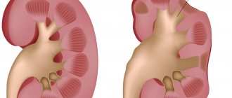
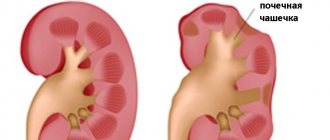
coral stone
A coral stone is a stone of the renal pelvis that has at least one spur into the calyx, or a stone that completely fills the renal pelvis. An incomplete staghorn stone fills only part of the kidney, a complete staghorn stone completely fills the renal calyces and renal pelvis.
Drawing. Coral stone of the right kidney.
The main method of treating coral stones is percutaneous endoscopic removal (percutaneous nephrolitholapaxy or percutaneous nephrolithotripsy).
Hakobyan Gagik Nersesovich – professor, doctor of medical sciences, oncologist, urologist in Moscow
The appointment is conducted by a doctor of the highest category, urologist, oncologist, doctor of medical sciences, professor. Author of more than 100 scientific papers.
Urological oncology experience – more than 15 years. Helps men and women solve urological and oncourological problems.
Conducts diagnostics, treatment and complex operations for such diagnoses as:
- tumors of the kidneys and upper urinary tract;
- prostate and bladder cancer;
- urolithiasis disease;
- BPH;
- hydronephrosis, ureteral stricture, etc.
During your consultation, the urologist will answer all your questions in detail.
If you are bothered by difficulty or frequent urination, pain in the lumbar region, blood in the urine, as well as other symptoms (read about what else should alert you here), seek help from a urologist.
Admission includes:
- familiarization of the doctor with the patient’s medical history;
- inspection;
- making a preliminary diagnosis, prescribing tests and necessary procedures.
*If you plan to get tested immediately after your appointment with your doctor, go to the clinic with a full bladder.
Do not delay your visit to the clinic - come for a consultation with a urologist at the State Urology Center in Moscow - the R. M. Fronshtein Urology Clinic of the First Moscow State Medical University named after I.M. Sechenov. Entrust your health to a competent specialist!
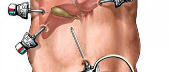
Target
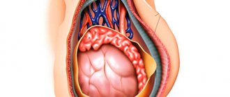
The goal of PCNL is to remove kidney stones to reduce pain, bleeding or urinary tract obstruction, and/or urinary tract infection. Kidney stones range in size from microscopic crystals to huge stones up to several centimeters (huge kidney stones occupy all the kidney cavities and begin to resemble corals - hence the term coral stone). Standard percutaneous nephrolithotomy is performed under general anesthesia and takes about 40 minutes - 4 hours. The surgeon makes a small incision, approximately 0.5 - 1.3 cm in length, in the patient's lumbar region. In percutaneous nephrolithotomy, the surgeon inserts a needle through the patient's skin directly into the kidneys using fluoroscopy and ultrasound guidance. The puncture tract is expanded and a nephroscope is installed in the kidney - an instrument that allows optical visualization of the renal cavity system. An ultrasound or laser probe is delivered through the nephroscope to crush large kidney stones. Pieces of stones are removed with instruments, and at the end a nephrostomy tube is installed to drain the kidney on the first day after surgery. In some cases, a ureteral stent is installed.
Drawing. Positioning the patient. Drawing. Creation of access to the renal cavity system.
Drawing. Nephroscopy.