doctor
Most often, women of reproductive age face the problem of inflammation of the organs of the reproductive system.
Since unprotected sexual intercourse has been considered the main method of transmission for a long time, inflammation occurs mainly in the sexually active part of the female population. The average age is 20 - 40 years.
It should be noted that the risk group for inflammation is occupied by girls and women with more than 3 sexual partners, in which case the frequency of development of pathology increases several times. The most common inflammations include vaginitis, cervicitis, endometritis, cervical erosion and, less commonly, adnexitis.
Inflammatory processes such as bartholinitis are quite rare. Very often, inflammation is associated with the presence of a sexually transmitted infection. When diagnosing and the presence of pathology, one should not forget about this type of lesion.
In severe cases, symptoms may be acute, with very high fever, severe abdominal pain and vomiting. Sometimes the symptoms can be mild and barely noticeable. If they appear, you need to contact a gynecologist without delaying the visit.
Women's diseases: types and symptoms
Gynecological diseases are conventionally divided into three large groups:
- Inflammatory diseases;
- Hormone-dependent pathologies;
- Diseases of hyperplastic, dystrophic and tumor nature.
Among the gynecological diseases of an inflammatory nature, the following are distinguished:
- Purulent-inflammatory Vulvitis, colpitis, endometritis, adnexitis, pelvioperitonitis, etc.;
- Sexually transmitted diseases Gonorrhea, trichomoniasis, chlamydia, ureaplasmosis, candidiasis;
- Viral Genital herpes, papillomavirus infections, cytomegalovirus infection, HIV infection.
Disturbances in the body's endocrine system lead to pathologies of puberty (delayed or premature sexual development, sexual hermaphroditism, menstrual irregularities, abnormal development of the genital organs). This also includes diseases such as pathologies of the menstrual cycle (premenstrual syndrome, amenorrhea, algomenorrhea), ovarian dysfunction, lack of ovulation, dysfunctional uterine bleeding, etc.
The third group includes such serious diseases as benign and malignant neoplasms in the genital organs, fibroids, hyperplastic and dystrophic changes in the cervix, in particular erosions and pseudo-erosions.
Diagnostics
Diagnostic measures are carried out by a gynecologist. If necessary, a sexologist, psychologist, oncologist, and other specialists are involved in the examination. During the interview, the doctor examines the obstetric and gynecological history, finds out how long ago and under what circumstances pain in the vagina first appeared, how the symptom changed over time, and what manifestations it was combined with. To clarify the nature of the pathology, the following methods are used:
- Gynecological examination.
It is possible to detect inflammation, space-occupying processes, prolapse of the internal genital organs, developmental anomalies, traumatic injuries, and cicatricial deformities. Sometimes a rectal-abdominal or rectal-vaginal examination is performed. - Colposcopy.
The doctor examines the vagina and cervix under a microscope, detects defects in the mucous membrane, and examines space-occupying formations. According to indications, performs a targeted biopsy for subsequent morphological analysis of a tissue sample. - Ultrasonography.
During a combined ultrasound, a comprehensive picture of the state of the pelvic structures is obtained, developmental defects, post-traumatic changes, and adhesions are determined. If there are signs of varicose veins, an additional ultrasound examination of the vessels is performed. If pathology of neighboring organs is suspected, an ultrasound of the rectum, urethra, and bladder is performed. - Lab tests.
A smear examination helps clarify the composition of the microflora in vaginitis. To determine the pathogen and its sensitivity to antibiotics, culture is carried out on nutrient media. For STIs, PCR tests are used. Biopsies are studied during histological or cytological examination. - Other methods.
To exclude damage to neighboring organs, rectoscopy, cystoscopy, urethroscopy, ultrasound of the kidneys and ureters, consultation with a proctologist or urologist may be required.
Symptoms of gynecological diseases
All gynecological diseases are accompanied by specific symptoms , the most characteristic of which are:
- pathological secretion;
- disturbances of menstrual and sexual functions (for example, pain during sexual intercourse, most often caused by the inflammatory process of the uterine appendages);
- reproductive dysfunction (for example, spontaneous miscarriages), infertility.
Menstrual dysfunction can manifest itself in a variety of forms: hypo- or hypermenstrual syndrome, amenorrhea, menorrhagia, algomenorrhea, anovulation (lack of ovulation).
Causes of gynecological diseases
The occurrence of gynecological diseases is promoted by:
- failure to comply with the rules of personal hygiene and sexual hygiene;
- alcoholism, drug addiction;
- active sex life with different sexual partners;
- use of irrational contraceptives;
- douching with various chemicals;
- temperature effects on the vaginal mucosa;
- sexual activity during menstruation;
- frequent abortions;
- endocrine, congenital, infectious diseases, especially in childhood;
- diseases of the urinary system, intestines;
- metabolic disorders;
- stress;
- eating disorders.
The consequences of early onset of sexual activity are especially unfavorable when defense mechanisms are not fully formed.
Types of inflammation
There are such types of vaginitis: specific and nonspecific, acute and chronic.
Nonspecific vaginitis
Nonspecific inflammation of the vaginal mucosa is provoked by conditionally pathogenic microflora. Microorganisms that are part of the normal flora begin to multiply under unfavorable conditions. The catalyst for the development of the pathological process are the following factors:
- failure to maintain intimate hygiene;
- frequent change of sexual partners;
- douching with antiseptic solutions;
- use of certain contraceptives;
- tendency to allergies;
- pregnancy;
- stress;
- viral infections that cause weakened immunity;
- overwork;
- trauma to the vaginal mucosa.
Nonspecific vaginitis can be bacterial and candidal. The most common pathogens of bacterial infection are staphylococcus, streptococcus, gardnerella, and E. coli.
Specific vaginitis
Its development is caused by sexually transmitted infections. Such diseases include trichomoniasis, gonorrhea, mycoplasmosis, ureaplasmosis, chlamydia.
Nonspecific inflammation of the vagina is treated with antibiotics; if a disease is detected, it is necessary to examine and treat the second partner.
Atrophic colpitis
This type of inflammation of the vaginal mucosa, which occurs in women during menopause, occurs due to hormonal dysfunction. In addition, this disease can be observed in patients of childbearing age who have had their ovaries removed, as well as while taking medications that affect estrogen production.
Atrophic colpitis is diagnosed in most women 3-4 years after the onset of menopause. It manifests itself as a feeling of dryness and discomfort in the vagina. Due to the lack of female hormones, its mucous membrane becomes thinner, this becomes a favorable factor for the development of opportunistic flora.
Prevention of gynecological diseases
Prevention of gynecological diseases includes:
- protecting the health of girls from the period of intrauterine development;
- timely treatment of infectious and other diseases;
- timely treatment of the consequences of birth injuries;
- healthy lifestyle, giving up bad habits, hardening;
- compliance with the rules of general hygiene and genital hygiene.
In gynecology, there are several mandatory principles for effective work for the benefit of women. Timely consultation in gynecology is necessary to avoid serious problems in the future, because female diseases, like all others, are treatable the better the earlier they are identified.
Diseases of the vulva: from inflammation to cancer
10Sep 2020
Chernova N.I., Ledina A.V. Diseases of the vulva: from inflammation to cancer. Infections in gynecological practice: an interdisciplinary approach // Women's health and reproduction: online publication. 2021. No. 11 (42) – No. 12 (43), 2021. URL: https://journal.gynecology.school/statyi/bolezni-vulvy-ot-vospalenija-do-raka/ (access date: dd.mm. yyyy)
Inflammatory diseases of the external genitalia, accompanied by itching, are a common reason for women to visit specialized medical institutions. Itching and irritation are widespread symptoms characteristic of a large number of vulvar diseases, which makes their differential diagnosis difficult. Often, painful itching not only significantly negatively affects the quality of life, leading to neurological disorders, but is often a marker of somatic diseases, including cancer. The delay in diagnosis and treatment from the onset of symptoms in some cases is several years.
In 1993, the International Society for the Study of Pathology of the Vulva, together with the International Society of Gynecological Pathology, proposed a classification of diseases of the external genitalia based on clinical and histological features. According to the latter, benign diseases of the vulva include lichen sclerosus (LS) (formerly lichen sclerosus and vulvar kraurosis) and squamous cell hyperplasia (formerly leukoplakia or hyperplastic dystrophy). Other dermatoses (lichen planus (LP), psoriasis, atopic dermatitis, allergic dermatitis, vitiligo, candidiasis, herpes, condylomas, molluscum contagiosum), related to benign diseases of the vulva, are less common.
LS and LP are the most common inflammatory chronic relapsing diseases of the vulva with the potential for atrophy, destructive scarring, functional impairment and malignant evolution. Almost 90% of cases of malignant transformation are represented by squamous cell carcinomas (VSCC), which develop from precancerous lesions - vulvar intraepithelial neoplasia (VIN) [1]. Previously, VIN was classified into three subtypes: VIN 1 (mild dysplasia), VIN 2 (moderate dysplasia), and VIN 3 (severe dysplasia). According to the modern classification, VIN terminology has been replaced by cytological one using the concept of squamous intraepithelial lesion (SIL), divided into three types: low-grade intraepithelial lesions (LSIL, VIN 1); high grade (HSIL, VIN 2, 3 or simply VIN) and very high grade, called dVIN, which are highly correlated with cancer risk over time [1, 2].
Currently, two pathogenetic pathways for the development of vulvar squamous cell carcinoma are being considered: HPV-independent and HPV-dependent [2]. HPV-related tumors are mainly represented by warty/basaloid tumors and occur in young women with HSIL. HPV-independent cancers more often develop in older women against the background of dVIN and have a high malignant potential [3]. They (HPV-independent tumors) are more often associated with inflammatory diseases of the vulva, such as lichen sclerosus [4] and chronic inflammatory dermatitis [5].
In general, the risk of developing squamous cell carcinoma in patients with LS ranges from 1% to 5% [6, 7]. With a long course of LS, one must always be wary of malignancy, since patients with benign diseases of the vulva are also at high risk of developing melanoma.
Therefore, correct diagnosis and early treatment of benign diseases of the vulva are extremely important, the most common of which is LS (lichen sclerosus, kraurosis) - a chronic inflammatory skin disease accompanied by itching, pathological changes in the dermis and epidermis, dystrophy, atrophy and sclerosis.
Data on the prevalence of LS in the population vary, which is due to the fact that patients are treated and observed by dermatologists, gynecologists, urologists and other specialists due to the wide variety of nonspecific manifestations and clinical picture, and this complicates correct diagnosis and subsequent routing.
In ICD-10 (WHO, 1995), lichen sclerosus is classified under the heading “Atrophic skin lesions” and is coded as “lichen sclerosis and atrophic” (L90.0), “kraurosis of the vulva” (N90.4).
Numerous observations have been described in which LS is combined with infectious and inflammatory diseases, often with candidiasis or other dermatoses.
Pathogmonic signs of lichen sclerosus are persistent painful itching, worsening at night, burning, dryness in the vulva, discomfort when urinating.
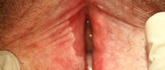
Clinically, damage to the labia minora, labia majora, interlabial grooves, clitoris, and clitoral hood is determined with the loss of the normal architecture of the vulva and perianal zone. Snow-white spots, porcelain-white papules or plaques, infiltration, ill-defined erythema, maceration are noted; erosion; petechiae and telangiectasia, lichenification.
Histologically, lichen sclerosus is represented by a thinned epithelium with hyperkeratinization, a layer of hyalinized collagen, devoid of blood vessels, and lymphocytic infiltration underneath [8, 9].
Another common benign disease is LP (lichen ruber planus) - a chronic inflammatory disease that can affect both the skin and mucous membranes. The term lichen ruber was first proposed in 1861 by F. Hebre, and the English dermatologist E. Wilson in 1869 first gave a clinical description of this disease. In Russian literature, the pioneer was V.M. Bekhterev (1881). There are neurogenic, viral and infectious-allergic theories of the origin of LP, the latter attaches great importance to the role of chronic infection in the pathogenesis of the disease.
In the vulva area, the most common erosive form of LP, in which the leading subjective symptoms are pain, burning and increased sensitivity (vulnerability). Objectively, well-demarcated erosions or glazed erythemas with a hyperkeratotic white border in erythematous areas (Wickham's mesh) and inflammation are noted. If treatment is not started in a timely manner, the vulva eventually loses its normal structure with the loss of the labia and clitoris, narrowing of the entrance and complete obliteration of the vagina occur. Numerous scars and adhesions lead to the impossibility of sexual activity. The term vulvo-vaginal-gingival syndrome is used when erosive lesions are present in these three areas.
Risk factors for developing the disease are age 40–60 years, female gender, hereditary predisposition, past stress, gastrointestinal diseases, diabetes mellitus, trauma to the mucous membranes.
Early diagnosis, adequate treatment and long-term follow-up of patients are mandatory. With timely initiation of treatment, consequences such as destruction of anatomical structures and the development of cancer pathology can be prevented. An important condition is interdisciplinary interaction and knowledge of the problem by dermatologists, gynecologists, urologists, and therapists. Diagnosis is based on medical history and assessment of the clinical picture of the disease. To identify concomitant processes (pathologies of the thyroid gland [10], autoimmune diseases [11], which are significantly more common in patients with lichen), as well as to take into account contraindications to long-term use of a number of drugs, consultations with a therapist and an endocrinologist, clinical, biochemical, and hormonal tests are required. blood and urine tests. Instrumental studies are also required, in particular vulvoscopy and extended vulvoscopy, ultrasound of the abdominal organs, kidneys, and thyroid gland. Currently, there is no diagnostic algorithm to predict the risk of malignant transformation in LS and LP of the vulva. In connection with these patients, who have foci of hyperkeratosis or new warty lesions, long-term non-healing erosive and ulcerative elements that do not respond to standard therapy, should be further examined using a biopsy and morphological examination of the obtained material to exclude a malignant process. In these cases, material for histology is obtained from the most altered areas [12]. Treatment of vulvar dermatoses is challenging. To achieve long-term remission, it is necessary to eliminate provoking factors: eliminate concomitant infections of the urogenital tract, compensate for deficiencies of hormones, vitamins, minerals, normalize the functioning of the thyroid gland and gastrointestinal tract [13].
Rational and effective treatment of chronic inflammatory diseases of the female genital organs is a difficult, but extremely urgent problem. Difficulties in therapy are often due to the presence of concomitant vulvovaginitis, most often caused by a mixed infection. In such a situation, a careful selection of drugs is needed that, on the one hand, can act on a wide range of opportunistic microorganisms, and on the other hand, do not irritate the skin damaged by a long-term inflammatory process.
Elzhina, a multicomponent drug for local treatment of vaginitis, including those associated with vulvar dermatoses, satisfies all the specified requirements. Adequate dosage of active ingredients, a combination of antifungal and antibacterial action, lack of toxicity, and ease of use allow Elzhina to be considered the drug of choice for the treatment of pathological vaginal discharge, which becomes an additional irritating factor that provokes or aggravates the course of the underlying chronic inflammatory disease. It should be emphasized that for a woman during treatment, a feeling of comfort is important, which is facilitated by the prednisolone included in the composition, which reduces irritation and itching. The ease of use of the drug provides the patient with a stable psycho-emotional state and improved quality of life.
According to modern recommendations, the basic treatment of patients with LS and LP should be the administration of topical glucocorticosteroids of high and medium strength or calcineurin inhibitors [14, 15].
The use of drugs is local, ointments are applied to the affected areas.
Recommended mode
Daily powerful or extra-potent topical steroids (0.5% clobetasol ointment) for 3 months, 1-2 times a day for the first month, then every other day for the second month, then 2 times a week for the third month of treatment.
Maintenance treatment
Twice weekly 0.1% mometasone furoate ointment is effective and safe for maintaining remission and helping prevent malignant changes (Ib, A). In this case, 30 g of a potent steroid should be enough for 3 months of therapy.
Treatment of superinfection
For secondary (concomitant) infections, most often candida and/or bacterial, heavy-duty or potent topical steroids in combination with antibacterial and antifungal agents are indicated. The domestic drug for external use, Tetraderm, meets the stated requirements - the first original combined glucocorticosteroid-containing drug, which simultaneously promotes skin regeneration. Tetraderm is a combination of highly active mometasone with antibacterial, antifungal agents and a component that promotes epithelial regeneration.
Mometasone belongs to the group of the most powerful glucocorticosteroids with a high safety profile; it has a pronounced anti-inflammatory, antipruritic and antiexudative effect. Econazole, which is part of the drug, is a highly effective antifungal component with a bactericidal effect, and gentamicin is an antibiotic used for the local treatment of primary and secondary bacterial skin infections.
The fourth active ingredient of tetraderm - dexpanthenol (a component that activates tissue regeneration with a metabolic effect) - helps restore the damaged stratum corneum of the skin.
Indications for use of Tetraderm are not limited to the treatment of LS and LP; it is used for dermatoses of inflammatory origin with concomitant bacterial and mycotic infection or a high probability of secondary infection; with simple, allergic, atopic dermatitis (including diffuse neurodermatitis), with limited neurodermatitis, eczema, dermatomycosis (dermatophytosis, candidiasis, lichen versicolor), especially if the process is localized in the groin area and in large folds of the skin.
However, it should be remembered that the duration of use of combined topical glucocorticosteroid drugs is limited, and these drugs should be used for 10-14 days, i.e., in the period when it is necessary to cope with concomitant infection (IV, C).
In general, the treatment of chronic inflammatory diseases of the vulva is a long, often lifelong process that requires not only the prescription of proven drugs and treatment regimens, but also patience.
The main goal of therapy is to stop the progression of the disease, reduce the activity of the pathological process, reduce the area of skin lesions and the severity of clinical symptoms of the disease, prevent the development of complications, and improve the quality of life of patients.
That is why one of the important components of therapy for LS and LP of the vulva is emollients (English emollient - softening) - softening and moisturizing agents. These agents increase the moisture content of the stratum corneum of the skin, strengthen weakened skin barrier function and reduce subclinical inflammation. Daily use of emollients along with topical corticosteroids increases the duration of remission.
Conclusion
Diseases of the vulva are characterized by a long, relapsing course with alternating periods of remission and exacerbation, when courses of therapy are required. This allows us to maintain the quality of life of patients at a fairly high level. At the same time, serious complications and malignancy can be prevented with the help of timely, early treatment using local forms of corticosteroids
Women's health and reproduction: online publication. 2021. No. 11 (42) – No. 12 (43), 2019
References 1. Dongre H., Ranaa N., Fromreidea S., Rajthalaab S., Engelsenc BI, Paradis J. et al. Establishment of a novel cancer cell line derived from vulvar carcinoma associated with lichen sclerosus exhibiting a fibroblast-dependent tumorigenic potential. Cell Res. 2019; 23: 111684. DOI: 10.1016/j.yexcr.2019.111684 2. Rogers LJ, Cuello MA Cancer of the vulva. J. Gynecol. Obstet. 2018; 143(suppl.2): S4–13. DOI: 10.1002/ijgo.12609 3. Cohen PA, Anderson L., Eva L., Scurry J. Clinical and molecular classification of vulvar squamous pre-cancer. J. Gynecol. Cancer. 2019; 29(4): 821–8. DOI: 10.1136/ijgc-2018-000135 4. Bleeker MC, Visser PJ, Overbeek LIH, van Beurden M., Berkhof J. Lichen sclerosus: incidence and risk of vulvar squamous cell carcinoma. Cancer Epidemiol. Biomarkers Prev. 2016; 25(8): 1224–30. DOI: 10.1158/1055-9965.EPI-16-0019 5. Nair PA Vulvar lichen sclerosus et atrophicus. Midlife Health. 2017; 8(2): 55–62. DOI: 10.4103/jmh.JMH_13_17 6. Hieta NK, Kurki SH, Rintala MA, Söderlund JM, Hietanen SH, Katri OJ Vulvar malignant pleomorphic adenoma in a patient with lichen sclerosus. JAAD Case Rep. 2019; 5(11): 994–6. DOI: 10.1016/j.jdcr.2019.09.013 7. Halonen P., Jakobsson M., Heikinheimo O., Riska A., Gissler M., Pukkala E. Lichen sclerosus and risk of cancer. J. Cancer. 2017; 140(9): 1998–2002. DOI: 10.1002/ijc.30621 8. Bunker CB Atopy, the barrier, urine and genital lichen. Br. J. Dermatol. 2013; 169(4): 953. DOI: 10.1111/bjd.12553 9. Fergus KB, Lee AW, Baradaran N, Cohen AJ, Stohr BA, Erickson BA et al. Pathophysiology, clinical manifestations, and treatment of lichen sclerosus: a systematic review. Urology. 2019; 9. pii: S0090-4295(19)30870-2. DOI: 10.1016/j.urology.2019.09.034 10. Stewart KMA Vulvar dermatoses: a practical approach to evaluation and management. JCOM. 2012; 19(5): 205–20. 11. Cooper SM, Ali I., Baldo M., Wojnarowska F. The association of lichen sclerosus and erosive lichen planus of the vulva with autoimmune disease: a case-control study. Arch. Dermatol. 2008; 144(11): 1432–5. DOI: 10.1001/archderm.144.11.1432 12. Kirtschig G, Becker K, Günthert A, Jasaitiene D, Cooper S, Chi CC et al. Evidence based (S3) Guideline on (anogenital) Lichen sclerosus. JEADV. 2015; 29(10): e1–43. DOI: 10.1111/jdv.13136 13. Fistarol SK, Itin PH Diagnosis and treatment of lichen sclerosus: an update. J. Clin. Dermatol. 2013; 14(1): 27–47. DOI: 10.1007/s40257-012-0006-4 14. Howard M., Hall A. Vulval lichen planus-lichen sclerosus overlap. Int J. STD AIDS. 2018; 29(10): 1017–23. DOI: 10.1177/0956462418758777 15. Federal clinical guidelines. Dermatovenerology-2015: Skin diseases. Sexually transmitted infections. M.: Business Express; 2021. 768 p.
When is it necessary to consult a gynecologist?
Ideally, a woman should have her first consultation with a gynecologist around the age of 18 or immediately after she becomes sexually active, whichever comes first. After this, many doctors recommend undergoing a gynecological examination twice a year and taking tests. Especially when a new sexual partner appears, because this increases the risk of bacterial vaginosis, chlamydia and trichomoniasis.
Such diseases in gynecology often pass without pronounced symptoms and, if neglected, can lead to infertility, ectopic pregnancy or premature birth. In addition, they increase the likelihood of contracting the immunodeficiency virus: after all, if you have at least one gynecological infection, the body's protective functions are reduced and you are more vulnerable to HIV.
A preventive examination by a gynecologist is just one of the reasons why you should go to a gynecologist.
Consultation required:
- If you haven't started menstruating by age 15.
- If during pregnancy your mother took the now banned drug diethylstilbestrol, previously used to prevent premature birth. It was found that the daughters of women who took diethylstilbestrol have an increased risk of developing uterine and cervical cancer
- If you have very painful periods.
- If your periods are very heavy, or they last more than 7-10 days, or you have had vaginal bleeding at other times.
- If you experience a burning sensation, itching, odor, or have unusual discharge or pain in the lower abdomen.
- If you feel pain during sexual intercourse, especially if you also have cold symptoms.
- If you are sexually active and have not had your next menstruation.
- If you abstain from sex and have not had two or more periods.
- If you experience a burning sensation every time or often when urinating.
- If you notice unusual papillary growths in the genital area, roughness, irritation, abrasion in the labia area.
Why does the vagina hurt?
Medical manipulations
Painful sensations are observed after abortion and diagnostic curettage. May cause concern after installation of an intrauterine device and hysteroscopy. Itching, mild or moderate, decreases and disappears within a few hours or days. The cause of periodic dull or nagging pain is cicatricial changes in the vagina after surgery, incisions and ruptures during childbirth.
Traumatic injuries
Minor injuries occur due to unsuccessful douching, the use of uterine rings and tampons that are not the right size. The latter option is more often observed in teenage girls and young girls. Sometimes the cause of superficial damage is careless sexual intercourse or an unphysiological position of the woman’s body during intercourse. There is no bleeding, the pain is not intense and disappears quickly.
The etiological factors of vaginal ruptures outside the period of labor are violent or overly aggressive sexual contacts, drug or alcohol intoxication of partners, rough use of intimate accessories or foreign objects to obtain satisfaction. The presence of a serious lesion is indicated by bleeding and sharp pain.
Severe combined injuries can be observed with birth ruptures and pelvic fractures due to high-energy impacts. Along with the vagina, the uterus, perineal area, and neighboring pelvic organs are affected. The clinical picture is determined by the nature and extent of the damage. Intense sharp cutting pain, shock, and massive blood loss are observed. The condition is life-threatening.
Soft vaginal foreign bodies and small foreign objects are usually not painful. The appearance of pain, which intensifies during urination and sexual intercourse, is possible with prolonged irritation of the mucous membrane and the development of inflammation. Sharp and hard objects injure the walls of the organ; the same clinical picture is observed as with vaginal ruptures.
Vaginitis
For acute colpitis, dull, low-intensity pressing or bursting pain in the vaginal area is typical. Urination and sexual intercourse are accompanied by an increase in pain, the pain becomes more acute, sometimes cutting or stabbing. Irritation, itching, swelling, hyperemia, local hyperthermia of the vagina and vulva are noted.
With coccal vaginitis, the discharge is copious, yellowish-white, with gardnerellosis - transparent, smelling of rotten fish. In patients with thrush (vaginal candidiasis), a heterogeneous, curd-like white discharge is noted. Along with infection by opportunistic microorganisms, colpitis develops with tuberculosis and the following STIs:
- gonorrhea;
- syphilis;
- chlamydia;
- trichomoniasis;
- mycoplasmosis and ureaplasmosis.
The downward spread of infection leads to the occurrence of vulvovaginitis, in which the above symptoms are combined with pain, irritation and redness of the labia, clitoris, perineum, buttocks and inner thighs. In patients with chronic vaginitis, pain occurs extremely rarely. They may appear during exacerbations of pathology after hypothermia and acute respiratory viral infections, during pregnancy or during menstruation.
Vaginal condylomas
Endometriosis
For patients with endometriosis, pressing, pulling and bursting pains are mainly bothered during sexual intercourse, defecation and urination, especially when the lesions are located in the vagina, rectouterine cavity, area of the uterosacral ligaments and rectovaginal septum. In approximately 20% of cases, there is a combination with localized or diffuse pelvic pain, in 40-60% of cases - with dysmenorrhea. Possible menorrhagia, posthemorrhagic anemia, infertility.
Volumetric formations
Nagging or contact pain is more often observed with true benign neoplasms of the vagina: lipomas, fibroids, fibromas, fibromyomas. They are supplemented by unpleasant sensations during sexual intercourse, a feeling of a foreign body, and disorders of urination and defecation. In rare cases, pain during sitting, walking, physical activity and sexual intercourse is observed in women with large cysts of the vagina and Gartner's duct.
Rapidly growing intense pain in the vagina, pubis, perineum, lower abdomen is detected with malignant neoplasia - cancer and sarcoma. There are purulent, bloody or bloody discharge, contact and spontaneous bleeding, urinary disorders, constipation or fecal incontinence, swelling of the lower extremities. Common symptoms are weakness, loss of appetite, weight loss, nausea, vomiting.
Dyspareunia
The cause of pain is the insertion of the penis into the vagina or friction. Sometimes pain occurs already at the stage of arousal or only after completion of intercourse. Painful sensations vary greatly and can be weak, barely noticeable, or painful, unbearable, stabbing, aching or burning. Covers only the vagina or vagina and vulva. Depends on the position and circumstances of copulation.
Dyspareunia can be combined with vulvodynia. Often accompanies vaginitis, vulvitis and vulvovaginitis. In addition, it is provoked by the following gynecological pathologies:
- bartholinitis;
- vaginal malformations;
- atrophic colpitis;
- uterine fibroids;
- retroflexion of the uterus.
It is found during adhesions and varicose veins of the small pelvis. Identified in interstitial cystitis and Sjögren's syndrome. Sometimes it is formed as a result of allergic reactions and microtraumas when using contraceptives. May be due to improper partner technique or psychological factors.
Prolapse of the genital organs
Discomfort and dull pressing pain bother women with prolapse of the vagina or uterus. They are supplemented by a sensation of a foreign body, nagging pain in the lower abdomen, and in a third of patients they are combined with dyspareunia. Possible bloody discharge from the vagina, mixed urination disorders, colitis, constipation, involuntary discharge of gases. When the uterus prolapses, infertility, algomenorrhea and hyperpolymenorrhea are often observed.
Hematocolpos
Hymenal atresia is asymptomatic until menarche. After the onset of menstruation, periodic spasmodic painful sensations are noted in the lower abdomen and lumbar area. A few months later, after a large amount of blood accumulates in the vagina, girls experience bursting pain in the vagina, caused by pressure on the walls of the organ. With hematocolpos against the background of vaginal aplasia and atresia, pain begins to disturb after the first or second menstruation, and sooner it becomes constant.
Fistulas
Painful sensations, itching and burning of the vagina are observed in patients with rectovaginal fistulas due to infection. The pain intensifies during sexual intercourse and forces the woman to refuse sex. There is the release of gases and feces through the vagina, and a constant smell of feces. With urethro-vaginal and vesicovaginal fistulas, the symptom is less disturbing; dysuric disorders and urine leakage through the vagina come to the fore.
Other reasons
Sometimes the symptom is provoked by diseases of nearby organs or has a psychological basis. The cause of pain is:
- Proctological pathologies:
hemorrhoids, proctitis, paraproctitis, perianal abscess, suppurating coccygeal tract, thrombosis of hemorrhoids, rectal tumors. - Diseases of the urinary system:
urethritis, cystitis. - Mental disorders:
hysteria, neurasthenia, obsessive-compulsive disorder, some psychotic states.
Colposcopy