Lung cancer is a pathology that begins with damage to the parenchyma or bronchi.
The main symptoms of this disease are:
- Dyspnea;
- Cough with blood and sputum;
- Chest pain;
- Temperature;
- Thinness;
- Pulmonary hemorrhage;
- Pericarditis;
- Pleurisy.
To find out the exact diagnosis, you need to do a computed tomography of the lungs, radiography, bronchoscopy, study sputum and pleural exudate, and do a biopsy.
Treatment usually involves complex therapy: surgery, radiation therapy and chemotherapy.
This type of cancer is of epithelial origin. It develops from the mucous membrane of the bronchi and their glands or from the pulmonary alveoli. The mortality rate with it is quite high.
The development of the disease will be directly influenced by the histological structure of the tumor:
- Differentiated squamous cell carcinoma progresses slowly;
- Undifferentiated cancer progresses rapidly and metastasizes;
- Small cell cancer also develops rapidly, but unnoticed, and metastasizes.
It was revealed that cancer forms more often in the right lung than in the left. It also forms more often in the upper part of the lung than in the middle and lower parts due to the entry of more carcinogens into the upper part.
Metastasis occurs in three ways:
- Lymphogenic;
- Implantation;
- Hematogenous.
The lymphogenous route is the most common. Metastases go to various types of lymph nodes:
- Pulmonary (affected first);
- Bronchopulmonary (affected after them);
- Tracheobronchial;
- Paraesophageal;
- Bifurcation.
With hematogenous germination through the blood, metastases occur:
- Into the adjacent lung;
- Brain;
- Kidneys and liver;
- Spine.
Metastases
Germination of an implantation nature occurs along the pleura.
The main causes of lung cancer are:
- Tobacco smoking;
- Bad air;
- Radiation.
Based on histology, lung cancer is of four types:
- Glandular;
- Squamous;
- Small cell;
- Large cell.
Treatment of lung cancer, treatment of stages 1, 2, 3. Symptoms, signs, metastases, prognosis.
Content:
- Causes of lung cancer
- Types of lung cancer
- Symptoms
- Diagnosis of lung cancer
- Additional examinations before treatment
- Treatment methods Surgical treatment
- Chemotherapy
- Radiation therapy
- Treatment of non-small cell lung cancer
- Treatment of small cell lung cancer
- Treatment of pleural mesothelioma
Causes of lung cancer
The causes of lung cancer can be divided into two groups: exogenous (external) and endogenous (internal).
Exogenous causes of lung cancer
- Tobacco smoking
- Harmful working conditions
- Ionizing radiation
- Environment
Endogenous causes of lung cancer
- Hereditary predisposition
- Chronic lung diseases
- Age over 50
- Hormonal shifts
One of the main factors in the development of lung cancer is active and passive tobacco smoking.
When smoking tobacco, more than 40 carcinogenic substances enter the body. These include benzopyrene, resins, and many others.
Active and passive smoking increases the risk of developing cancer by more than 5 times.
Mining workers face great risks when extracting radioactive minerals. Ionizing radiation plays an important role in the development of lung cancer.
Forecast
The Israel Cancer Society provides statistics showing how many people can expect to live another 5 years or more after being diagnosed with cancer at each stage.
NSCLC
The average chances of surviving 5 years or more with NSCLC are:
- 60% when it is localized
- 33% when it is regional
- 6% when he is far away
- 23% overall
MRL
The chances of surviving another 5 years or more are equal:
- 29% when it is localized
- 15% when it is regional
- 3% when he is far away
- 6% overall
Find out if the treatment is prescribed correctly
Symptoms
Lung cancer is often asymptomatic. There are no pathognomonic symptoms of lung cancer.
General symptoms are caused by tumor intoxication:
- weakness;
- hyperthermia;
- paraneoplastic syndromes (joint pain, Cushing's syndrome, etc.).
Local symptoms are caused directly by the tumor:
- chest pain (tumor growth into the chest wall);
- hemoptysis (disintegration in the tumor);
- shortness of breath (lung atelectasis, tumor pleurisy).
Secondary symptoms are caused by the spread of the tumor to neighboring organs and structures;
- hoarseness (ingrowth into the recurrent nerve);
- dysphagia (ingrowth into the esophagus);
- superior vena cava compression syndrome (ingrowth into the superior vena cava).
What happens during radiation therapy?
Radiation therapy involves the use of focused, high-energy X-rays, gamma rays, or other charged particles. Radiation targets rapidly dividing cells, such as cancer cells. Many cancers, including lung cancer, are made up of cells that divide much faster than healthy lung cells. This allows you to eliminate cancer formation with virtually no harm to surrounding healthy cells. Radiotherapy has a detrimental effect on the genetic material, that is, DNA, of tumor cells, which disrupts the cycle of their growth and division. Healthy tissues are also damaged (albeit much less), but have the ability to restore their structure and function. Therefore, radiotherapy is based on daily irradiation of the tumor with doses high enough to destroy the bulk of rapidly dividing malignant cells, but not sufficient to significantly damage healthy cells with a slow division cycle.
Up
Diagnosis of lung cancer
Non-invasive examination methods are used to diagnose lung cancer:
- inspection,
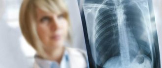
- chest x-ray,
- computed tomography,
- magnetic resonance imaging,
- ultrasound examination of the abdominal organs
and invasive methods:
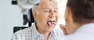
- bronchoscopy;
- thoracoscopy;
- mediastinoscopy;
- transthoracic needle biopsy of the tumor.
Digital chest radiography
Contrast-enhanced computed tomography of the chest allows one to assess the extent of the tumor to large vessels
Additional examinations before treatment
When diagnosing lung cancer before starting treatment, additional examination is necessary to solve several problems:
1.Assessment of tumor localization in the lung and its spread.
Based on the location of the tumor in the lung, central and peripheral cancer are distinguished. Central lung cancer grows from large and medium bronchi (in the region of the root of the lung), and peripheral cancer grows from small bronchioles and the respiratory tissue of the lung (alveoli) and is located peripherally. For this purpose, computed tomography is performed. Computed tomography of the chest is a highly accurate diagnostic method. It allows you to study in detail and with high resolution the size and location of the tumor, its growth into surrounding organs, the presence of metastases (secondary tumor lesions) in the surrounding lung tissue, in the lymph nodes of the mediastinum, in the bones of the thoracic spine, ribs and sternum. Another main and mandatory research method for lung cancer is bronchoscopy. In this case, a thin flexible instrument with a lens and a light source at the end is inserted into the lumen of the trachea and bronchi. With the help of bronchoscopy, you can not only visually evaluate changes in the organ wall, but also, if necessary, take a scraping or piece of tissue (perform a biopsy) for examination under a microscope (cytology and histology, respectively) to determine the presence of cancer cells and determine its shape. Microscopic confirmation of any malignant tumor is mandatory and lung cancer is no exception.
2. Exclusion/confirmation of metastatic lesions of regional lymph nodes.
In most cases, metastases initially appear in regional lymph nodes (bronchopulmonary lymph nodes, mediastinal lymph nodes), and subsequently metastases appear in other organs. Damage to regional lymph nodes affects the stage of the tumor process and the choice of treatment tactics. Non-invasive diagnostic methods such as chest computed tomography and positron emission tomography, as well as invasive methods such as endobronchial ultrasonography (EBUS) and esophageal ultrasonography (EUS), are used to assess lymph node involvement. The essence of the methods is similar to bronchoscopy and esophagogastroscopy. The difference is that at the end of the flexible endoscope there is an ultrasound sensor, through which the walls of the esophagus, trachea and large bronchi are scanned, which allows visualization of the lymph nodes and their puncture biopsy through the wall of the esophagus/trachea. These studies take longer and often require general anesthesia. In difficult clinical situations, diagnostic minimally invasive operations are performed: videothoracoscopy and mediastinoscopy. This requires hospitalization.
3. Exclusion of the presence of metastases in other organs.
Lung cancer most often metastasizes to the liver, adrenal glands, bones, brain, and lungs. An examination of these organs must be performed before starting treatment. The minimum examination includes a computed tomography scan of the chest and ultrasound of the abdominal organs. It is advisable to conduct an MRI study of the brain and positron emission tomography (PET). PET scanning is one of the newest modern techniques in the field of cancer detection. With PET-CT, a CT scan of the entire body is performed, and metabolic activity in tumor foci is also assessed, which makes it possible to accurately determine the extent of the spread of the tumor process. The results of the scan play a huge role in the process of making a diagnosis and studying secondary manifestations of the disease.
4. If necessary, study of tumor markers in the blood:
carcinoembryonic antigen (CEA); neuron-specific enolase (NSE); fragment of cytokeratin 19 (Cyfra-21-1), however, they play only an auxiliary role in the diagnosis of lung cancer and are not recommended for routine use.
5. In the presence of distant metastases
It is necessary to conduct a molecular genetic study of tumor tissue to determine activating mutations: EGFR, ALK, ROS-1, BRAF, KRAS in order to determine the possibilities of targeted therapy.
6. Assessment of the general condition of the patient, the degree of risk of the operation.
These examinations are prescribed individually and usually include ECG, spirometry, consultation with a therapist, ECHO-CG, gastroscopy, ultrasound of the veins of the lower extremities, ultrasound of the arteries of the lower extremities, ultrasound of the brachiocephalic arteries.
Targeted drugs and immunotherapy
Targeted drugs and immune drugs are modern, relatively new groups of drugs that can be used for non-small cell lung cancer. Targeted drugs block substances that activate angiogenesis, the process of growth of new blood vessels that supply oxygen and nutrients to tumors.
Two drugs are used: Bevacizumab and Ramicirumab. Among the immunotherapy drugs used for lung cancer are checkpoint inhibitors: Atezolizumab and Nivolumab.
Normally, the immune system uses certain substances for self-control - so-called checkpoints . They help curb the immune response and protect the body's own tissues from attack. Sometimes cancer cells use this mechanism to protect themselves, to “hide” from the immune system. Checkpoint inhibitors help remove the blockage and “free up” the immune system so it can attack the cancer.
Sometimes targeted drugs and immunotherapy drugs are prescribed as first-line therapy in combination with chemotherapy drugs.
Lung cancer treatment methods
The main treatment for lung cancer is surgery. The extent of the operation is determined after examination and depends on the location of the tumor in the lung and its extent.
Surgical treatment may include the following operations
- Mediastinoscopy: a small incision in the neck, special instruments, optics, intrathoracic lymph nodes are taken;
- Thoracoscopy: small incisions, special instruments, optics, removal of intrathoracic lymph nodes, removal of a section of a lung, removal of a section of a rib(s), removal of a mediastinal tumor, etc. can be performed;
- Atypical lung resection – removal of a section of the lung;
- Lobectomy – removal of a lobe of the lung;
- Pneumonectomy – removal of the lung. The Center’s departments also provide treatment as part of scientific research if there is no effect from the standard treatment.
For peripheral cancer, removal of a lobe of the lung (lobectomy) is most often performed, and for central cancer, removal of the lung (pneumonectomy).
In some cases, for central cancer, bronchoplastic operations are performed, in which it is possible to preserve a lobe of the lung with the imposition of an interbronchial anastomosis.
If the tumor involves the chest wall or anatomical structures of the mediastinum, combined operations with resection of these structures are performed.
The development of anesthesiology and surgery has now made it possible to perform complex surgical interventions on the trachea, as well as on the lungs and heart using artificial circulation. The use of video thoracoscopic technology allows for lung resection or removal of a lobe of the lung through small incisions in the chest wall and reduces the invasiveness of the operation.
Video-assisted thoracoscopy
After surgery for lung cancer, additional chemotherapy or radiation therapy may be required.
After treatment, follow-up is required: for the first year, every 3 months, examination, x-ray of the chest organs in 2 projections, ultrasound of the abdominal organs.
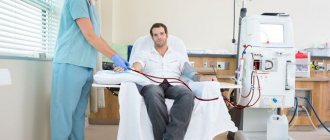
Chemotherapy
Anticancer drugs are used for this treatment method. In most cases, a combination of two chemotherapy drugs (polychemotherapy) is prescribed to achieve maximum effect. In weakened patients, monochemotherapy can be used. Most chemotherapy drugs are given intravenously as drips of varying lengths, depending on the drug used. Chemotherapy is carried out in cycles (courses), which means that when prescribing drugs there is a pause, which can last several weeks. Chemotherapy is the main treatment for patients with advanced lung cancer (with distant metastases).
The goal of chemotherapy is to damage the cells of a malignant tumor and thus slow down the development of the tumor. However, chemotherapy drugs also have a toxic effect on healthy cells, without distinguishing them from tumor cells. Therefore, this method of treatment is accompanied by side effects. Side effects of chemotherapy may vary depending on the drugs used and their combination. The most common are nausea and vomiting, hair loss and attacks of weakness, and decreased levels of white blood cells and platelets. When carrying out chemotherapy, it is imperative to carry out therapy aimed at minimizing side effects. The occurrence of such phenomena must be reported to your doctor. In most cases, your doctor can relieve side effects with appropriate treatment.
Radiation therapy
Radiation therapy uses radiation to damage tumor cells. Radiation therapy is based on the fact that the tumor is more sensitive to radiation than the healthy tissue surrounding the tumor. External beam radiation therapy is most often used for lung cancer. This means that the tumor is irradiated using an external radiation source (without contact with the patient).
Before starting radiation therapy, the patient is invited to the radiation therapy department, where radiation therapy planning takes place. It is used to determine the location where the radiation will be directed during treatment. This procedure usually lasts up to 40 minutes, but in some cases it may take longer. During the simulation, markings are applied to the patient's skin and then a CT scan is performed. Markings on the body ensure the correct positioning of the patient during radiation therapy. On the day of radiation therapy planning, the patient is given a start date for radiation therapy. The radiation oncologist takes time to calculate the dose and plan the radiation regimen to ensure the most effective and safe treatment. Radiation therapy is usually carried out once a day, 5 times a week (on weekdays) and lasts several weeks. One radiation therapy session lasts approximately 15-20 minutes.
Currently, thanks to the use of modern equipment, high-precision irradiation of a tumor is possible with minimal damage to healthy tissue and minimization of side effects. In lung cancer, radiation therapy is used as an independent method at an early stage of lung cancer in patients with severe concomitant pathology in the presence of contraindications to surgery as an alternative treatment method, as well as when the patient refuses surgical treatment.
Radiation therapy is also used in combination with chemotherapy in patients with locally advanced lung cancer when surgical treatment is impossible or impractical.
Treatment for non-small cell lung cancer:
- Stage I-II – standard surgical treatment, in the absence of contraindications; if there are contraindications, radiation or chemoradiotherapy is performed. Some patients undergo postoperative chemotherapy;
- Stage IIIA-IIIB – complex (surgical and chemoradiotherapy) or chemoradiation treatment is carried out;
- Stage IV – chemoradiotherapy.
Treatment for small cell lung cancer:
- Stage I-II – complex (surgical and chemoradiotherapy) treatment is carried out;
- Stage III-IV – chemoradiotherapy.
Treatment of pleural mesothelioma
- Stage I-III – in the absence of contraindications, complex treatment is carried out (intrapleural thermochemotherapy, pleuropneumonectomy and 4 courses of polychemotherapy), if complex treatment is intolerable, then chemoradiotherapy is carried out;
- Stage IV – chemoradiotherapy.
Intrapleural thermochemotherapy - performed thoracoscopically or by thoracotomy (a wide lateral incision of the chest wall with the ribs separated), using special equipment, the pleural cavity is washed with Ringer's solution heated to 42 °C with the addition of chemotherapy drugs (cisplatin, vinorelbine).
Pleuropneumonectomy
– removal of the pleura, lung, diaphragm and outer membrane of the heart on the side
Is the effect of radiation on healthy organs great?
Unfortunately, radiation energy in general is destructive and can damage not only cancerous tissue, but also normal, healthy tissue. As radiation penetrates the body - that is, first through the skin, then through layers of tissue towards the lung - it can partially dissipate into healthy tissue located near the affected organ. Very important organs are located near the lungs. This includes the esophagus, the heart, and normal lung tissue. All of them are at risk of minor damage. Usually the harm is relatively minor and almost unnoticeable, but some patients sometimes experience, for example, difficulty swallowing - especially if they are nearing the end of their course of radiotherapy.
Prevention
Prevention of lung cancer includes the following recommendations:
- Quitting bad habits, primarily smoking;
- Maintaining a healthy lifestyle: proper nutrition rich in vitamins and daily physical activity, walks in the fresh air;
- Treat bronchial diseases in a timely manner so that there is no transition to a chronic form;
- Ventilation of the premises, daily wet cleaning of the apartment;
- It is necessary to reduce contact with harmful chemicals and heavy metals to a minimum. During work, be sure to use protective equipment: respirators, masks.
If you experience the symptoms described in this article, be sure to see a doctor for an accurate diagnosis.
TNM classification
Another staging method is the TNM system.
TNM focuses on:
- Tumor size (T): How big is the tumor and has it spread to other tissues or areas?
- Lymph nodes (N) : Has the cancer spread to nearby lymph nodes?
- Metastasis (M) : Has it reached other parts of the body such as the other lung, liver and other organs?
Doctors use TNM stages to describe SCLC and NSCLC.
The stages of cancer can be complex. A consultation with a doctor at the Ichilov Cancer Center can help a person understand how cancer affects them.
Get exact price
Advantages of lung cancer treatment at the Republican Scientific and Practical Medical Center named after. N.N. Alexandrova:
- More than 55 years of experience in diagnosis and treatment;
- The Center employs 12 professors, 19 doctors of science, 78 candidates of science, 10 associate professors, 1 corresponding member of the National Academy of Sciences of Belarus;
- Experienced qualified specialists;
- The best diagnostic and treatment base in the Republic of Belarus;
- Providing medical services directly, without intermediaries;
- Initial appointment without prior appointment;
- Providing a full range of services within one clinic;
- Individual approach
Price lists for foreign citizens
Basic medical services related to the diagnosis of lung cancer:
*Note: Prices are for foreign citizens. Citizens of the Republic of Belarus can receive medical services without charging a fee. Read more...
| Type of medical research | Amount, BYN | Amount, USD | Amount, RUB |
| X-ray computed tomography of the chest cavity (lungs and mediastinum) without contrast enhancement | 364,39 | 180,00 | 11 352,42 |
| X-ray computed tomography of the chest cavity (lungs and mediastinum) with contrast enhancement | 576,95 | 285,00 | 17 974,64 |
| CT angiography | 688,30 | 340,00 | 21 443,70 |
| Tracheobronchoscopy | 141,71 | 70,00 | 4 414,92 |
| PET/CT with “2-[l8F]-Fluorodeoxyglucose” whole body | 1 315,86 | 650,00 | 40 995,08 |
| Sequencing of exons 19, 21 of the EGFR gene (each subsequent) | 232,81 | 115,00 | 7 253,10 |
| Determination of ALK gene rearrangement using fluorescence in situ hybridization (FISH) | 657,93 | 325,00 | 20 497,54 |
*Prices are indicative, because Payment for services is made in Bel. rub. *You can view all types of services and exact prices on the page
Calculate an exact estimate