Flat feet are an extremely common pathology among adults. It accounts for 25% of all visits to an orthopedic doctor. Among all foot deformities, flat feet account for 80% of cases. The disease progresses slowly but constantly without treatment. Over time, it leads to severe pain, limits a person's physical activity and leads to complications in the foot, spine and large joints of the lower limb. Let's talk about whether it is possible to cure flat feet in adults without surgery and when there is a need for surgical treatment.
Causes and symptoms
All children have flat feet at birth. Typically, by the age of 5, arches are formed: longitudinal and transverse. In childhood, flat feet are considered normal. It does not require correction with insoles or orthopedic shoes.
But as an adult, the foot should not be flat. However, flat feet develop in at least 15% of the adult population. It can be congenital or acquired. Congenital is extremely rare. Acquired by origin can be:
Useful tips
Simple preventive measures will help prevent the development of flat feet:
– the right choice of shoes, which should be made taking into account the growing child’s foot. Don’t press or rub anywhere, but don’t be too loose;
– each child has his own shoes: due to shoes worn out by a brother or sister, the load on the feet may be incorrectly distributed;
– the presence in the diet of foods rich in phosphorus and calcium, which prevent the development of rickets;
– in summer – sunbathing, in winter – preventive intake of vitamin D as prescribed by the pediatrician;
– moderate physical activity. Avoid activities with axial load - gymnastics, dancing and weight lifting;
– walking barefoot on pebbles, sand, earth, grass, which causes the feet to constantly strain. But you only need to walk on a flat and smooth floor in orthopedic shoes.
flat foot
flat foot
- static – due to prolonged standing;
- rachitic – a consequence of previous rickets (develops due to a disorder of calcium metabolism, mainly caused by a lack of vitamin D);
- paralytic - the result of damage to peripheral nerves or diseases of the central nervous system;
- traumatic – the result of a foot injury (damage to bones, joints, muscles, tendons).
The most common type is static flatfoot. Its symptoms depend on the stage of the pathology:
- Stage 1 – patients complain of pain and fatigue of the calf muscles of the legs, which appear only after a long walk;
- 2nd degree – pain with minimal loads, the first signs of foot deformation;
- 3rd degree – severe deformation of the foot and its expansion.
Static flat feet are the result of constant overload of the feet. It leads to weakened muscles and sprained ligaments. It usually develops in people who stand for a long time, lift and carry heavy objects.
All according to plan
An orthopedic examination is required:
– immediately after birth, this is necessary to exclude congenital deformities and skeletal diseases, including congenital flat feet; also at this age it is necessary to conduct an ultrasound examination of the hip joints;
– at 3 and 6 months;
– at 1 year, when the child already sits, crawls, and walks independently;
– at 3 years of age to check posture, gait, condition of the feet and measure the length of the limbs.
flat feet
flat feet
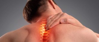
Flat feet reduce the spring (shock-absorbing) function of the foot. As a result, the vibration load on the spine increases and joints suffer. Patients with flat feet have a higher risk of scoliosis, osteochondrosis, and deforming osteoarthritis of large joints of the lower limb.
Thus, if signs of foot deformation appear, you should consult a doctor immediately, since flat feet need to be cured as quickly as possible. The disease is prone to progression. In addition, it causes many complications that are difficult to treat.
Hallux valgus becomes a frequent complication of transverse flatfoot. This is a deformity of the first (big) toe, which is popularly called a “bunion.” The disease is 20 times more likely to develop in women than in men. A predisposing factor is considered to be congenital weakness of the ligamentous apparatus of the foot. The situation is aggravated by wearing tight high-heeled shoes. Severe deformities may require surgical treatment.
Why does it appear
In 90% of this, incorrectly selected shoes are to blame, for example, due to the lack of shock absorption and instep supports. Parents also often select models for growth, which also deforms the foot. The obesity and high weight of the child create more pressure on the legs, the growing body cannot withstand this, and this disease occurs.
Therefore, it is important to choose good orthopedic and preventative summer, winter and demi-season shoes from us - Ortopanda has a large selection of comfortable models made from corrective materials.
How to treat flat feet?
There are conservative and surgical treatment methods. In the vast majority of cases, conservative methods are used.
How flat feet are treated depends on the stage of the pathology and the presence of concomitant foot deformities. Typically, at stage 1, massage, gymnastics, and arch supports in shoes are used. At stage 2, individual insoles, physical therapy, and myostimulation are required to strengthen muscles. At stage 3, conservative treatment can be the same as at stage 2. But sometimes they resort to surgical intervention if a radical improvement cannot be achieved without surgery.
Before treating flat feet in adults, the doctor must conduct an examination. Basic diagnostic methods:
treat flat feet
treat flat feet
- plantography - obtaining foot prints with subsequent decoding;
- radiography – to visualize the bones of the foot;
- podometry – change in the nature of walking;
- computer pedobarometry is the most accurate method that measures pressure during standing and walking on different parts of the foot (allows you to ideally select orthopedic insoles).
In conservative treatment, the most important role is played by the use of orthopedic insoles or shoes. At stage 1 of flat feet, patients can buy ready-made insoles. However, they do not take into account the individual configuration of the foot, and therefore have only a symptomatic and not a corrective effect. Pre-made insoles relieve pain, but over time they can further weaken the muscles. Therefore, the disease can progress with their use.
The best option is individually selected and custom-made insoles. They not only reduce symptoms, but also help eliminate foot deformities.
Drug therapy
Medicines are used to relieve unpleasant symptoms. They cannot cure the disease.
A doctor should prescribe medications, as they may have contraindications. In addition, prescription medications may be prescribed if necessary. The doctor may prescribe:
- non-steroidal anti-inflammatory drugs (Diclofenac, Ibuprofen) – relieve pain;
- muscle relaxants (“Mydocalm”, “Sirdalud”, “Tolisor”) – relieve muscle spasms and eliminate cramps.
Can flat feet be treated with insoles?
Flat feet in the initial stages, without accompanying foot deformities and without arthrosis of the joints, can be cured with the help of insoles or orthopedic shoes. Insoles should only be made to order for a specific person. They are worn at least 5-6 hours a day. The course of treatment lasts from 2 to 4 years or more. The desired result is achieved in 60% of patients. The rest show improvement, but the disease is not completely cured.
Important conditions for the effectiveness of insoles:
- ideal conformity to the topography of the plantar surface of the foot;
- the absence of areas of increased pressure on the foot, which over time can lead to structural changes in soft tissue structures;
- creating conditions for training the muscles of the arch when walking.
Custom insoles affect the degree of muscle tension, joint position, and tendon tension. They also have an effect on proprioceptive sensitivity and contribute to better control of the dynamics of the human body. Good insoles not only relieve symptoms and correct foot deformities, but also improve posture and form a correct gait.
Right choice
The inventor of shoes with arch support was Salvatore Ferragamo (1898–1960), an Italian who became the favorite shoemaker of Hollywood stars. He made shoes for Gloria Swanson and Marlene Dietrich. It was Ferragamo who first created “flat” shoes for Greta Garbo, “stiletto” heels for Marilyn Monroe, and platform shoes for Carmen Miranda, whose height was only 155 cm.
One of the most common causes of flat feet is “wrong” shoes. As soon as your child starts to stand up, it's time to buy him his first shoes.
– high and rigid heel for fixing the ankle joint and longitudinal arch in the correct position
– lacing or Velcro that gently secures the leg
– natural materials used in the production of shoes
– insole with arch support
– perforated “breathable” surface
– stable, hard and non-slip sole with a small heel (0.5 cm)
– wide, round toe that prevents pinching of the toes
Making insoles
Making insoles
- custom size;
- reconstruction of the arch of the foot;
- energy-saving depreciation;
- optimal thermoregulation.
Methods for planning the production of insoles are mechanical and computer. Although mechanical methods were historically the first, today the computer method is increasingly used in modern clinics.
The mechanical method involves obtaining a cast of the foot. In the future, orthoses are made on its basis. However, the technique has one huge drawback: it does not take into account the load on the foot when walking. The impression is taken in a static body position. However, when walking, areas of overload shift, and in different ways for different people. The cut is adjusted subjectively, based on the patient’s feelings, not on objective measurements.
The computer method is more accurate. Pedobarography is performed using a special apparatus. Many parameters are assessed: the position of the direction of the patient’s center of body mass, his movement when walking. There are 5 zones of greatest load on the plantar surface:
- finger;
- metatarsal (metatarsophalangeal joints, rollover axis);
- medial middle region;
- lateral medial region;
- the heel area, which is the axial support.
Based on the results, the structure of the integral load graphs is assessed. A model of the insole for manufacturing is formed. After its production, the orthosis is adjusted to the patient, based not on his sensations, but on the data of pedobarography and photoplantometry.
The use of individual insoles allows obtaining positive treatment results after 1.5 years in 85% of patients. Their pain and muscle fatigue when walking decrease or disappear. Measurements show an increase in arch height. Further treatment can be carried out using other insoles. The examination and orthoses are made again, as the configuration of the foot changes. In a few years, in this way, a person can completely recover from flat feet, after which the need for further use of insoles will disappear.
Diagnosis of the disease
Identifying flat feet begins with taking a medical history to find out what foot complaints the patient has and when they began. Next, they proceed to the inspection, where special attention is paid to the following points:
- foot skin color;
- presence of thickened skin, calluses;
- position of the big toes when the foot is tightly closed;
- the presence of deviations of the foot inward or outward;
- changes in the anterior and calcaneal regions.
Even without instrumental diagnostics, based on complaints and examination, flat feet can be suspected based on the following points:
- the presence of a protruding “bone” near the thumb;
- wear on the inside of shoes;
- rapid fatigue in the legs during prolonged walking or standing;
- an increase in the volume of the foot - the inability to wear your favorite shoes that previously fit;
- swelling, aching pain in the legs, feeling of heaviness at the end of the day;
- discomfort when wearing high-heeled shoes;
- violation of posture and gait;
- Difficulty maintaining balance when squatting.
To make an accurate diagnosis, determine the degree of deformation, and select the most effective treatment tactics, patients are required to undergo a number of studies.
Computer plantography
An improved and accurate method for diagnosing foot diseases using foot prints. The technique is simple, painless and highly informative. Before the examination, the doctor enters all information about the patient into the computer system. The patient stands barefoot on a special platform that looks like glass. The device scans, and in order to obtain more accurate information, the examination is carried out in different planes.
Podometry
A method for diagnosing flat feet with determination of the Friedland index of the longitudinal and transverse arches. It represents the ratio of the height of the foot to its length as a percentage. Normally, the longitudinal arch index is 29-31%. If the indicator decreases, this indicates flat feet. The transverse arch index should not exceed 40%.
Podography
A research method for studying the biomechanics of walking. The procedure is carried out using special equipment - shoes with metal plates and a metal-coated track. During podography, the duration and width of the step, turn of the foot, and gait characteristics are recorded.
X-ray examination
It is necessary to identify changes in the bones of the foot, determine the degree of the disease, and monitor dynamics during treatment. Using X-ray images, you can accurately determine the height of the arch, the angle of the longitudinal arch, and the intermetatarsal angles.
treat flat feet
treat flat feet
You can often find orthopedic insoles and shoes on the Internet, orthopedic salons, pharmacies and medical equipment stores. They are much cheaper than those performed individually and do not require preliminary diagnostics. You can start using these insoles immediately after purchase. Sellers help you choose them, taking into account the degree and type of flat feet.
The only problem is that such insoles do not treat flat feet. In them, a person can feel more comfortable, pain and fatigue in the leg muscles are reduced. But such treatment is only symptomatic. Purchasing ready-made insoles always creates areas of increased pressure on the foot. In addition, the arch muscles are not trained. On the contrary, they are unloaded and gradually atrophy.
The relief of the vast majority of mass-produced insoles does not at all correspond to the relief of the foot of an individual person. It does not take into account the characteristics of walking.
Thus, store-bought insoles are unlikely to be a good treatment, since flat feet in adults cannot be cured with their help. Even minor positive changes are unlikely. The disease will progress more quickly with regular use of these medical products.
Kinds
Common types of foot deformities in children include flat feet, hallux valgus, and varus deformities. Much less common: clubfoot, hollow foot, equine foot, calcaneal foot.
Each type is described in more detail below.
Flat feet
Flat feet are the most common type of foot deformity in children, which consists of flattened arches. The less developed the arches, the worse they perform the function of shock absorption or mitigation of shock loads. This happens most often due to weakening of the muscles and ligaments involved in supporting the arches. A decrease in their tone leads to the fact that they do not hold joints and bones in the correct position.
Depending on which arch is flattened, flat feet occur:
- Longitudinal. The longitudinal arch descends, the foot increases in length, and tends to touch the support with its entire surface. According to the height of the longitudinal arch, there are 3 degrees of flat feet: 1st degree - arch height 20 - 30 mm, 2nd degree - 15 - 20 mm, 3rd degree - less than 15 mm;
- Transverse. The transverse arch lowers, the foot becomes wider and shorter. The thumb deviates outward. Depending on the size of the angle between the thumb and the base of the middle finger, 3 degrees are distinguished: 1st degree - an angle of 20 - 25 °, 2nd degree - an angle of 25 - 35 °, 3rd degree - an angle of more than 35 °;
- combined or longitudinal-transverse, This is the name for cases when a child simultaneously has both longitudinal and transverse flatfoot.
Most often, longitudinal flat feet occur in children.
What does a foot print look like with flat feet, depending on the severity, look at the photo:
Parents may suspect flattening of the arches of the feet based on the following signs:
- child's complaints about tired legs;
- reluctance to walk for a long time, the baby asks to be held or in a stroller after a half-hour walk;
- complaints of pain when lightly pressing the fingers on the inside of the foot;
Up to 3 years of age, flat feet are not a cause for concern. It is physiological, due to the presence of a thick fat pad on the feet, fragile muscles and ligaments that are not yet able to support the weight of the child. Over time, it disappears on its own.
Valgus and varus deformity
Hallux valgus deformity accounts for an average of 60% of all orthopedic abnormalities in a child. Most often occurs in children over 5 years of age. In rare cases, hallux valgus deformity refers to a congenital pathology associated with incorrect (vertical) position of the talus.
With this type of deformation occurs:
- curvature of the vertical axis of the feet;
- the heels and phalanges of the toes deviate outward;
- the main support goes to the inner edge of the sole;
- the ankle moves inward;
- the arch of the foot becomes flatter.
Such changes are usually immediately noticeable. Parents notice that something is wrong with the child’s legs. The distance between the ankles increases and the distance between the knees decreases. The position of the legs becomes X-shaped.
Valgus can be determined by measuring the distance between the ankles with your straightened legs pressed tightly together. If it is 5 cm or more, then there is deformation. Characteristic signs of deviation include shoe trampling from the inner edge.
If valgus deformity is combined with flat feet, then such a disorder of the structure of the child’s foot is called flat-valgus. About 18% of all forms of flat feet in children are accompanied by such a deviation.
With varus deformation of the feet, external changes opposite to valgus are observed:
- the heels deviate inward from the vertical axis;
- the main support goes to the outer edge of the foot;
- the distance between the ankles is reduced;
- knees spread apart;
- O-shaped leg position;
- shoes wear down more on the outer edge.
Varus deformity develops slowly. It is caused by congenital abnormalities of bone structure, joint pathologies, endocrine disorders, infections, rickets, ankle injuries, excess weight, and other factors.
How hallux valgus and varus deformities of the feet differ in children, look at the photo:
Clubfoot
Clubfoot is a severe, in most cases, congenital defect when the foot turns inward and downward. It is manifested by subluxation of the ankle joint with a subsequent decrease in the length of the foot. In approximately 50% of cases, the pathology affects both legs. The prevalence rate is 1 case per 1000 newborns. This deviation occurs more often in boys than in girls.
Congenital clubfoot is usually detected during fetal development during a routine ultrasound at the 20-21st week of pregnancy. But sometimes the diagnosis is made at birth.
What feet look like with clubfoot, look at the photo:
Acquired clubfoot is caused by spastic paralysis, leg injuries, and polio. Until the child begins to walk, the pathology does not cause pain or other medical problems.
But the lack of treatment in the future leads to serious consequences:
- severe pain when walking or even inability to walk;
- the formation of calluses on those parts of the foot that act as a support when walking;
- shortening of the limb.
Clubfoot is a serious aesthetic defect that can cause a child’s complexes, fear, and lack of self-confidence. Therefore, treatment of congenital clubfoot should begin immediately, 1 to 2 weeks after birth. Depending on the severity of the pathology, treatment takes from 2 months to several years.
Hollow foot
A hollow foot is the reverse form of flat feet, when the arch of the foot is not flattened, but, on the contrary, is too curved. This condition is caused by congenital abnormalities, diseases of the neuromuscular system, and trauma. Possible causes also include a hereditary factor, then the hollow shape of the foot is considered not as a pathology, but as a constitutional feature.
Changes in pes cavus and flat feet look like this:
As with flat feet, with a hollow foot there is an incorrect distribution of the load, deterioration of shock absorption during walking, jumping, and running. In a vertical position, the support falls on the heel and the heads of the metatarsal bones (the area under the toes). The middle part of the foot does not come into contact with the surface and does not have the ability to smoothly roll from heel to toe during a step, which causes overload of the forefoot, fatigue, pain in the joints, and deformation of the toes. If the change in the arches on the right and left sides is asymmetrical, then this leads to a relative shortening of one of the limbs, which can cause protrusions and herniated intervertebral discs.
Heel and cauda foot
Foot equinus or pes cauda equina is a deformity in which the angle between the axis of the tibia and the axis of the calcaneus is more than 125°.
With such a deviation:
- the child walks only on tiptoes;
- the toe of the foot is pointed towards the floor, the child cannot pull it towards himself;
- in a vertical position of the body or walking, the fingers and joints act as support;
- Due to the shortening of the calf muscle, it is impossible to lower the leg onto the heel.
In appearance, feet with this deformation resemble horse hooves. It can be bilateral or unilateral. With the unilateral variant, the affected leg rises higher when walking than during a normal gait.
The pathology is more often acquired than congenital. One of the main reasons for its occurrence is damage to the sciatic nerve, which causes a violation of muscle tone in the foot and lower leg. Deformity can occur due to muscle injuries, tendon rupture, bone damage, and inflammatory processes in the tissues of the foot.
Heelfoot is a disorder of the structure of the foot in which the angle between the axis of the tibia and the axis of the calcaneus is less than 90°.
Noted:
- excessive extension, valgus (less often varus) position of the foot;
- increase in the longitudinal arch, lowering of the tubercle of the calcaneus;
- shortening of the foot;
- smoothing the contours of the Achilles tendon.
The disease is caused by damage to the long flexors of the foot due to congenital pathology, birth injuries, neurological disorders, poliomyelitis, injuries to the foot, the front of the leg, paralysis of the triceps surae muscle.