Causes and mechanism of development of hemorrhagic stroke
The factors that cause rupture of the vascular wall and hemorrhage may be primary and secondary diseases of the cerebral arteries. Primary diseases initially develop in one or more vessels of the brain; they are not a consequence of any other diseases and often represent congenital changes. Secondary pathologies of the cerebral arteries appear against the background of other diseases, such as diabetes mellitus or systemic lupus erythematosus.
The most common primary factors may be:
- aneurysmal dilatation - an area where, due to the weakness of the vascular wall or the consequences of an injury, the artery becomes wider and adapts less well to increased blood pressure or other changes in vascular tone;
- vascular malformation is a congenital disorder of the structure of a vessel, which is manifested by the formation of “vascular glomeruli” with cavities of various sizes;
- other congenital anomalies of the arterial vessels of the brain.
Secondary causes that can lead to changes in cerebral vessels and their rupture include:
- arterial hypertension, especially difficult to treat;
- atherosclerotic damage to the vascular wall;
- vasculitis - inflammatory changes in the wall of blood vessels;
- thrombosis of intracranial veins, which lead to hemorrhage;
- diseases manifested by pathology of the coagulation system (blood and liver diseases);
- uncontrolled use of drugs that affect blood clotting (anticoagulants, antiplatelet agents, fibrinolytics);
- pronounced metabolic disorders that develop during long-term severe illnesses.
The most common cause of hemorrhagic stroke in elderly patients is arterial hypertension - this is about 80% of “vascular accidents”. The second place is occupied by atherosclerosis.
In age groups under 40 years, congenital local dilations of blood vessels (aneurysms) and malformations predominate among the causes of hemorrhagic stroke.
Due to the differences in the causes of the disease in different age groups, early and most accurate diagnosis of the pathology comes first. On our website you can find comprehensive information about methods for identifying risk factors for stroke, as well as find out in which centers they are carried out.
Diagnostics
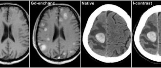
To accurately identify the disease and differentiate hemorrhage from ischemia, an instrumental examination is performed:
- MRI or CT scan - the images show a limited area of the hematoma. The safety of magnetic tomography allows for multiple examinations and monitoring of neuronal recovery.
- Angiography - shows the condition of blood vessels. Well displays arterial rupture, signs of atherosclerosis and congenital anomalies.
- Lumbar puncture - taking cerebrospinal fluid in the lumbar region for analysis. The diagnosis of hemorrhagic stroke is made when red blood cells are detected in the cerebrospinal fluid in high concentrations.
Types of intracranial hemorrhages
Depending on the location of the vessel from which the blood leaked, this type of stroke is divided into:
- parenchymal hemorrhage, characterized by the formation of a hematoma inside the brain or hemorrhagic impregnation of the nervous tissue;
- subarachnoid, which occurs when blood accumulates between the arachnoid and pia mater of the brain.
With parenchymal hemorrhage, depending on the caliber and location of the vessel, blood may break through into the ventricles of the brain. Such strokes are often characterized by an extremely severe course and loss of a relatively large amount of blood.
What happens during a stroke
Regardless of the cause of hemorrhagic stroke, the mechanism of damage to brain cells is the same. The pathological effect has several directions:
- Impaired tissue nutrition - due to the fact that the brain receives blood from several main vessels, this effect is partially compensated.
- Mechanical damage due to the fact that blood leaves the vessel under pressure and “pushes” the tissues apart.
In the case of parenchymal damage and hematoma formation, the second pathway is more pronounced, resulting in a disruption of communication between brain cells. Hemorrhagic permeation is a relatively uniform distribution of blood between the tissue structures of the brain, therefore the clinical course of such hemorrhage is more favorable.
In subarachnoid hemorrhage (SAH), there is a space where blood gets trapped. For this reason, mechanical damage is less than during the formation of a hematoma, but blood loss and malnutrition of brain tissue can be pronounced.
Cerebral hemorrhage (no matter how mild it may initially seem) is a condition that requires urgent diagnostic and therapeutic measures. Prescribing the correct treatment is possible only after neuroimaging, that is, magnetic resonance or computed tomography.
On our website you can get reliable information about the current discounts and promotions at various diagnostic centers for brain research this month. And also find out where it is possible to conduct an inspection at night, if necessary.
Manifestations of hemorrhagic stroke
Symptoms and signs of hemorrhagic stroke are very diverse, they depend on:
- process localization;
- prevalence of the lesion;
- volume and duration of blood loss;
- the degree of swelling of brain tissue and increased intracranial pressure.
The shorter the period from the onset of the disease to the provision of qualified care to the patient, the fewer complications and the better the prognosis. In order not to waste time, you need to know the general signs of a stroke:
- sudden weakness in the arm/leg;
- numbness of half the face, arms/legs on one side;
- new facial asymmetry;
- speech disorder;
- a sharp decrease in vision in one eye or blindness;
- causeless loss of consciousness, especially in combination with previous symptoms.
If any of the symptoms occur against the background of complete well-being, it is necessary to carry out an accurate diagnosis as soon as possible in a specialized medical institution.
All available information about the nearest medical centers where urgent computer or magnetic resonance imaging of the brain can be performed can be obtained by calling 8 (812) 317-00-37.
Different types of hemorrhagic stroke may occur in different ways, but they also have common features. The disease is often characterized by:
- abrupt onset;
- rapid deterioration of the condition;
- loss of consciousness;
- development in the afternoon, at the peak of physical or emotional stress.
Taking into account the peculiarities of the course of the disease, the most important factor for a successful outcome is time. This means that you should not wait for the condition to improve; you should urgently seek qualified medical help.
It is possible to carry out a timely and accurate diagnosis of this disease by dialing a phone number. Our employees will help you quickly select a medical center that meets your needs: the nearest location, the qualifications of specialists, the quality of diagnostic equipment, the cost of the study and the availability of current promotions.
How does a stem lesion differ from other types of stroke?
Depending on the location and extent of the pathological process, disruption of the blood supply to the trunk can have a variety of clinical manifestations.
The first signs of the disease may be severe pain in the back of the head, dizziness, and in 70-80% of cases loss of consciousness occurs. The clinical feature of vascular damage in this particular department is expressed in the appearance of alternating syndromes - damage to the cranial nerves on one side in combination with motor and sensory disorders on the opposite. The functioning of the cardiovascular and respiratory systems and thermoregulation are disrupted, and paralysis of the muscles of the face, pharynx, and muscles of the limbs develops. The development of “locked-in person” syndrome is possible. All symptoms are severe and, if timely medical care is not provided, lead to death in the first two days of the disease. An imbalance of blood circulation in this part of the brain brings serious consequences associated with a person’s life. Basic health parameters are affected. In particular, the following symptoms result from a stroke:
- dysfunction of the heart muscle. There is a change in the heartbeat rhythm, which causes bradycardia, arrhythmia or fibrillation. A distinctive feature of damage to the stem structure is a poor prognosis for rehabilitation;
- failure in the respiratory system. Shortness of breath and inability to take in air appear. In such a situation, the presence of an artificial respiration apparatus is of great importance, otherwise death is possible;
- speech and swallowing problems. The first is more harmless in nature due to the inability to pass saliva. The latter leads to profuse salivation, and some body positions allow fluid to enter the oxygen-receiving organs, thereby provoking the onset of pneumonia.
In addition to all of the above, there is a possibility of problems with vision and coordination, and a decrease in muscle activity.
If timely assistance is not provided, the outcome can be fatal. In ICD-10, brainstem stroke is designated by code G 46.3. According to WHO statistics, stroke occupies a leading position in the structure of morbidity and mortality. Every year, cerebrovascular accidents are detected in 3 people per 100 thousand population. Death at the onset of the disease occurs in 15–30% of cases. With a second episode of stroke, the mortality rate is 70%. According to statistics, the five-year survival rate after a stroke is 40–60%. The prognosis is influenced by the form of cerebrovascular accident. Brainstem stroke affects the brainstem. Doctors consider this type of disease to be the most dangerous.
To diagnose stroke, the Yusupov Hospital uses modern equipment. CT and MRI are considered the most informative ways to determine the localization of a pathological focus in the brain. Therapy includes methods of drug treatment and surgical intervention if indicated. The drugs used meet quality and safety standards. The duration of rehabilitation is determined by the severity of the condition.
Thanks to an individual approach to each patient, positive results are achieved in the shortest possible time.
Expert opinion
Author: Andrey Igorevich Volkov
Neurologist, Candidate of Medical Sciences
Brainstem stroke is the least favorable type of cerebral stroke. The structures of the trunk are responsible for providing the following life support functions: breathing, blood circulation and cardiac activity, vital reflexes. Patients who have had this stroke have a poor prognosis, which is further worsened by hemorrhage. Mortality reaches 75%.
Diagnosis is based on clinical symptoms and MRI data. Among the clinical signs, disturbances of consciousness and bulbar syndrome prevail. The standard for diagnosis is MRI, which gives an idea of the topography and volume of the lesion.
At the same time, rehabilitation and partial restoration of lost functions are possible with timely diagnosis and immediate initiation of therapy. Patients with brainstem stroke are less likely to lose interaction and communication skills, allowing them to participate in their own recovery.
The Neurology Clinic of the Yusupov Hospital is equipped with a ventilator, necessary during the acute period of a stroke, and simulators for restoring motor skills. The effectiveness of treatment most of all depends on the timing and quality of the treatment measures.
General cerebral symptoms
General cerebral symptoms include:
- Headache of great intensity, which occurs suddenly and is accompanied by dizziness. Some patients describe it as “a blow to the head.” It does not stop on its own and is not relieved by painkillers. Often SAH is accompanied only by this symptom.
- Vomiting without previous nausea, caused by cerebral edema and increased intracranial pressure.
- Loss of consciousness followed by the development of a coma. This symptom may be the only manifestation of a hemorrhagic stroke. It is important that there is a person with the patient who saw what happened.
- Stunnedness, causeless drowsiness. However, often at the initial stage of development of hemorrhagic stroke there is increased emotional and motor excitability, which is then replaced by inhibition.
- Signs of dysfunction of the autonomic nervous system: cold sweat, feeling hot, dry mouth.
- An increase in body temperature to high values can be observed in severe cases.
Characteristic symptoms
The fact that the pathological process has affected the brainstem can be understood in 95% of cases from examination data.
Focal neurological manifestations can be observed in the first hours of the disease and during the recovery period. The clinical picture of brainstem damage appears suddenly and develops at lightning speed, compared to other parts. The prognosis for recovery is usually worse. Signs of brain stem pathology are directly related to its purpose. It belongs to the nervous system and is its central part. Responsible for perspiration, heart function, body temperature, swallowing and chewing. Therefore, brainstem strokes are extremely dangerous, even fatal. The defeat of the department manifests itself sharply. The state of health quickly deteriorates, temperature fluctuations begin, nausea, vomiting, tachycardia, loss of consciousness, and coma often occurs.
In medicine, indicators for a disorder of this type are divided into two groups, depending on the form of the ailment, but there is a more general list suitable for all types:
- speech dysfunction;
- failure to regulate body temperature;
- poor coordination;
- muscle paralysis;
- change of voice after swallowing;
- change in pulmonary ventilation;
- inflammation of the respiratory tract;
- bradycardia or tachycardia;
- deterioration of vision of space;
- clouding of consciousness;
- poor sensitivity of the limbs;
- loss of consciousness;
- coma.
Hemorrhages occur suddenly, and the speed of assistance plays a big role. As soon as the severity of symptoms becomes obvious to others, you need to immediately call an ambulance. Most often, loss of consciousness or complete paralysis occurs. Particular attention should be paid to the quality of breathing.
Dizziness and loss of coordination
The harbinger is a feeling of pain in the skull or dizziness, accompanied by deterioration in coordination of movements and loss of balance.
This is often the earliest symptom of a pathological process in the trunk. Due to a persistent feeling of dizziness and pain in the back of the head, it is difficult for a person to stand on his feet and maintain balance. In such cases, the patient may suddenly fall or be forced to take a horizontal position. Coordination of movements suffers: the patient may not understand the position of body parts, an unsteady gait develops, hand movements are uneven and slow, and handwriting changes. All manifestations have a similar picture, but there are signs that are more noticeable than others or appear earlier. Unfortunately, a symptom such as dizziness will not allow you to accurately determine the cause of its occurrence, but will help you prepare for all possible outcomes. If the patient exhibits the described signal, place him in a lying or sitting position and wait for improvement. If none are noted, call a team of medical workers.
Make an appointment
Movement disorders
A brainstem stroke causes motor problems that become noticeable in the period following the stroke.
It occurs due to damage to the area of the brain responsible for cognitive activity. At the initial stage, programmatic decay manifests itself, which is expressed in changes in gait, frequent motor errors and focusing increased attention on arbitrary objects. Morphological damage in the nervous tissue of the trunk leads to impaired motor activity. This defect can progress and provoke structural disintegration of the statolocomotor system, which leads to disorganization of dynamic control of movement. At the final stage of development, the basic characteristics of the central generator of the step rhythm are affected. In this case, the patient experiences asymmetry in movements of both legs and arms, as well as freezing in place while walking. In other cases, while consciousness is preserved, a person is struck by paralysis of one half of the body (hemiparesis) or spreads to all limbs (tetraparesis). The prognosis for the restoration of motor functions is more favorable in the first 2-3 months of the disease.
Swallowing disorders - dysphagia
This is a severe manifestation of an acute disorder of cerebral blood supply.
It is observed in 65% of patients with brain stem stroke. When the nerve center is damaged, vital parameters, for example, the swallowing of saliva, are significantly affected. These tasks involve a significant number of muscles that can fail due to illness. Imperfect swallowing is called dysphagia. The danger of this complication is associated with a high risk of developing respiratory disorders and aspiration pneumonia. Most of these patients require tube feeding. The prognosis is usually unfavorable. In approximately 85% of cases, the ability to swallow is restored on its own within 2-3 weeks after the incident. Rarely does this defect remain for the rest of one's life. In this case, complications will follow:
- poor nutrition will lead to a deterioration in overall health;
- constant inflammation of the lungs due to unwanted microorganisms entering the respiratory tract;
- suffocation.
Timely treatment will allow you to get rid of all possible consequences and recover. To do this, it is recommended to contact a speech therapist, he will determine the severity and prescribe appropriate complex therapy.
Speech disorders - dysarthria
The cause of this disorder is a violation of the motor functions of the muscles of the tongue, facial muscles, and pharynx.
It is observed in approximately 30% of cases and manifests itself in changes in sound pronunciation, timbre, and intonation. Speech is distorted, losing intelligibility and articulation. In severe cases, speaking skills are completely lost. Dysfunction is accompanied by paralysis of the facial muscles located on the face and responsible for the ability to speak. This phenomenon is medically called dysarthria. The ability to communicate is lost due to disturbances in the brain stem. Associated symptoms are as follows:
- poor facial mobility;
- distorted, unintelligible speech;
- uneven speed of pronunciation;
- stops in the middle of a phrase;
- lack of emotional coloring.
To restore speech functions, the supervision of a speech therapist is required. Based on clinical information about the patient, he will select a personal plan of classes and exercises, after which he will begin rehabilitation. It is worth noting that the speed of recovery directly depends on the moment you contact a specialist.
Eye symptoms
If the part where the oculomotor center is located is damaged, the patient loses the ability to control the movements of one or both eyeballs.
This is reflected in the inability to fixate on an object, double vision, strabismus, outward deviation of the eyes and drooping eyelids. Sometimes fields of vision may disappear. Vision plays one of the main roles in the perception of surrounding information. It allows you to distinguish colors, objects and significantly affects the quality of life. Damage to critical areas leads to deterioration of visual perception. The described disadvantage can be determined by the following characteristics:
- black spots before the eyes;
- difficulty concentrating on one object;
- impossibility of recognition.
A problem with seeing the environment can haunt the patient throughout his life and cause disability, but it is not uncommon for him to be restored through medical practice.
Alternating syndrome - paralysis and distorted face
This symptom complex occurs as a result of dysfunction of the cranial nerves on the side of the lesion and conduction disorders on the opposite side of the body. Due to paralysis of the facial muscles, the patient on the affected side experiences sagging skin, drooping of the corner of the mouth and upper eyelid. In this case, the upper and lower limbs from the opposite area of the body remain immobilized. Paralysis or distortion of the face is one of the main symptoms of the disease. This has a significant impact on the ability to speak, causing dysarthria or dysphagia. To confirm, ask the victim to smile. If asymmetry is observed, then specialists should be called immediately.
Disorders of breathing, heartbeat and thermoregulation
Damage to the respiratory, vasomotor centers and thermoregulation center in a large percentage of cases ends in death.
Impaired respiratory function during a trunk stroke manifests itself in the form of severe shortness of breath or complete loss of the ability to breathe independently. Such patients are completely dependent on a ventilator. In other cases, when the center is not completely affected, the period of wakefulness may be accompanied by spontaneous breathing, but during sleep periods of apnea are observed. A change in the functioning of the cardiovascular system is expressed in an increase in heart rate, which is then replaced by bradycardia and a drop in blood pressure. Arrhythmias and fibrillations may develop. Taking traditional antiarrhythmic drugs in such cases is ineffective. Unstable thermoregulation is observed in rarer cases and is characteristic of the most severe degree of the disease. It manifests itself in the form of a sharp and persistent increase in body temperature to 39ᵒC and above, and cannot be corrected with medication.
In other cases, critically low temperatures may occur. Both options have a disappointing prognosis for recovery.
Focal symptoms
Focal manifestations are a group of symptoms characterized by a number of specific neurological signs that occur depending on the location of the lesion.
The most common focal manifestations are:
- weakness in the arm/leg/half of the body, up to paralysis;
- violation of the movement of facial muscles, due to which the eyelid does not rise, the corner of the mouth drops and the cheek “sails” when exhaling;
- impaired skin sensitivity;
- pathology of speech, vision, hearing;
- exotropia;
- swallowing disorder;
- spatial disorientation of varying severity - staggering, tilting and turning to the side when walking, inability to assume a vertical body position.
Sometimes, based on characteristic focal manifestations, the localization of the pathology can be assumed:
- When the lesion is located in the frontal lobe, characteristic symptoms may be a forced rotation of the patient’s head and eyes in the direction of the hemorrhage. The so-called “frontal psyche” often develops, which manifests itself in an inadequate assessment of the state of health and a decrease in criticism of one’s actions.
- The localization of the hematoma in the temporal lobe may be indicated by early epileptic seizures.
- Hematomas of the parieto-occipital localization can sometimes have scant symptoms and are detected by chance during CT or MRI of the brain, which indicates the importance of neuroimaging methods.
- Hemorrhagic stroke in the brainstem often manifests as breathing and heartbeat disturbances.
- Damage to the cerebellum is characterized by loss of consciousness, vomiting, severe dizziness and loss of coordination.
It should be noted that these clinical manifestations can be seen with small hematomas. With its significant size, cerebral edema rapidly increases and the patient’s condition becomes so severe that it becomes impossible to determine focal symptoms.
Clinical picture
At the first symptoms of a hemorrhagic stroke, it is important to begin treatment immediately. This is an acute condition that does not go away without symptoms. In most cases, the patient experiences a sharp deterioration in health, and loss of consciousness may occur.
The first symptoms of hemorrhagic stroke
The first manifestations of a stroke may differ, depending on the volume of the hematoma and its location. The task of loved ones is to identify them at an early stage and call a medical team for urgent hospitalization of the patient. Experts from the Clinical Brain Institute advise you to familiarize yourself with the main symptoms that may indicate acute cerebrovascular accident:
- tingling sensation of the skin, numbness of part of the face;
- nausea and vomiting;
- headaches, as well as soreness in the eye area and behind the eyes;
- impaired motor coordination;
- rapid pulse;
- paresis and paralysis of muscles in any part of the body, most often the process is one-sided.
These symptoms are typical if the patient remains conscious. Fainting is a common sign of a hemorrhagic stroke, and it is strictly forbidden to try to revive the person. There are several phases of consciousness regression - the prognosis for each of them will also be different:
- stunning - minor changes in which the victim is poorly aware of what is happening and practically does not react to others;
- somnolence - lack of response to external factors, while the eyes remain open, breathing and heartbeat are normal;
- stupor - a condition that resembles sleep, with preservation of the swallowing reflex and the reaction of the pupils to light;
- coma is a complete lack of reaction to what is happening; the patient’s vital activity is supported by special equipment.
Various disturbances of consciousness are present in almost all patients, say experts from the Clinical Brain Institute. Based on their condition, it is already possible to preliminarily predict their chances of recovery. Thus, if consciousness is maintained, the probability of death remains within 20%, with stunning - up to 30%, with somnolence - up to 55%, with stupor - up to 85%. With the development of coma, the prognosis is questionable - survival rate is no more than 10%.
Signs of the acute phase of stroke
The clinical picture of stroke includes several main syndromes. They are a consequence of acute cerebrovascular accident and occur when neurons die. These syndromes suggest a hemorrhagic stroke, even if the patient is conscious:
- anisocoria - the patient’s pupils are dilated unevenly and have different sizes;
- decreased severity of reflexes, including a slower reaction of the pupils to bright light;
- the appearance of an oculocephalic reflex - if a patient in a coma turns his head to the side, his pupils shift in the opposite direction;
- bulbar syndrome - decreased tone of the chewing, swallowing muscles and tongue, which is manifested by corresponding symptoms;
- pseudobulbar syndrome - a violation of swallowing, chewing and speech while maintaining the tone of the corresponding muscles.
Immediately after a hemorrhagic stroke, it is difficult to predict the likelihood of full recovery. The patient’s condition can only be determined dynamically, so the patient spends the first time under 24-hour medical supervision. The fact is that during the course of the disease there are often several critical periods during which the likelihood of relapse is increased. The first of them occurs on the second to fourth day after the attack, the next one appears 10-12 days later.
Meningeal symptoms
Signs of irritation of the meninges can occur with parenchymal hemorrhage due to cerebral edema, but they are more characteristic of subarachnoid hemorrhage.
The most typical sign is a stiff neck, when the patient is unable to press the chin to the chest when lying down.
If the patient bends his knees while checking for rigidity, this is another sign of irritation of the meninges, which is called Brudzinski's sign.
Complications of hemorrhagic stroke
The most common complication of non-traumatic hemorrhage is epileptic seizures. They are divided into early, which arose in the first week after a stroke, and late, which developed 7 days after a cerebrovascular accident.
To a serious life-threatening illness, the human body responds with a systemic inflammatory response - an increase in body temperature, changes in blood tests. Such a complication may indicate a deterioration in the patient’s condition. Then the doctor assumes an increase in the source of hemorrhage or the addition of an infection.
The consequence of maintaining a recumbent position may be the development of deep vein thrombosis of the legs, which will entail the development of other, no less dangerous, diseases.
Complications also include cardiac decompensation, which can manifest itself in the development of various arrhythmias, cardiomyopathies, and congestive pneumonia.
Due to damage to brain tissue, memory and mental performance may be impaired.
Prevention and treatment of complications
Stroke patients are susceptible to many complications. These people typically have underlying medical conditions such as hypertension, diabetes, heart disease, or other illnesses that increase the risk of systemic medical complications during stroke recovery.
However, some complications may occur as a direct consequence of the brain injury itself, immobility, or stroke-related treatment. These events significantly impact the final outcome of stroke patients and often impede neurological recovery.
Brain swelling
Swelling or inflammation is part of the body's natural response to injury. Edema refers to swelling due to accumulated fluid. However, if swelling occurs in the brain, it can cause serious complications. Brain swelling can restrict blood supply to the brain, causing brain tissue to die.
Treatment:
- decrease in body temperature;
- elevation of the head position;
- relief of pain and cramps;
- sometimes a surgical operation in which part of the cranial bone is removed to avoid compression of the nerve tissue.
Inflammatory processes in the lungs
Pneumonia is one of the common complications of stroke. In the case of a stroke, problems swallowing can cause aspiration, the entry of food or liquid into the airways, which can lead to pneumonia. Therefore, working with a speech therapist and breathing exercises are the main methods of preventing inflammation in the lungs.
Treatment:
- Therapy is prescribed - antibiotics and other medications.
Urinary tract inflammation and urinary incontinence
After a stroke, the patient may experience problems with urination in the form of urinary retention or incontinence.
Treatment:
- antibiotics;
- anticholinergic drugs such as oxybutynin chloride, which are used to reduce urges and frequency;
- an external catheter (uro-sheath) in men or diapers in women;
- use of catheters.
Thrombosis
If blood moves through the veins too slowly, a blood clot can form in the blood vessels. A case of thrombosis can become life-threatening if the clot breaks off and travels to a vital organ.
Thrombosis can occur from 2 weeks after a stroke due to prolonged immobility, previous surgery and other chronic diseases.
Treatment:
- exercise therapy;
- using elastic bandages or elastic stockings;
- antithrombotic therapy.
Stiffness in the joints
Due to forced immobility after a stroke, contractures may occur in the joints. Contracture is a loss of mobility over time due to abnormal shortening of soft tissue structures covering one or more joints. These include skin, ligaments, tendons, muscles and joint capsules.
Prevention:
- physiotherapy;
- massage.
Diagnosis of hemorrhagic stroke
In order for a person without medical education to be able to promptly suspect the initial signs of a stroke in a relative or friend, special recommendations have been developed.
The “Face-Hand-Speech” test involves detecting the most typical signs of pathology:
- Detection of facial asymmetry.
- The patient needs to extend both arms in front of him. If one of them involuntarily descends, then there is a possibility of a vascular accident.
- The patient is asked to give his full name, address, time of year to determine speech impairment and the person’s orientation in space and time.
There are simplified versions of the tests, for example, UZP - smile, speak and raise your hands. If there are noticeable deviations in these actions, emergency measures should be taken to transport the patient to the hospital, that is, call an ambulance.
Before providing effective treatment, the doctor must answer two questions:
- Is there a stroke?
- If yes, what type of stroke developed?
Determining the nature of brain damage is only possible using CT or MRI - without these highly accurate research methods, the diagnosis of stroke is incomplete.
There are signs that are more likely to indicate the occurrence of a hemorrhagic stroke:
- The patient was previously diagnosed with arterial hypertension. In this case, the medications did not have the desired effect or the person did not take them.
- Deterioration of the condition is associated with a peak in physical activity or emotional stress.
- A sharp and significant deterioration in health with pronounced cerebral and focal symptoms.
For subarachnoid hemorrhage, slightly different signs have been identified, including:
- Young age of patients.
- Often the primary symptom is a severe headache.
- Insignificant severity of focal symptoms.
Often an important distinguishing feature of hemorrhagic stroke from ischemic stroke is the increase in impairment of consciousness with unchanged motor and sensory pathology. This may be a sign of an enlarged hematoma.
Differential diagnosis is vital because treatment for different types of stroke can vary dramatically.
Symptoms of hemorrhagic stroke
Subarachnoid hemorrhage is accompanied by severe headache, vomiting, convulsions may begin, consciousness is impaired, Terson syndrome is observed, and meningeal symptoms appear. Parenchymal stroke is characterized by unexpectedly occurring severe headache, pallor or redness of the face, asymmetry of facial features, impaired coordination of movement, impaired respiratory function, and agitation. Ventricular stroke, the most severe form of the disease, is characterized by rapid deterioration of the patient's condition; bloody vomiting, loss of consciousness, fever, convulsions, and coma may occur. Ventricular stroke most often leads to the death of the patient. In most cases, hemorrhagic stroke occurs during the daytime, and the patient suddenly loses consciousness.
At the first examination, doctors note a change in complexion, high blood pressure, impaired respiratory function, slow pulse, decreased muscle tone, and tendon reflexes. Such symptoms are characteristic of the first hours after a stroke, then an increase in muscle tone and tendon reflexes occurs, the function of the pelvic organs is disrupted, the condition worsens, and the patient may fall into a coma.
Make an appointment
Instrumental diagnostics
Computed tomography is the gold standard for detecting cerebral hemorrhage. This study is distinguished by its accessibility, information content and short scanning time.
A CT scan performed in the first hours of the disease will help the doctor establish an accurate diagnosis and prescribe the correct treatment.
If the cause of the vessel damage is a malformation, then to identify it using CT, it is necessary to administer a contrast agent. Magnetic resonance scanning without any problems will allow you to localize pathologically altered vessels without paramagnetic enhancement.
MRI is more sensitive to small foci of hemorrhage and helps to more accurately determine the stage of the process. However, in order to clarify the hemorrhagic nature of the stroke, an MRI scan should be done 24 hours after the onset of the disease. Even many years after a vascular accident, an MRI of the brain will reveal pathology.
Since MRI shows the structures of the brain in more detail, this type of tomography is preferable to determine the root cause of a hemorrhagic stroke.
Each method has its own advantages and disadvantages, so before using them, it is recommended to examine a specialist - a neurologist.
Since the patient’s further well-being depends on the time of initiation of treatment, the primary diagnosis is carried out using computed tomography. Later, after starting the necessary therapy or surgical treatment, an MRI will help clarify the cause of the stroke.
Diagnosis of the disease
During the initial examination, the doctor collects anamnesis and conducts a neurological examination. In some cases, this is enough to make a diagnosis and classify the disease. If localization cannot be established, additional examinations are prescribed.
What diagnostic methods are used:
- emergency general and biochemical blood test;
- ECG;
- coagulogram to assess blood clotting;
- CT;
- MRI.
The images allow you to determine the location of the damaged area, its size and damage to the brain.
If it is necessary to identify the source of hemorrhage, CT angiography is prescribed.
Treatment of hemorrhagic stroke
After identifying signs of a stroke, relatives or friends need to provide emergency assistance to the victim, which consists of:
- positioning the person on the right side (especially when vomiting) to prevent stomach contents from entering the respiratory tract;
- raising the upper half of the body by approximately 30 degrees;
- measuring blood pressure and helping to take medication for high blood pressure (in this case, you should not give medications that need to be washed down with water, because a stroke may be accompanied by a swallowing disorder);
- organizing optimal air access, that is, loosening the collar, tie, belt and freeing from restrictive clothing.
Upon arrival of the ambulance team, any information about the disease will help in correct diagnosis. To do this, you need to tell doctors about:
- time of onset of the disease;
- the nature of the development of the pathology: the condition worsened sharply or gradually;
- the symptoms with which the disease began;
- signs of a stroke that occurred later;
- chronic diseases (especially those that could be the cause of the development of a vascular accident);
- constant use of medications;
- blood pressure numbers (if it was measured).
The medical professional should tell everything that happened to the victim.
Causes of brainstem stroke
The following factors contribute to the development of the disease:
- Hypertonic disease. It is the most common cause of vascular damage to the brain stem. With high blood pressure, the walls of the arteries become brittle, which in turn leads to their rupture and hemorrhage.
- Atherosclerosis, thrombosis. When the lumen of a vessel is blocked by an atherosclerotic plaque or thrombus, the blood supply to the surrounding tissue is disrupted, which leads to the development of ischemia.
- Aneurysms, collagenosis, vascular malformations. Pathological changes in the structure of the vascular wall increase the risk of its rupture. This anomaly can cause a stroke at a young age.
- Diabetes mellitus, rheumatological diseases, heart pathologies, blood clotting disorders, etc. Features of the course of some diseases lead to the development of atherosclerosis, blood clots, macro- and microangiopathy.
- Lifestyle. Smoking, alcohol abuse, physical inactivity, poor diet, stress, and chronic fatigue increase the risk of vascular accidents.
- Age. Each subsequent 10 years increases the likelihood of developing ischemia or hemorrhage by 5-8 times.
- Genetic predisposition. Cohort studies have revealed that the risk of stroke increases by 30% if a person has a history of this disease in their family.
Make an appointment
General principles of treatment
After admission to the hospital, the neurologist assesses the severity of the condition, and then determines whether the patient needs to be operated on or whether it is better to treat him conservatively.
Many doctors agree that the most dangerous are the 3rd day after a vascular accident, and stabilization of the condition occurs on days 5-7-14, depending on the severity of the pathology.
Regardless of the type of stroke, treatment begins with:
- Bed rest.
- Elimination of any physical stress. For this purpose, laxatives and other symptomatic drugs may be prescribed, for example, antitussives for severe dry cough.
- Organization of patient care for the prevention of infectious complications and bedsores.
- Adequate nutrition depending on the level of consciousness: if the patient can swallow on his own, he eats gentle food through the mouth, if he cannot, through a tube.
- Prescription of drugs that regulate blood clotting. This is necessary to stop bleeding or prevent its recurrence.
- The use of neuroprotectors - medications that reduce the “suffering” of brain cells from oxygen starvation.
Medicines are prescribed only when they are necessary and not contraindicated.
Features of treatment
Treatment of hemorrhagic stroke today is more in the purview of neurosurgeons than neurologists. The blood has already been shed, and the possibilities of medical assistance are limited. Removal of an intracerebral hematoma can save lives and maintain an acceptable level of health. Surgical removal is more successful the earlier it is performed. Optimally, the patient should be operated on on the first or, in extreme cases, on the second day after the incident.
The main intervention is aspiration (suction) of the hematoma and clipping of the aneurysm. It is only possible when the artery is in an accessible place. There is no way to help with an aneurysm deep in the medulla. In a typical case, the blood is aspirated, a clip is placed at the base of the aneurysm, and the aneurysm is isolated from the bloodstream.
Malformations are excised whenever possible. These are complex operations that require not only the skill of a specialist, but also first-class equipment.
Management of patients with subarachnoid hemorrhage
If the diagnosis of subarachnoid hemorrhage is confirmed, the patient must remain in the hospital for at least a month. Treatment consists of:
- Monitor blood pressure and lower it with medications if necessary.
- Prescriptions of drugs that relieve headaches and agitation.
- Normalization of breathing and metabolic disorders.
- Reduce intracranial pressure by administering osmotic diuretics and elevating the head of the bed for several weeks.
- Carrying out therapeutic measures to prevent complications in the form of spasm of cerebral vessels, that is, the prevention of ischemic disorders.
The danger of SAH lies in the fact that many patients consult a doctor only some time after the onset of the pathology. Therefore, treatment may be less effective.
Prognosis after brainstem stroke
The type of stroke, the extent of the pathological process, and the timeliness of medical care are of great importance for the prognosis.
The ischemic type is more favorable - mortality is about 25%. In case of hemorrhage, according to statistics, every second patient dies within a month. The main reasons: cerebral edema with entrapment in the foramen magnum, death of stem structures. Even if life is preserved, the majority of such patients remain deeply disabled. According to research, a negative outcome is more often observed in cases of respiratory failure, persistent bradycardia, changes in thermoregulation, and loss of speech. An uncertain prognosis is given if the patient has dysphagia, movement disorders, or impaired eye movement functions. In any case, treatment of patients with brainstem stroke requires competently selected therapy and the use of all possible rehabilitation methods.
The prognosis of brainstem stroke and further condition is determined by the degree of manifestation of symptoms. Doctors see the most favorable recovery in patients with less pronounced signs of the disease, but, of course, there are always exceptions. The most skeptical view of complete recovery is captured by the following observations:
- problems with speech and breathing;
- dysregulation of blood pressure and body temperature.
A more positive prognosis can be heard when:
- weakness of swallowing;
- complete or incomplete paralysis of the limbs;
- problems with eye rotation;
- frequent dizziness.
Important factors when forming a prognosis are the speed of referral to specialists and the age of the patient. The older a person is, the more difficult the recovery process is.
Management of patients with intracerebral hemorrhage
Hemorrhagic stroke is a condition that requires emergency diagnosis, adequate treatment and observation.
To do this you need:
- Conduct constant monitoring of the patient’s condition, especially in the first days after the onset of the disease: monitoring breathing and indicators of the state of the cardiovascular system.
- Monitor blood pressure numbers. They are often not reduced to normal levels, so as not to impair blood supply to the brain and provoke vasospasm.
- Prescription of hypoglycemic drugs - in almost half of patients, blood glucose levels increase.
- Use of antipyretic drugs. An increase in body temperature entails worsening cerebral edema, increased intracranial pressure and can lead to a significant deterioration in the patient’s general condition.
- Carrying out symptomatic therapy and treatment of complications.
Forecast
The prognosis for hemorrhagic stroke depends on the severity of brain damage and the time of seeking first aid. If the stroke was not extensive and did not affect the vital centers of the brain, timely assistance was provided - there is a chance to return to normal life. It is impossible to predict the patient’s life expectancy - the prognosis depends on many factors:
- Patient's age.
- Severity of brain damage.
- Concomitant diseases.
According to statistics, more than 30% of patients die within a few weeks after a stroke, more than half of patients die within a year, and more than 60% of patients become disabled. No more than 20% of patients can recover completely within a few years.
Effective rehabilitation programs have been developed and tested at the Yusupov Hospital. The recovery process after a hemorrhagic stroke is unpredictable, lengthy and difficult. You can get advice on the treatment and rehabilitation of a patient by calling the hospital.
Make an appointment
Rehabilitation after hemorrhagic stroke
Recovery of post-stroke patients is an important stage in improving a person’s quality of life. Carrying out these activities helps eliminate existing movement and sensitivity disorders.
Medical rehabilitation should be:
- individual - developed for a specific patient;
- timely - start as early as possible;
- adequate - depend on the patient’s condition and his capabilities;
- staged - gradual, slow complication of exercises and an increase in the variety of rehabilitation methods;
- long-term;
- comprehensive - for greater efficiency, the maximum possible rehabilitation methods are used to influence the human body from different angles;
- successive - when a patient moves from one medical institution to another, recovery measures must continue each other.
The main criterion for starting rehabilitation measures for hemorrhagic stroke is not only normal hemodynamics (blood pressure, heart rate), but also the reverse development of changes in the brain (for example, signs of a decrease in cerebral edema).
The most important factor influencing the quality of recovery is the active participation of the patient in the rehabilitation process.
The transition to more complex exercises is made only if the condition of the patient’s cardiovascular and respiratory systems is monitored.
Why is a stroke not always fatal?
With hemorrhage, the risk of mortality is very high - about 70%. The survival of the remaining patients is explained by the following points:
- Area of damage - much depends on where the hematoma is located (vital centers, peripheral areas or lining of the brain). In some cases, a person’s lungs may fail, in others, coordination, vision or hearing will only decrease.
- Features of cerebral circulation - intracranial vessels are designed in a unique way, they reduce pulsation and ensure uniform blood flow. This slightly reduces the trauma to neurons caused by the growing hematoma.
- The presence of anastomoses is a bypass for blood circulation. When one vessel ruptures, some neurons receive nutrition through collaterals.
In most cases, the stroke clinical picture is the same, but with varying degrees of symptom severity. This is explained by the localization of hemorrhage and the death of specific neurons.
Rehabilitation in the intensive care unit
12-24 hours after a vascular accident, it is already possible to carry out restoration measures. Before they begin, a test is performed to assess independent swallowing, after which the patient’s feeding method is selected.
The following rehabilitation methods are used during the acute period of a stroke:
- Treatment by position. The position of the patient's head/arms/legs should be changed approximately every two to three hours, as well as turned to the side (if the condition allows it). In order to hold a person in a certain position, bolsters or pillows are used.
- Verticalization - giving the patient an upright position. This can happen passively (with the help of a functional bed or table), passive-active and active. However, to carry out this rehabilitation method, it is necessary to ensure the stability of the circulatory system. More often, verticalization begins in the neurological department.
- Breathing exercises can be carried out in various ways depending on the patient’s condition: vibration with the hands while exhaling, in a position that facilitates breathing, contact breathing (stimulation by touching the hands to the chest) and others.
- Therapeutic gymnastics includes assistance in restoring not only the large muscles of the limbs, but also the muscles of the neck and eyes. At the initial stage, passive gymnastics is performed, when flexion/extension of the patient’s fingers, arms and legs is performed by a qualified physical therapy nurse.
Also during this period, physiotherapeutic treatment is actively used: magnetic therapy, magnetic laser therapy, reflexology, electrical stimulation, darsonvalization and other methods.
Rehabilitation in the acute period
At this stage (from 24 hours to 3 weeks), maintaining the achieved effect and improving exercise tolerance plays a significant role. For this purpose, methods such as:
- treatment by position;
- massage, which is equivalent to passive gymnastics;
- breathing exercises;
- active verticalization in the absence of contraindications (if necessary, using additional support);
- mechanotherapy;
- training on cyclic simulators.
During this recovery period, active correction of speech disorders is carried out with the involvement of speech therapists.
Rehabilitation in the early recovery period
This stage begins 21 days from the onset of the disease and lasts up to six months.
Early recovery can take place in rehabilitation centers, clinics or at home. Self-execution of exercises is possible only if they are performed correctly and there is no need for constant monitoring of the patient. Therefore, all exercises at home are selected strictly individually.
The period of early rehabilitation should be a continuation of the inpatient recovery phase. Nowadays, more attention is paid to everyday skills: renewing them or teaching possible alternatives. Equally important is assistance in a person’s social adaptation.
Rehabilitation in the late recovery period
This stage lasts from six months to a year after the stroke, and sometimes longer (with persistent changes). Rehabilitation in the late recovery period involves preparing the patient for the maximum independence that he can achieve.
In addition to physical rehabilitation, work continues on social adaptation and restoration of professional skills or reorientation.
Most experts point to the positive effects of occupational therapy, especially work related to the earth.
During this period, a person may need psychological support, so psychotherapeutic consultations are carried out.
Recovery after a stroke
The rehabilitation period after a hemorrhagic stroke is long, especially in old age. It depends on the lost functions and does not guarantee their complete rehabilitation. Lost abilities are restored most quickly in the first year after a stroke, then this process is slower. The neurological deficit that remains after three years will most likely remain for life.
Neurologists and rehabilitation specialists are ready to help restore lost functions as much as possible. For this:
- classes are held with a psychologist or psychotherapist;
- if reading/writing skills are lost, classes are held to restore them;
- hydrotherapy is carried out (massage in the pool, light exercises in the water);
- classes on special simulators;
- if speech reproduction is impaired, the person will have to work with a speech therapist; for paresis or paralysis, physiotherapy is carried out (for example, on the Myoton apparatus), massage and exercise therapy are performed with an instructor;
- drugs are prescribed that will help restore lost neural connections (“Cerakson”, “Somazina”), reduce high blood pressure (“Enalapril”, “Nifedipine”), antidepressants and sedatives;
- color therapy - treatment with visual images.
The prognosis for recovery depends on how large the area was covered by the hemorrhage, as well as on how qualified the actions of doctors and rehabilitation specialists were. Hemorrhagic stroke is a very complex pathology, the consequences of which are unlikely to be completely eliminated. Maintenance treatment and rehabilitation continue for a very long time.