Soft sky
- This is the muscular part in the upper back part of the mouth. It is located behind the hard palate, which is the bony part of the oral cavity. The palate plays an important role in swallowing, breathing and speech.
The hard and soft palates make up the upper part of the oral cavity. The soft palate has no bones and is a fleshy area that ends at the uvula. The uvula hangs from the soft palate and is visible when a person opens his mouth. The function of the tongue is to block the nasal cavity when a person eats or drinks.
The soft palate consists of muscles and tissues that make it mobile and flexible. When a person swallows, the soft palate completely separates the mouth from the throat to prevent food from entering the airways.
Anatomy
The anterior part is called the hard palate, it occupies two-thirds of the total volume, the posterior part, the soft palate, forms the remaining third.
The color of the soft palate is red with a pink tint, the color of the hard palate is pink and pale.
The hard palate is a bone formation that separates two cavities of the skull - the nasal and oral. For the oral cavity, the hard palate is the roof, and for the nasal cavity it is the base. The palatine processes of the upper jaws form its anterior part, and the horizontal plates of the palatine bones form the posterior part. The most common is a pronounced dome-shaped shape. The degree of curvature changes with age. In infancy, the dome is almost flat, towards adulthood its steepness becomes greater, and in old age the bend decreases again.
The mucous membrane contains glycogen in large quantities, so it is not susceptible to keratinization, as indicated by the pink color of the tissue. However, where the surface is subject to the greatest friction with food, the superficial layers of the epithelium are in various stages of keratinization.
The mucous membrane performs a protective function; it prevents microorganisms from penetrating into tissues. Receptors located in the mucous membrane perceive temperature, pain, and tactile stimulation and participate in the formation of taste sensations.
Rudimentary transverse folds of the hard palate are located in its anterior third, they are best expressed in children, and are practically smoothed out in old age.
On both sides of the midline, closer to the posterior edge of the hard palate, there are palatine dimples. Dentists use these grooves to determine the placement of removable dentures.
Injections for local anesthesia are made into the tissue surrounding the nasopalatine nerves, which pass through the incisive canals. The location of the canals is the incisive fossa of the hard palate.
The soft palate looks like a fold of mucous membrane that separates the pharynx from the oral cavity. At rest, it hangs freely down almost vertically, and during eating it regularly rises in response to swallowing efforts and blocks the upper part of the pharynx. The fold consists mainly of ligaments of tendons and muscles. The fibrous plate in front is attached to the hard palate; it is covered with mucous membrane on both sides.
The small uvula of the soft palate prevents food bolus or liquid from entering the nasal canals; in addition, it is involved in the formation of sounds, for example, the sound r. The uvula of the palate is located above the root of the tongue and has its own muscle fibers.
The two arches, which form a continuation of the ends of the velum palatine, are named in accordance with the organ to which they are closer - the pharyngeal and lingual.
Muscles of the soft palate
- the uvula muscle, it raises and shortens the uvula;
- palatoglossus muscle, lowers the palate and narrows the pharynx;
- the velopharyngeal muscle lifts the larynx, pharynx and tongue, narrows the swallowing space, pulls the soft palate to the back wall of the pharynx;
- The levator muscle is involved in lifting the velum palatine and separating the mouth and nasal cavity;
- The tensor muscle stretches the palatine aponeurosis and the soft palate.
Due to the action of the muscles, when swallowing, the lower palate blocks the posterior openings of the nose and the entire upper area from the rest of the pharynx.
Causes and risk factors for palate cancer
Malignant tumors of the oral cavity occur under the influence of the following provoking factors:
- Irritating effects of aggressive substances contained in cigarettes, alcohol, smoking mixtures;
- Constant consumption of too hot dishes, which burn the mucous layer and change the structure of cells;
- Chronic injury to the palate due to poorly installed dentures.
A tumor in the palate develops against the background of precancerous conditions of the oral cavity - leukoplakia, papillomatosis. They often degenerate into a cancerous tumor under the influence of provoking factors.
Risk factors for the development of malignant neoplasms of the palate include hereditary predisposition, periodic inflammatory diseases of the oral cavity, vitamin A deficiency, which occurs with poor nutrition or in smokers due to a disruption in the process of its absorption in the body. Palate cancer can be a secondary disease - metastases of malignant neoplasms of the neck and head.
Diseases
Pain in the palate is caused mainly by inflammatory processes.
Causes of inflammation
- Mechanical damage. Injury can occur from cuts from sharp edges of chipped teeth, improperly fitted dentures, or chewing hard food.
- Caries, pulpitis or osteomyelitis cause pain from cold or hot food or chewing it.
- An incision in the gum and removal of the dental nerve sometimes lead to disruption of the temporomandibular joint, which causes pain.
- Inflammation of the tonsil or trigeminal nerve.
- Stomatitis as a consequence of damage to the mucous membrane.
If the reasons described above are not corrected in a timely manner, the inflammatory processes will progress, which will only worsen the situation.
Diagnosis of cancer of the soft and hard palate
The resulting cancerous tumor of the palate is difficult to determine independently in the early stages. If the pathological process has affected large areas of the soft or hard palate, a preliminary diagnosis can be made after a visual examination of the oral cavity.
To confirm the diagnosis, oncologists at the Yusupov Hospital perform the following diagnostic procedures:
- X-ray – finds pathological changes in bone tissues adjacent to the oral cavity;
- Biopsy – taking a piece of tissue for histological examination (analysis is necessary to identify altered tumor cells and its type);
- Blood tests - signs of anemia;
- Radioisotope examination - allows you to examine the structure of the neoplasm.
Ultrasound examination is carried out to detect cancer metastases in distant organs. Patients of the Yusupov Hospital can undergo complex diagnostic procedures in partner clinics and receive advice from leading dental oncologists in Moscow.
Treatment
The consequences of mechanical damage to the mucous membrane of the palate can be eliminated by rinsing the oral cavity with recommended compositions, for example, decoctions of various herbs; a solution based on iodine and salt, propolis works well. During treatment, you should refrain from eating solid food, there is a possibility of re-damage to the problem area. In such cases, doctors prescribe the use of targeted medications.
Treatment of tonsils does not involve independent actions. You should definitely consult a doctor and follow his instructions.
Ordinary stomatitis is a consequence of neglect of oral hygiene, so treatment of its mild form consists of following a diet, eliminating too cold and hot foods from the diet and regular rinsing with antiseptic agents.
Severe forms of stomatitis: aphthous, ulcerative, herpetic are characterized by massive damage to areas of the mucous membrane. In such cases, the help of dentists and therapists is required.
It is impossible to cure caries and pulpitis on your own. The sooner a patient seeks help from a dental clinic, the more successful the treatment will be and the less likely there are serious complications.
Injuries and diseases of the soft palate
Some diseases affect the palate and the person experiences difficulty speaking, hearing, swallowing and breathing.
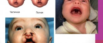
Cleft lip and palate
Among the most common congenital anomalies that occur in children are cleft lip and cleft palate. Often people have both. A cleft lip occurs when the tissue that forms the lip does not come together between the fourth and seventh weeks of pregnancy. A cleft palate occurs when the top of the mouth does not close, which occurs between the sixth and ninth weeks of pregnancy. In some children, only part of the palate remains open, while other children may have an opening across the entire palate.
A cleft palate can cause problems with speech and swallowing because the palate cannot separate the airway from the digestive tract. Some children with cleft palates may have ear infections, hearing problems, or dental problems.
Surgeons usually operate on cleft lip and palate in the first year of a child's life. However, additional surgeries will be required for complete recovery.
Tumor of the soft palate
In some cases, people may develop a tumor in the soft palate. Doctors usually find tumors on the front part of the soft palate, which is closer to the hard palate.
Injury
People can injure the palate with hot food. If the soft palate is burned, ulcers may appear in the mouth. Burns and ulcers can become infected, so doctors prescribe a 0.12% chlorhexidine solution as a mouth rinse. To help the injury heal faster, they also recommend avoiding smoking, drinking alcohol, and very hot or acidic foods or drinks. Rinsing your mouth with cold water may relieve discomfort. It is also important to be careful when brushing your teeth to avoid further injury to the roof of your mouth. Depending on the injury, doctors may prescribe antibiotics if there is a risk of infection and recommend nonsteroidal anti-inflammatory drugs (NSAIDs) for pain and inflammation.
Scientific article on the topic: Scientists have identified an important gene that is associated with the development of cleft lip and palate.
Surgery to treat oral cancer
If the results show signs of early stage oral cancer, your doctor may recommend surgery. Surgery is the most common treatment for precancerous lesions (cancers that have not yet developed into cancer) and early-stage cancers (cancers that have not yet spread to other organs). Your doctor may decide to remove precancerous lesions to prevent them from developing into cancer.
Treatment goals for early stage oral cancer include:
- cure cancer;
- maintaining your appearance and oral function;
- preventing the spread of cancer.
The type and scale of surgery depends on the location of the lesion. Your healthcare provider will tell you which area is affected, and you can then read below about your treatment options.
Before the operation, you will receive anesthesia (medication that will make you sleep). All these operations are performed through an open mouth.
Lip
The lesion and surrounding skin will be removed. The incision (surgical cut) will be closed with stitches.
Sutures used inside the mouth and on the lip will dissolve (disintegrate and fall off) on their own. Another type of suture that does not dissolve will be used on the skin. You will have a follow-up appointment to have them removed.
Language
The lesion will be removed. The scale of the operation depends on the size of the lesion. Surgery to remove part of the tongue is called a partial glossectomy.
The incision may be closed with absorbable sutures. Sometimes a flap of artificial (synthetic) skin may be used instead to temporarily cover the area from which the lesion was removed. This flap will be sutured with absorbable sutures; it will fall off on its own in a few weeks.
Hard palate and upper gum
The scale of the operation will depend on the size of the lesion and the depth of its penetration. Small lesions located on the surface (superficial tumors) are removed without subsequent suturing. New tissue will grow in this place and heal the wound.
If the lesion is large and deep, part of the roof of the mouth may need to be removed. After this, the area will be covered with a flap of natural or artificial leather. If using a natural skin flap, it will be covered with gauze secured in place with a dental plate. The dental plate will be made by your MSK dentist, who you will meet with before your surgery. The gauze and dental plate will be removed after 5-7 days.
If artificial leather is used, it will be sewn on with absorbable sutures; it will fall off on its own in a few weeks.
Soft sky
The lesion will be removed. The incision will be covered with absorbable sutures or a skin flap. If using a natural skin flap, it will be covered with gauze, secured in place with stitches. The doctor will tell you when to come for your next appointment to have your stitches removed.
Floor of the mouth
The scale of the operation depends on the size of the lesion and the depth of its penetration. Small lesions located on the surface are removed without subsequent suturing. New tissue will grow in this place and heal the wound.
Larger lesions will be removed and covered with a flap of natural or artificial skin. If using a natural skin flap, it will be covered with gauze, secured in place with stitches. The gauze will be removed after 5–7 days. If artificial leather is used, it will be sewn on with absorbable sutures; it will fall off on its own in a few weeks.
Buccal mucosa
The lesion will be removed. The incision will be closed with a flap of natural or artificial skin. If using a natural skin flap, it will be covered with gauze, secured in place with stitches. The gauze will be removed after 5–7 days. If artificial leather is used, it will be sewn on with absorbable sutures; it will fall off on its own in a few weeks.
Lower gum and area behind wisdom teeth in the lower jaw
The lesion will be removed. Depending on the depth of the lesion, the doctor may also remove a small piece of bone tissue from the lower jaw. Surgery to remove a small piece of the lower jaw is called a marginal mandibulectomy.
The incision will be closed with a flap of natural or artificial skin. If using a natural skin flap, it will be covered with gauze, secured in place with stitches. The gauze will be removed after 5–7 days.
If artificial leather is used, it will be sewn on with absorbable sutures; it will fall off on its own in a few weeks. The jawbone will not need reconstruction and the shape of the jaw will not be affected.
to come back to the beginning