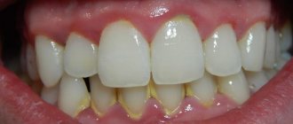
Oral leukoplakia is a disease of the oral mucosa in which plaques of whitish, gray or yellowish shades form. They (plaques) are localized on the tongue, the inner surface of the cheeks, the palate, and also in the corners of the lips. The keratinized epithelium rises slightly above the mucous membrane and brings a lot of physical and aesthetic discomfort to the patient. Plaques can form from 7 days to several months. The risk group for this disease is men aged 30-40 years, but recently the disease has also been diagnosed in young children. The main threat of oral leukoplakia is the prospect of transformation into a cancerous tumor. That is why, in case of alarming symptoms, it is necessary to urgently contact an experienced dentist.
Causes of oral leukoplakia
- taking strong medications (in particular, antibiotics, as well as constant use of drugs of various spectrums of action in large quantities);
- smoking, as harmful components penetrate through cigarettes and deplete the mucous membrane;
- poor nutrition and abuse of spicy, salty and sour foods;
- chronic gum disease in an advanced stage;
- abundant carious lesions of teeth;
- incorrectly placed fillings;
- incorrectly selected dentures that cause an allergic reaction;
- alcoholism;
- hormonal changes;
- bite problems;
- diabetes;
- HIV infection;
- avitaminosis;
- diseases of the stomach and intestines;
- stress;
- hereditary predisposition;
- unfavorable environmental conditions;
- harmful production activities.
Typically, at the initial stage, oral leukoplakia can be completely asymptomatic. That is why it is very important for people who are susceptible to risk factors to undergo regular dental examinations for prevention. The 32 Dent dental network will help you, even if the disease is in an advanced stage.
Causes of the disease
Doctors do not have a consensus on why leukoplakia develops [1, 2]. Among the main factors are external stimuli and internal disorders in the body.
- Mechanical effects on the oral mucosa are too frequent and systematic:
- malocclusion and other defects in the location of teeth;
- caries poor-quality or incorrectly placed fillings;
- destroyed crowns, chipped teeth;
- improperly manufactured and fixed dentures;
- galvanic current if the prostheses are made of dissimilar metals;
- bad habits: constant biting of lips, gums, inner surface of cheeks, tongue, as well as the habit of chewing a pen or pencil;
- too hard, rough food
- Smoking and chewing tobacco.
- Abuse of strong alcohol.
- Hot and spicy foods - in large quantities.
- Household chemicals and harmful factors in production that irritate the mucous membranes.
- Elevated temperatures: too hot food, inhaling hot air in workshops, burning the lips with a cigarette for those who like to finish smoking.
- Unfavorable climate: too dry and hot, with frequent winds.
- Disturbances in the gastrointestinal tract (for example, chronic gastritis and colitis), due to which the mucous membrane becomes more sensitive to external factors.
- Vitamin A deficiency.
- Hereditary predisposition to keratinization processes and tumor diseases [6]
- Male gender: studies show that the development of leukoplakia is influenced by sex steroid hormones, namely high levels of free testosterone [2, 3, 8].
- Human papillomavirus.
- Chronic inflammation of the oral mucosa.
- Weakened immunity, including HIV infection [4].
Types of oral leukoplakia
- Verrucous - the epithelium has a pronounced keratinized layer, the plaques rise noticeably above the mucous membrane, the plaques acquire a gray tint, sometimes yellowish. This type of disease is considered one of the most threatening, as it can transform into an oncological process.
- Soft – a form in which the top layer of plaques peels off. The disease belongs to the category of benign tumors.
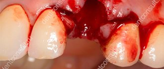
- Erosive - accompanied by erosions, deep cracks in the plaques. Gradually, there are more plaques, they spread over the entire surface of the mucous membrane, the person feels great discomfort, even breathing problems.
- Flat - characterized by the presence of flat plaques that do not rise above the mucous membrane. Erosions have a rough surface, the patient feels pain and discomfort.
What it is
It is believed that leukoplakia occurs against the background of chronic irritation of the oral mucosa and is a kind of protective reaction of the body. This is a local reaction and is not transmitted through contact with a sick person.
The disease is not so harmless, since it most often occurs shortly before the development of oral cancer. Many people mistakenly believe that leukoplakia itself is cancer, however, this opinion is incorrect. This disease affects the epithelial covering of the oral cavity in response to constant external irritants; vitamin deficiency, a decrease in the level of immunity and the presence of chronic foci of inflammation in the oral mucosa serve as an additional impetus for its development.
Leukoplakia of the oral cavity is a significant lesion of the mucous membrane of the lips, tongue, cheeks, upper and lower palate, manifested by keratinization (hardening) of the epithelial cover to varying degrees. Doctors usually classify leukoplakia as a disease that precedes the development of a tumor.
The greatest risk of developing this disease are men in the age group from 30 to 70 years, who abuse alcohol, smoke and wear dentures. Leukoplakia does not occur overnight, and its development can last for years.
Symptoms of oral leukoplakia
- plaques located on the mucous membrane of the palate, the inner surface of the cheeks, on the tongue, along the edges of the lips;
- erosions, cracks from which liquid may ooze;
- the plaques have uneven, sharp edges, first acquire a white and then a grayish, yellowish color;
- pain, burning, tingling in the mouth;
- increased sensitivity to cold, hot, salty, sour foods;
- problems with chewing food, swallowing;
- temperature increase;
- bad breath;
- general malaise, weakness, irritability.
To diagnose the disease, the doctor must perform a visual examination. It may be necessary to perform a cytological examination of scrapings from the mucous membrane to exclude a malignant tumor process.
Although leukoplakia can often occur with minimal symptoms, it is very dangerous. In medicine, it is generally accepted that this condition is precancerous. If these symptoms appear, you should immediately contact your dentist. The doctor will assess the patient’s health condition and prescribe adequate treatment.
If you have a problem similar to that described in this article, be sure to contact our specialists. Don't diagnose yourself!
Why you should call us now:
- We will answer all your questions in 3 minutes
- Free consultation
- The average work experience of doctors is 12 years
- Convenient location of clinics
Single contact phone number: +7
Make an appointment
Types and forms
The classification of typical signs of the disease is divided into the following types of leukoplakia:
| Verrucous | In the absence of timely treatment and elimination of external irritants, verrucous leukoplakia develops from flat leukoplakia. Due to the acceleration of the process of keratinization of epithelial cells, the affected dense area begins to stand out strongly above the level of healthy tissue and acquires a characteristic white color. In this case, the patient may complain of constant discomfort in the oral cavity, since the plaques have a hard, warty surface. This type of leukoplakia often develops into a malignant form. |
| Erosive | Often, patients develop erosion on the lateral surface of the tongue or in the corners of the mouth; leukoplakia in such cases passes from the verrucous to the erosive stage. In this case, the patient has difficulty opening his mouth, and eating becomes very difficult and painful for him. The erosive form of the disease ends in the formation of cancerous tumors more often than other forms. |
| Smoker's leukoplakia (Tappeiner) | It occurs in people who are prone to regular smoking. A characteristic manifestation is a white coating with red dots. Such formations rarely develop into a malignant tumor; the best method of treatment is complete cessation of cigarettes. |
| Soft leukoplakia Pashkova | It is characterized by a benign lesion of the epithelial surface of the oral cavity (most often in the lips and cheeks), associated with heredity or with frequent neuropathic biting of the affected areas. Most often, Pashkov's soft leukoplakia is observed in children and young people under 30 years of age and is not accompanied by unpleasant or painful sensations. It is diagnosed during a simple routine examination at the dentist. If the seals are frequently bitten, roughness and peeling appear, the damaged area may have a grayish color, and erosions may occur. |
| Flat | Flat leukoplakia is the most common. In most cases, it does not cause a person any inconvenience or pain, so it is diagnosed only during a routine preventive examination by a doctor. Sometimes the patient may complain of constant dryness of the oral mucosa, which leads to increased fluid consumption. This type of mucosal lesion is determined by one or several white spots with clearly defined edges, and the disease itself can accompany a person for many years without causing any inconvenience or developing. |
| Simple leukoplakia without atypia | It is diagnosed by the presence on the oral mucosa of a thin whitish film or dense plaques with pronounced contours. By removing the keratin layer, you can see the actual size of the lesion in simple leukoplakia. In most cases, the simple form is asymptomatic and does not lead to tumor formation, however, patients are recommended to be observed by a specialist and periodically undergo histological tests to exclude cellular activity and atypia of the affected area. |
How to properly treat oral leukoplakia?
- urgently eliminate provoking factors (alcohol, cigarettes, salty, spicy foods, sour);
- perform applications with solutions of vitamins A and E to extinguish inflammation;
- treat plaques with special healing solutions and ointments;
- take vitamin complexes;
- adjust your diet (fill your diet with foods high in fiber, eat fruits and vegetables).
Among non-invasive techniques, modern dentistry offers laser therapy, as well as cauterization of plaques with nitrogen. As practice shows, such safe and affordable methods are effective. You will receive more information about the effective treatment of oral leukoplakia during a personal consultation with an experienced 32 Dent dentist. Make an appointment at a time convenient for you!
Leukoplakia is a keratinization of the oral mucosa (OM), accompanied by inflammation of the stroma and usually occurring in response to chronic exogenous and endogenous irritations [15, 35].
The term “leukoplakia” was first used in the works of Schwinmer (1977), although detailed clinical characteristics of this process were proposed in the second half of the 19th century under different names: ichthyosis, psoriasis, keratosis, etc. [15, 41].
Epidemiology of leukoplakia of the oral mucosa
The overall prevalence of leukoplakia SOP, according to American authors, is 0.5-3.46%, and the incidence of malignancy is 0.7-2.9% [108]. Leukoplakia SOP is more common in India and the southern states of America, where smoking and other use of tobacco and betel nut are more common than elsewhere [81, 96, 116].
Leukoplakia SOP is usually diagnosed in middle age, and the prevalence of the disease increases over the years. Idiopathic leukoplakia of the oral cavity accounts for 10% of cases, and leukoplakia caused by exogenous and endogenous factors accounts for 90% [125]. Men are more often affected than women. The mucous membrane of the cheeks is affected in 25% of cases, the gingival edge of the jaw - in 20%, the tongue - in 10%, the floor of the mouth - in 10%; other areas account for the remainder of the lesions [77, 78, 121, 126].
Etiology and pathogenesis of leukoplakia SOP
There are many studies devoted to this disease [6, 36, 40, 48, 54, 80, 94], but the question remains open about the causes of abnormal keratinization of the oral cavity. Many authors [30, 49, 12, 123] consider tobacco smoke or chewing nas, hot food and alcohol to be one of the main exogenous factors. However, in some sources you can find a description of leukoplakia in patients who never smoke or rarely smoke [118, 134]. Hot, hot, spicy foods can also cause the development of the disease. With strict dietary restrictions, it is possible to prevent the development of leukoplakic plaques, and sometimes even cause their complete disappearance.
The reasons for the development of leukoplakia can be chronic traumatization of the oral cavity by sharp edges of decayed teeth, improperly manufactured dentures, or galvanic current that occurs in the presence of dentures made of dissimilar metals [19].
A certain role in the etiology of leukoplakia of the lower lip belongs to glandular cheilitis in combination with unfavorable meteorological conditions, as a result of which the red border is subject to constant irritation [7, 127]. In addition, when leukoplakia is localized on the red border of the lips, chronic injury from a mouthpiece, cigarette, pipe or cigarette is of great importance.
Leukoplakia SOP can occur as an occupational disease in people working in chemical production or in workshops with high temperatures [122].
Considering leukoplakia as a reaction of impaired keratinization of the oral cavity to various external stimuli, one cannot deny the influence of endogenous factors, which, in turn, disrupt the resistance of the mucous membrane to external stimuli [93].
The decrease in resistance, apparently, may be associated with pathology of the gastrointestinal tract - GIT [12]. According to other authors [13, 85], chronic gastritis, gastric and duodenal ulcers were observed in 90.3% of patients observed for leukoplakia, but the clinical picture of leukoplakia and gastrointestinal pathology did not reveal significant parallels between them.
Lack or disturbance of the metabolism of vitamins A and E, the role of which in the process of keratinization is well known [45, 58, 99], may be one of the causes of leukoplakia.
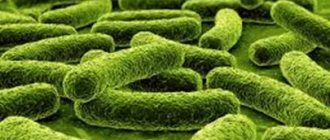
Some authors trace a connection between the development of leukoplakia and chronic candidiasis infection [72, 132]. During its life activity, Candida albicans
forms N-nitroso-benzymethylamine (NBMA), which can promote malignancy of leukoplakia lesions [62, 67, 70, 111].
Ya.L. Makarov (2004), R.I. Stryuk (2008) assigns a certain role in the occurrence and malignancy of the disease to endocrine disorders. Thus, when examining patients with leukoplakia, diabetes mellitus types 1 and 2 were detected in 15% of cases. And in women, in some cases, there was a connection between changes in progesterone levels and the occurrence of leukoplakic plaques in the mouth.
Genetic factors occupy a certain place in the pathogenesis of leukoplakia SOP, which is confirmed by the development of this disease in patients with congenital and hereditary dyskeratoses. The risk of developing leukoplakia was associated with p53 protein expression and the simultaneous presence of the GSTM1o and GSTT1o genotypes.
Currently, the question of whether there is a connection between the occurrence of oral leukoplakia and the detection of human papillomavirus (HPV) remains controversial [46, 47, 114].
HPVs that infect the oral cavity are divided into low-risk viruses for malignancy (HPV 6, 11, 13, 32) and high-risk (HPV 16, 18, 31, 33 and 35) [53, 97].
This division of HPV is universal for studies of oncogenic significance in all areas of the upper digestive tract [56, 68].
According to foreign authors [79, 110], high-risk viruses, especially HPV 16, were detected in verrucous leukoplakia. The probability of the presence of HPV in the intact mucosa in patients with simple leukoplakia and leukoplakia verrucous was 10, 20.2 and 26.2%, respectively [129].
In addition to the above, in patients suffering from various forms of leukoplakia, an imbalance of the immune system was sometimes detected [43, 102], characterized by the appearance of cytotoxic lymphocytes and macrophages in the peripheral blood. Interleukin-2 alone or in combination with monocytes exhibited increased cytotoxicity, especially in verrucous leukoplakia. When studying immune complexes in people chewing tobacco, compared to controls, their number was slightly higher, and the activity of natural killer (NK) cells decreased. Thus, it is possible that T-cell-mediated cytotoxic reactions may occur in leukoplakia, which is apparently explained by the general immunological response to the oncogenic form of the inflammatory process in the mouth [113].
Summarizing the data on this issue, it should be noted that this information is contradictory and fragmentary.
Clinical and morphological characteristics of leukoplakia
The clinical picture of leukoplakia depends both on the form of the disease and the factor causing it, as well as on the location. The course of leukoplakia from the moment of its appearance to the onset of malignancy is characterized by several stages [32, 115].
The process usually begins with the so-called pre-leukoplakic stage, which is characterized by inflammation of the oral cavity, and extremely rarely this stage can be observed on the red border of the lips [16].
Then flat leukoplakia develops, which is a uniform keratinization of a limited area of the mucous membrane, which is the result of epithelial hyperplasia and is accompanied by chronic inflammation of the stroma. More often, foci of flat leukoplakia are located on the mucous membrane of the cheeks, in the corners of the mouth, sometimes on the floor of the mouth, having the appearance of a plaque of color: from grayish to grayish-white; the plaque does not rise above the level of the surrounding mucous membrane and is not removed by scraping.
As the process progresses, foci of leukoplakia begin to rise above the level of surrounding tissues and can take on a lumpy appearance. Such a lesion is characteristic of verrucous leukoplakia.
Verrucous leukoplakia comes in two types: plaque and verrucous. In both of these forms, the plaques are grayish-white or milky in color, sharply protruding above the surrounding mucosa, and have a dense consistency upon palpation. Localization of lesions is most often the dorsal and lateral surface of the tongue, the buccal mucosa, rarely - the lingual surface of the alveolar process and the floor of the mouth. Malignancy is observed in 20–23% of cases [91, 130].
With the verrucous form of leukoplakia, cracks and erosions may appear, which are systematically exposed to thermal or mechanical irritation. This is the so-called erosive form, the most prone to malignancy (up to 25-27% of all cases of other forms of leukoplakia) [34].
Tappeiner's smokers' leukoplakia, or nicotine stomatitis, occurs mainly on the mucous membrane of the hard palate in patients who smoke regularly and frequently use a pipe. The mucous membrane of the hard palate is whitish or white, against which reddish dots are clearly visible - the gaping mouths of the excretory ducts of the minor salivary glands. This disease was described by Grutz in 1928, and in 1941 Thoma described a similar clinical picture called “nicotine stomatitis”. But in 1949, it was Tappeiner who gave a detailed clinical and histological description of this form of leukoplakia, noting that this pathology is based on damage to the excretory ducts of the minor salivary glands, and not the salivary glands themselves.
A characteristic feature of leukoplakia, found in all its forms, is diffuse inflammation, often accompanied by significant infiltration of the upper part of the stroma [103].
In the flat form of leukoplakia, parakeratosis is observed, the thickness of the spinous layer is 40-50 rows of cells, and acanthosis is noted. There is no granular layer. Thus, parakeratosis in leukoplakia is often combined with epithelial hyperplasia.
With verrucous leukoplakia, severe hyperkeratosis is determined, rarely combined with small foci of parakeratosis, sometimes the stratum pellucida is involved in the pathological process; the granular layer consists of 4-5 rows of cells with well-defined granularity, the spinous layer - of 8-12 rows of cells. Sometimes atypical cells are detected. In some cases, acanthosis is expressed, accompanied by elongation and expansion of epithelial processes.
Much more often, such discomplexation of the cells of the spinous layer and cellular atypia are expressed in the erosive form of leukoplakia, in which the inflammatory reaction in the stroma reaches a maximum (dilated lymphatic and blood vessels, a sharp change in the walls of deep-lying stromal vessels) [92].
When malignancy of leukoplakia begins, in addition to the elongation of epithelial processes far beyond the physiological border, discomplexation of the cells of the lower rows of the spinous and basal layers, destruction of the basement membranes is observed, and an increase in the number and size of nucleoli in the cells of the lower rows of the spinous layer occurs.
Thus, the features of malignant transformation of the epithelium of the oral cavity can be difficult to assess, so it is necessary to look for additional diagnostic criteria that allow more accurately determining the degree of dysplasia of the epithelium of the oral cavity [18, 42, 103].
Currently, to diagnose dysplasia of the epithelium of the oral mucosa, an immunohistochemical (IHC) method is used, which makes it possible to more accurately determine the degree of dysplasia (SIN1, SIN2, SIN3). Markers of malignant transformation are the Ki-67 protein, the acanthosis marker - p53 protein, the cytoskeletal proteins - cytokeratin-8 and -19, E-cadherin and β-catenin, as well as the basement membrane protein - type IV collagen and the enzyme that breaks it down - matrix metalloproteinase-9. Histological and IHC research methods are necessary for the diagnosis of various forms of leukoplakia.
One of the main trends in modern medicine is the improvement of non-invasive methods for diagnosing pathological processes accompanied by changes in tissue structure. The standard method, as already mentioned, is histological examination of tissue biopsies. Taking a biopsy from the periphery of the tumor, where all stages of the process from cancer in situ to dysplasia of various degrees can be observed, according to G. Ghurani (2001), is the most common cause of false negative biopsy results. Therefore, information about the internal structure of biological tissues is important both for diagnosing diseases and for adequate monitoring of treatment results.
Routine imaging methods - computed tomography and magnetic resonance imaging - allow one to assess the structural features of tissues with a spatial resolution of no higher than 100-1000 µm [60, 128]. Recently, attempts have been made to bring the resolution of methods closer to the cellular level (≈10 μm), which became possible for nuclear magnetic resonance, confocal optical microscopy and optical coherence tomography (OCT) [39, 51, 52, 57, 65, 66, 105 ].
The essence of the OCT method is to display the structure of biological tissues of the body (in particular, the SOP) in a cross section with a high level of resolution, which provides intravital morphological information at the microscopic level. The operation of OCT is based on the principle of low-coherence interferometry [17, 20, 28, 71].
Possessing all of the listed characteristics, OCT in the coming years may find wide application in dentistry as a non-invasive method for diagnosing various degrees of oral dysplasia.
Treatment of leukoplakia SOP
Treatment of leukoplakia is a difficult task, since this disease is a precancerous condition [37]; it is necessary to talk about a differentiated medical approach to different forms of the disease, because each of them has a different potential for malignancy. It follows that treatment methods can be divided into: medical, surgical and combined [74].
However, an indispensable condition for the treatment of various forms of leukoplakia is, first of all, the cessation of exposure to exogenous stimuli. This primarily applies to smoking, chewing tobacco, drinking alcohol and eating irritating, hot and spicy foods.
No less important in the treatment of leukoplakia is sanitation of the oral cavity, removal of decayed teeth, grinding of sharp edges of teeth, orthopedic treatment, including rational prosthetics and replacement of dentures made of dissimilar metals.
Local treatment consists of prescribing keratoplasties (vitamin A, E, β-carotene), which normalize the processes of keratinization and improve metabolism in the affected tissue.
A number of authors note a pronounced effect of topical application of antifungal agents (10% borax in glycerin, Clotrimazole cream, etc.). J. Epstein, F. Wong [64] used the cytostatic drug bleomycin locally as an additional treatment for severe forms of leukoplakia SOP, when surgical excision for one reason or another was impossible.
General drug therapy includes the administration of large doses of vitamin A. It is known that vitamin A helps normalize the synthesis of DNA and RNA of epithelial cells, accelerating the proliferation and differentiation of keratinocytes.
Currently, vitamin A derivatives—retinoids—are widely used [64]. By influencing the genetic apparatus of the cell, they normalize the processes of proliferation and differentiation of the epithelium and mesenchyme, and affect the condition of cell membranes.
In medical practice, tigazone is used, isatretinoin (13-cis-retinoic acid) in a daily dose ranges from 25 to 50 mg; course of treatment up to three weeks.
In addition to vitamin A and its analogues, some authors [38, 117] prescribe vitamin E and C in combination with B vitamins and folic acid. It is well known that vitamins B and C are natural antioxidants, regulate redox reactions, regenerate tissues, normalize hypoxin and regulate cell proliferation processes.
As mentioned above, work in recent years indicates the role of some biotypes of Candida albicans in the processes of malignancy and a decrease in local immunity, which contribute to the development of leukoplakia, therefore the inclusion of anti-candidiasis drugs in the treatment process is justified [100].
A number of authors [89, 95] include general antimycotic agents (Flucostat, Diflucan, Nizoral, etc.) in complex treatment. The drugs are prescribed orally for 7-10 days, the dose varies from 50 to 100 mg/day.
Some domestic and foreign authors [11] in their works in the treatment of leukoplakia SOP mention drugs called “adaptogens,” in particular the phytomixture Fitomix-40. This drug contains 40 components that have anti-inflammatory, diuretic, choleretic, and sedative effects.
According to the work of I.K. Evseeva [10], drugs such as clanin and phytolan were included in the complex treatment of leukoplakia SOP. Belonging to the group of dietary supplements, they are made from extracts of pine needles and kelp and have pronounced antioxidant properties. The drugs are taken 2 tablets 3 times a day or 50-60 drops 2-3 times a day after meals.
Speaking about the occurrence of leukoplakia in men and women at a certain age, L.V. Petrova et al. (2004) used the drug spironolactone, which is a competitive antagonist of aldosterone. The drug was prescribed at 200 mg/day with a gradual dose reduction to 50 mg/day. General course - 4 months.
The works of some authors [24], as indicated above, are devoted to the detection of changes in the gastrointestinal tract and diabetes mellitus in patients with oral leukoplakia; therefore, treatment of these background types of pathologies is necessary. B.M. Pashkov (1969), A.L. Mashkilleyson [15], V.M. Abramova (1984) recommend treatment of patients with leukoplakia SOP by a gastroenterologist, endocrinologist, general practitioner with the prescription of drugs panzinorm, pyridoxine and their analogues.
Based on the results of studies on disorders in the immune system in patients with leukoplakia SOP, a number of authors [98, 107, 131] used interferon-α, which, in their opinion, controlled the course of leukoplakia and prevented its malignancy, which can probably be associated with its effect on NK activity.
Thus, the data presented indicate that the elimination of exogenous factors and the use of local therapy gives a positive result in the treatment of flat leukoplakia and Tappeiner’s leukoplakia. In severe forms of leukoplakia, in addition to local therapy, the prescription of general medications and surgical treatment methods is required. Surgical intervention, especially for verrucous and erosive forms of leukoplakia, is a common method of treatment today [2-5], especially when the size of the lesion allows it to be excised within healthy tissue and at the same time it is possible to conduct a thorough histological examination to identify the onset of malignancy of leukoplakia [133] .
According to some authors [69], the main method of surgical treatment is excision of the area of leukoplakia with a scalpel. It should be noted that this method is indicated when the size of the leukoplakic lesion clearly contours to the healthy mucosa and access to them is not difficult.
At one time, electrocoagulation and cryodestruction, which can be classified as physical methods of treating leukoplakia, were widespread [84, 101, 106].
A number of authors [73] have successfully used helium-neon laser in combination with cryotherapy on lesions. L. Gaspar, G. Szabo (1990) used laser surgery in 126 patients with leukoplakia SOP and clinical cure was obtained in 118. But subsequently, in 98 patients, within 5 years, a relapse of the disease occurred at the previous sites of the lesion. A relatively high relapse rate was observed by M. Schoelch et al. (1999): 41 out of 70 patients had a relapse, and 5 had malignancy of the process.
Nevertheless, laser surgery is widely used in the treatment of this pathology of the oral cavity [44]. The use of modern laser methods [50, 104, 109] makes it possible to achieve high-quality results in the treatment of severe forms of leukoplakia (verrucous and erosive).
The rapid development of laser technologies in the 90s of the last century made it possible to introduce into widespread practice gentle, minimally invasive methods of treating leukoplakia SOP, one of which is photodynamic therapy (PDT) [8, 33, 60].
PDT is based on the ability of a photosensitizer to selectively accumulate in tumor tissue [21, 22, 26, 29]. In the presence of oxygen, under the influence of laser light with a wavelength corresponding to the type of absorbed photosensitizer, a photochemical reaction develops, which leads to the formation of singlet oxygen and other oxidants, followed by selective death of tumor cells. Healing of the tissue defect occurs with the formation of a gentle scar, which preserves the surrounding healthy tissue [55, 120].
In Russia, the domestic drug Photogem is used for PDT [23, 25], developed at the Moscow State University of Fine Chemical Technologies named after. M.L. Lomonosov, as well as the photosensitizer Photoditazin, which has several absorption bands at wavelengths of 400, 504, 534, 608 and 662 nm.
A number of authors used Photosan, administered intravenously at a dose of 2.0 mg/kg in a darkened room. The PDT session was performed 3 hours after drug administration; The irradiation time for 1 field was 10 minutes, the power did not exceed 167 mV/cm2. As a rule, a delicate whitish scar formed at the site of the resorbed tumor.
Currently, PDT is used as a stand-alone method or in combination with chemoradiotherapy or combined treatment for cancer of the oropharynx, oral cavity, tongue and lower lip [59, 60, 81]. PDT has shown positive results in the treatment of leukoplakia SOP as a precancerous disease [27, 83, 87, 88].
A number of American scientists [75, 86, 119] attempted to treat verrucous leukoplakia using local PDT with the photosensitizer Photofrin and ALA.
Today, PDT is approved by oncologists as a method of polytherapy for the initial stages of cancer, “inconvenient” localizations of lesions on the face, ears, as well as for combined and complex treatment in cases of recurrent cancer of the skin, lower lip and tongue [124].
Laser and cryodestruction
One of the safest and most effective is the freezing method . Cold cauterization is performed using nitrous oxide. This method allows leukoplakia to be removed as painlessly and accurately as possible, without damaging adjacent tissues.
Laser treatment is also very popular, although it is quite expensive, since it is bloodless and its use, like cryotherapy, does not damage healthy tissue of the mucous membrane. This method does not require preliminary anesthesia and can be used in ordinary well-equipped dentistry. For uncomplicated cases of leukoplakia, only one method of therapy is suggested, however, for more serious lesions, laser treatment or cryotherapy is recommended to be combined with a course of antibiotics, hormonal drugs, and immunomodulators.
Is it possible to cure leukoplakia with the same methods if there is severe deformation of the mucosal surface? , a surgical scalpel is most often used . In this case, before starting treatment, the doctor must conduct a biopsy and find out whether atypical cancer cells are present in the sample. Depending on the result of the analysis, further postoperative therapy is prescribed.