Many people ask what an orthopedic surgeon does, or what an orthopedic surgeon treats. Orthopedic doctors use both surgical and non-surgical treatments to treat musculoskeletal disorders, which include sports injuries, spinal disorders, degenerative diseases, infections, tumors and congenital disorders. In this article, we will answer these questions and clearly show what orthopedic doctors do in detail. There are many surgical and non-surgical treatments that the surgeon/orthopedic doctor uses to relieve a patient's pain and here we will briefly review these treatments. After answering the questions “who is an orthopedic surgeon/doctor?” and “what does a surgeon/orthopedist do?” We will check surgical methods and non-surgical treatment methods in detail. These topics and techniques include: arthroscopy, fusion, internal fixation (implant fixation), joint replacement, osteotomy, soft tissue reconstruction, injections and pain management. These topics practically cover all the activities that an orthopedic doctor does to treat a patient's painful part of the musculoskeletal system of the body.
Who is an orthopedic doctor?
An orthopedic physician is a physician's specialist in the diagnosis, treatment, prevention and rehabilitation of disorders, injuries and diseases of the body's musculoskeletal system. The musculoskeletal system includes bones, nerves, joints, ligaments and tendons. In other words, when you have pain in your hand and wrist, foot and ankle, knee, shoulder and elbow, neck, back and hips, you should see a podiatrist. Orthopedic doctors use both surgical and non-surgical means to treat the musculoskeletal system of your body. Non-surgical treatment may include the use of medications, exercise and other types of rehabilitation or alternative treatments. Orthopedic doctors are specialized to treat all aspects of the musculoskeletal system, however many orthopedic doctors specialize in specific areas, such as the arm, shoulder, elbow, spine, hip, knee or foot and ankle.
When to ask for help
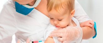
Visiting an orthopedist should become a regular procedure. Get a general medical examination 1-2 times a year to monitor your own body and prevent illnesses in time. Bone disorders can develop over years without causing much discomfort or pain to the patient. That is why frequent diagnosis is the key to the health of the musculoskeletal system.
An unscheduled consultation with an orthopedist may be needed unexpectedly. Have you or your child been injured while playing sports, are experiencing discomfort in the limbs for no apparent reason, or have simply been out in the cold a little longer than expected? Consult an orthopedist to rule out serious illnesses or treat them immediately.
Best materials of the month
- Coronaviruses: SARS-CoV-2 (COVID-19)
- Antibiotics for the prevention and treatment of COVID-19: how effective are they?
- The most common "office" diseases
- Does vodka kill coronavirus?
- How to stay alive on our roads?
Sprains, bruises, animal bites, deformation of the limbs/spine/chest/functionality of small or large joints should also prompt a person to undergo urgent diagnosis.
The main rule is that you cannot tolerate pain. Crunching in the joints, numbness or swelling of the hands, painful movement, occasional aching pain throughout the body, rapid fatigue or poor posture significantly affect the quality of human life. Protect your own comfort, do not wait for the pain to go away on its own or develop into chronic pain - visit an orthopedist.
People who engage in active sports, enjoy extreme recreation, or work in exhausting conditions (for example, in production) should visit an orthopedist more often, as they are at risk.
What does an orthopedic doctor do?
Musculoskeletal pain is the main reason why people visit doctors every year. There are many musculoskeletal conditions and injuries that can cause pain, and often orthopedic doctors can help reduce or eliminate the pain. As you may know, orthopedic doctors treat broken bones and replace painful joints, but they also treat patients with problems such as:
- Sports injuries
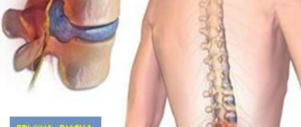
- Back pain, ruptured discs and spinal stenosis
- Bone tumors
- Carpal tunnel, hand arthritis and hand injuries
- Club foot, nasal and hip dysplasia
- Orthopedic trauma
- Limb lengthening
- Achilles tendon injuries, joint injuries and foot and ankle injuries
- Osteoporsis
- Arthritis
As you can see, orthopedic doctors repair torn tendons and ligaments, as well as treat sprains and sprains. They may also deal with fractures and dislocations. They often counsel patients on how to strengthen weakening muscles and perform surgeries. Some of these surgeries repair broken bones and soft tissue, while others correct birth defects and injuries that develop due to disease or infection. But what orthopedic doctors do is not limited to these subjects. What does an orthopedic surgeon/doctor do? Here's the short answer:
- arthroscopy : a process using a camera to visualize inside a joint
- Fusion : A process using internal devices or rods in which bones are fused together.
- Internal fixation : holding broken pieces of bone together with plates, screws, or pins.
- Joint Replacement : A process in which a damaged joint is removed and replaced with an artificial joint, partial, complete, or augmented.
- Osteotomy : Correction of bone deformities by cutting and repositioning the bone.
- Soft tissue repair : repair of tendons or ligaments.
- Injections and pain relief. In this list you can find a short answer about each activity. As you can see, there is not much detail here, and the tasks and activities of orthopedic doctors are not limited to these subjects.
Orthopedics
The meaning of the word Orthopedics according to Efremova: Orthopedics is a branch of medicine that deals with the study, treatment and prevention of diseases of the organs of movement and support: arms, legs, spine.
The meaning of the word Orthopedics according to Ozhegov: Orthopedics is a branch of medicine dealing with the prevention and treatment of deformities of the spine, limbs, as well as deformations of the body in general
Orthopedics in the Encyclopedic Dictionary: Orthopedics - (from the Greek orthos - direct - correct and paideia - education), a section of clinical medicine that studies congenital and acquired deformations and dysfunctions of the musculoskeletal system and develops methods for their prevention and treatment. Together with traumatology, it forms a single medical specialty.
The meaning of the word Orthopedics according to the dictionary of medical terms: orthopedics (orthopaedia; ortho-Greek paideia education, training) is a field of clinical medicine that studies diseases and deformations of the musculoskeletal system and develops methods for their diagnosis, treatment and prevention.
The meaning of the word Orthopedics according to Ushakov’s dictionary: ORTHOPEDICS
orthopedics, g. (from Greek orthos - correct and paideia - education) (med.). Department of surgery dealing with the treatment of painful curvatures of the torso and limbs.
The meaning of the word Orthopedics according to the Brockhaus and Efron dictionary: Orthopedics
(definition and history) - the science of recognizing, preventing and treating distortions of the human body, i.e.
persistent deviations of individual parts of the latter from their normal form and direction. It, unlike teratology (see), is limited to the study of only disfigurements, expressed by anomalies in the position and shape of parts of the skeleton. Hippocrates already left several works with a detailed description of the curvatures of the spinal column, which he treated with specially adapted devices, clubfoot, congenital dislocations of the hip and ankle joints, etc. Galen used various types of hunchbacks by bandaging the chest; Antillus (at the end of the 3rd century AD) cut tendons for ankylosis and contractures. Then O. was completely forgotten and only Arab doctors (especially Albukazem at the beginning of the 12th century) continued to study it. It was revived again only in the 16th century, thanks mainly to French surgeons who brought it out of oblivion. The famous Ambroise Paré, in his book on deformities, not only describes deformities, but also offers ingenious prosthetics and apparatus for club feet. He was the first to publish an essay on the causes and treatment of spinal curvatures and recommended corsets made of perforated tin to keep the body in a straight position. At the same time, the Spanish surgeon Arceus treated clubfoot with special devices. At the beginning of the XVII century. Fabricius Gildanus eliminated curvatures of the spinal column with special machines he invented. The success of studying the causes of curvatures was greatly influenced by the work of the English physician Glisson on rickets (see), which appeared in 1660, which not only describes all rachitic changes, but also methods of treating them with gymnastics and supporting devices. Before him, in 1652, Minius, in one case of torticollis, (see below) cut the sternocleido-mamillary muscle (musc. sterno-cleido-mostoideus) - an operation that had not been performed since the time of Antillus. But as an integral science, the doctrine of deformities has existed only since 1741, when the Englishman Andry grouped all the material that had accumulated before him, added to it what he personally collected and published the book: “The Art of Preventing and Improving Body Disfigurements in Children.” He was the first to give this science the name O. (from όρθός - straight and παϊς - child), a term that has remained in practice to this day (names have been proposed: orthomorphy, orthozomatia, orthporaxia). The first orthopedic institution was founded in 1780 by the Swiss doctor Venel. The most famous was the Heine Orthopedic Institute in Würzburg (in 1812). Heine, based on the fact that only a mechanic could find use in O., improved and himself invented all kinds of apparatus and mechanical devices, so that, thanks to him, O. more and more passed into the hands of toolmakers, which caused a reaction from others doctors who successfully proved that neither massage, nor gymnastics, nor various surgical techniques should be forgotten in orthopedic treatment. In 1825, the first special body on O. arose: “Journal clinique sur les difformité s dans le corps humain est susceptible.” Almost at the same time, operative O. began to develop successfully with the introduction of subcutaneous transection of tendons and muscles, which some doctors were carried away to the point of impossible abuse. In 1826, the American doctor Rea-Borton performed the first osteotomy for ankylosis: in 1827, Esterlen improved osteoclasia (see). In 1837, the Royal Orthopedic Hospital was founded in London, which provided rich material for the scientific improvement of O. Our famous surgeon N. I. Pirogov rendered enormous services, recognized by all, to scientific O. with his important histological studies of tenotomy, which appeared in 1840. Currently, orthopedic surgery is flourishing especially in America, where there are numerous orthopedic hospitals and where it has achieved extraordinary technical perfection. In the same way, brilliant successes have been achieved in Germany, England and everywhere where the level of modern surgery is very high. In Russia, the first orthopedic institution arose in 1859 in St. Petersburg. It was widely developed by Dr. K. K. Reyer and a number of his students (Prof. Velyaminov, Turner, Dr. Horn, etc.). O. deals with various curvatures of the human body: such deviations occur very often, but in mild degrees of development they do not attract attention and therefore elude medical observation. So, for example, of those examined during 1893-94, 2254 pupils of St. Petersburg. of women's institutes, 825 (36.6%) turned out to be so incorrectly built in relation to the spinal column that they were prescribed medical orthopedic gymnastics (“Medical Report on the Department of Emperor Mary”, St. Petersburg, 1896, p. 34). To Moscow institutes in the same year, out of a total number of 1684 pupils, curvatures of various kinds were found in 481 (28.5%; ibid., p. 44). But very few seek medical help. So, for example, out of 67919 patients who applied during 1879-1839. to the Munich surgical clinic, there were 1444 (2.13%) with curvatures. At the same time, a curious fact was noted, which is of great importance for school hygiene, that the vast majority of cases of curvature occur in the first decade of life (almost 42% of the total number of patients); persons aged 10-20 years are already much less affected (33.3%), and in general the frequency of curvatures decreases with age. From the same data, it turned out that the majority of curvatures (88.7%) were acquired during life and only a ninth part were congenital. As for the distribution of curvatures according to the reasons for their origin, the Würzburg prof. Goffa offers the following classification of them: I. Congenital curvatures.
A. Primary congenital curvatures: a) Caused by disorders of the embryonic rudiment;
b) Caused by developmental delays. B. Secondary congenital disfigurements: 1) The fetus itself is normally developed.
Curvatures are caused by: a) Injuries;
b) Pathological conditions of the aqueous membrane: 1. Fusion of the embryo with the aqueous membrane; 2. Insufficient separation of amniotic fluid (intrauterine curvature due to burden). c) Pathological change or position of the umbilical cord. 2) The fetus presents pathological changes:
a) Injuries;
b) Intrauterine rickets; c) Diseases of the central nervous system. II. Acquired curvatures.
A. Primary extrauterinely acquired curvatures: Traumatic curvatures due to improperly healed fractures and dislocations.
B. Secondary extrauterinely acquired curvatures: a) Contractures: 1. Dermatogenic; 2. Desmogenic; 3. Myogenic; 4. Neurogenic; 5. Arthrogenic. b) Curvature due to weight: 1. Habitual; 2. Vestimentary; 3. Inflammatory-osteopathic; 4. Arthropathic; 5. Static. c) Ankyloses: 1. Connective tissue; 2. Cartilaginous; 3. Bone. Congenital, caused by internal or external causes, are divided into primary
or idiopathic congenital, and
secondary.
Primary ones represent such developmental anomalies, the causes of which lie in the embryo itself, without an external reason, and lie either in a disorder of the fertilization processes, or in the abnormal properties of the seed or egg cell;
This includes, for example, a clubbed foot or a clubfooted hand, resulting from the congenital absence of certain bones (scaphoid, tibia, radius, etc.). Such anomalies sometimes occur repeatedly in some families, hereditarily or atavistically (acquired deformities are not hereditary). Secondary congenital disfigurements occur under the influence of external forces during the development of a fetus with an initially normal embryo. External violence can affect both a normally developed fetus and a fetus that has been exposed to disease during any period of intrauterine development. Any concussions, and even more so injuries, can have a harmful effect on the development of the embryo. Sometimes the latter is normal, but its relationship to the surrounding parts, especially to the aqueous membrane, is abnormal: the fetus can grow together with it; if there is insufficient amniotic fluid, the uterine walls are closely adjacent to the fetus; when they appear late, the first growth of the embryo is delayed. The uterine cavity can be narrowed and, therefore, affect the growth of the embryo due to various neoplasms in it. The result may be torticollis, congenital dislocations, clubfoot, intrauterine fractures of members, etc. When bridges or cords form in the aqueous membrane, individual members of the embryo may become unlaced. Sometimes abnormal pressure on the fetus is produced by the umbilical cord knots. Curvatures acquired during extrauterine life can also be primary
or
secondary.
Primary injuries include predominantly traumatic injuries - bone fractures, incorrectly fused, dislocations, incorrectly or completely unreduced, etc. Much more often observed are
secondary
forms of extrauterinely acquired curvatures, the causes of which are much more diverse and complex.
Their signs are revealed gradually, and they are always preceded by some kind of primary suffering. Usually some external force must be added to it, most often the force of gravity, pressure, attraction, in order to cause disfigurement, and, it goes without saying that all these forces can cause certain distortions when the part of the body on which they act , maintains a constant position conducive to the development of disfigurement. Thus, it is customary to divide the latter into curvatures due to aggravation, i.e., the action of gravity, reduction (contracture) and ankylosis (fusion of joints). Most often, we observe curvatures due to weights, which is determined by the laws of statics of the human body, while the bones involved in the formation of joints change. According to the so-called According to the law of transformation
developed by the surgeon Wolf, the internal architecture of the bones corresponds to the laws of statics in both normal and pathological states, namely, it exactly corresponds to the lines of the strongest pressure in traction to which the organ is subjected.
The bone crossbars of the cancellous substance of the bone pass only in the directions of the strongest pressure and traction, thereby achieving the greatest possible strength with minimal consumption of material. Wolf believes that curvatures are nothing more than functional adaptations of the bone shape to pathologically altered static conditions. But the cause of curvature can also be the joint itself, since its shape is precisely designed for its inherent functional activity and for the burden that it must withstand under normal conditions. The mutual pressure of the joints on each other is proportional for all possible positions of a given member, and therefore any deviation from normal pressure will entail uneven growth of the articular ends. In that part of them where the pressure is increased, growth is retarded; where it is reduced, growth occurs more freely. Where the cartilages do not touch, they die. The traction force to which the articular ends are subjected through the ligamentous apparatus also acts similarly to intra-articular pressure. If during the growth period the normal articular pressure changes for some reason, if the heaviness of the body, i.e., the burden of the joint by the overlying parts, is distributed unevenly at the articular ends, then the result is uneven, asymmetrical growth of the articular ends and, moreover, the easier the younger the subject is and the faster it grows. In this case, curvatures are formed not without the participation of the muscles and ligaments surrounding the joint, which also provide resistance to various weights acting on the bone. If for some reason the muscles quickly become tired, then their ability to resist disfiguring violence is significantly reduced; Only the bones are burdened, and the so-called bones develop. habitual
curvatures.
These also include the so-called. “professional distortions”, such as among porters, shoemakers, woodcutters, and especially among students. Subjects whose muscles are not fully capable of functional activity instinctively turn off the muscle groups involved in this act, giving their joints a position in which the joints are fixed in the appropriate direction only through physiological brakes of movement. In this case, the transforming force is directed entirely to the ligamentous organs and the joint capsule. The ligaments stretch and, due to increased work, become significantly thicker. Vestimentary
disfigurements are those that arise as a result of burdening any part of the body with inappropriate clothing and shoes (see, for example, Corset).
Inflammatory-osteopathic disfigurements
are those caused by primary inflammatory diseases of the bones (for example, humpback after an inflammatory process in the vertebrae, see Pott's disease).
arthropathic
disfigurement
may result static
deserve special attention , a typical example of which is curvature of the spinal column and pelvis, with unequal leg lengths.
For curvatures from contractures, see Information, from ankylosis - Joints. Curvatures cause subjective and objective seizures; the former may sometimes even be absent; but with a more significant development of the abnormality, patients complain of rapid fatigue or a feeling of tension in the affected area, and sometimes the sensitivity here is increased, and sometimes there is real pain, often increasing with pressure or appearing when the weather changes. This pain sometimes causes reflex contracture of the joints, and, consequently, characteristic functional disorders, which may also depend on a certain weakness or paralysis of the deformed part. In other cases, the curvature itself causes a corresponding dysfunction. So, for example, when a joint is disfigured, the normal brakes on its movements are displaced or completely destroyed, and as a result of this, movements in this joint are either delayed too early, or, conversely, they go beyond normal limits. Objective
signs are expressed in a violation of the normal external form, determined by inspection, palpation and measurement, and in disorders of the whole organism.
For example, disorders of the digestive and respiratory organs due to direct displacement or pressure on them are not uncommon. Rarely developed curvature can be cured without medical intervention. On the contrary, over time it becomes more and more intensified until it reaches a significant degree and becomes persistent. But in the initial stages, with proper treatment, all curvatures, with the exception of those caused by developmental anomalies, are curable. But even more important is prevention, i.e. preventing their development. So, for example, when treating fractures and dislocations, it is necessary, immediately giving the member the correct position and shape, to take care of maintaining them until complete recovery. If the development of ankylosis is inevitable, then from the very beginning this member must be given a position in which it turns out to be most useful for the patient in a functional sense. Children affected by the English disease should be freed from any abnormal burden at the first sign of it. Fast-growing, weak children who have a tendency to contort themselves should be protected from inappropriate positions when writing and sitting for too long (in the St. Petersburg women's institutes of the department of Empress Maria, such children lie on the floor for 5-10 minutes every day between lessons - reception worthy of complete imitation). With congenital deformities, the tasks of prevention are limited to preventing their further strengthening and starting treatment as early as possible. When using, you do not always have to be content with local treatment alone; Often it is necessary to take care of the proper nutrition and regimen of patients: clean air, skin care, methodical gymnastics, hydrotherapy, and sometimes internal medicines also play a significant role. Local treatment, which is a feature of O.; must strive to restore normal static conditions, using mainly the transformational power inherent in the body. Having restored the form, you need to take care of restoring the functions of the disfigured parts. Restoring the correct static conditions and, at the same time, the normal shape of a curved part of the body is called straightening or redressing them. It can be achieved in a bloody or bloodless way. Of the non-bloody or orthopedic methods of treating curvatures, massage, gymnastics and retraining techniques constitute mechanotherapy.
It plays a dual role in O.: 1) it strives to achieve known therapeutic effects and 2) it aims to prevent the bad consequences that occur when using other methods of treatment.
See Massage, Gymnastics. Redressing manipulations refer to movements performed by a doctor on deformed parts of the body in order to correct the shape. At the same time, the known violence is used and instant restoration of the correct form is trying to interfere with the adaptation of ligaments and bones to an abnormal installation. Such techniques are gradually overcome moments that counteract the rabbits and, if treatment is started early enough, they sometimes manage to achieve healing. But with sharply pronounced curvature, they are only a preparatory tool for mechanical devices, to preserve the correct static conditions, known by the common name of the mechanical Xupypiu.
The latter uses
orthopedic bandages
and
orthopedic devices.
Orthopedic dressings are applied accordingly to the general rules of desmurgey (see).
For orthopedic purposes, ordinary bandages are used, sticky patch bandages and hardening (gypsum, liquid glass, glue, wooden shakes, cardboard, gutta -perch, felt). The main value of the last, motionless dressings imposed in various ways, depending on the needs of this case. They got particular importance due to the fact that they began to make them removable. In addition to the retention of the generated part in the normal position, the bandage often serves for stretching, which is achieved through weights. The latter lies in the fact that a loop from a sticky patch is glued to a stretched member, to which a cord with a load attached to it is attached. Extension can also be achieved by means of tires and devices, of which some are very complex, since along with elimination, anti -discharge is also carried out. So, for example, in the treatment of the congenital dislocation of the hip joint, a tire-gilase apparatus is used. As for orthopedic devices, they are distinguished: 1) reduction,
returning their normal position to curved parts of the body;
2) holding,
which are designed to maintain one or another member;
either parts of the body, or to free them from burden, as well as for obstruction to the adoption of an abnormal form or position; 3) replacing,
i.e., prostheses (see artificial members).
All these devices can be portable,
i.e., the patient can wear them on the go, or for using them it is necessary to stay in bed (
laid
apparatus).
For no matter what purpose they serve, they must satisfy well -known requirements: they are neatly attached to curvature, they are equally simply arranged, so that the patient himself and his surrounding are familiar with the action and overlapping of him; the nickelly insignificant weight so that natural movements delay as little as possible; Firmly fit a member, without preventing, however, blood circulation in it and without producing pressure on the nerves. It must be adapted to all the improved installation of a penis each time. The patient should only get used to him gradually. In the types of this earlier preparation, it must be an exact model of it. In order for the device to allow the known movements of the penis, its individual tires must be connected with each other with the help of articulated and spherical joints with such a device that there are devices to limit these joints to the known limits. Finally, O. uses for therapeutic purposes various surgical operations ( RAM
O.), bloody and necrovous, produced both on soft parts and on the skeleton.
So, for example, with the information due to extensive scars, the latter are cut out, and the skin transplants according to one of the ways offered by surgery. With contractures of muscle origin, a subcutaneous myotomy (muscle interruption) or tenotomy (tendon interruption) is often performed by means of a tenotoma, i.e., a small, narrow, stupid, direct or slightly curved knife. Following the operation, after some time, the ends of the tendon or muscles are soldered by the newly developed scar. Brisement Forc é, that is, forcibly stretching and breaking out of the ankylose parts, after which the bandage is applied, from operations on the skeleton; Osteoclazia (see), osteotomy (see), orthopedic resections, in which parts of it are removed from the bone in the form of a wedge or a circle segment. Sometimes you have to resort to arthrodisa,
i.e., artificial artificial ankylization.
For this purpose, the joint is opened, the articular ends are refreshed and directly interconnected through silver wire or in the circulation of long nickelled pin or ivory nails. With orthopedic surgery, compliance with general surgical rules in relation to aseptics and antiseptics must be observed. Separate forms of curvatures see spine, foot, hand, etc. G. G.
Definition of the word “orthopedics” by BSE: Orthopedics - [French. Orthopydie, from Greek. Orthus-direct, correct and Paidia-upbringing (from PBIS, born PADEZH PAIDUS-child)], medical discipline that studies the recognition, prevention and treatment of deformation and damage to the human musculoskeletal system. In the USSR and some other countries, together with traumatology, a single medical specialty is, although each of them has its own historical and specific features. The beginning of the scientific O. was laid by the French doctor N. Andri (1658 - 1742), who, under this name, published a two -volume labor dedicated to the prevention and treatment of body deformations in children. Even in the works of Hippocrates there are classic descriptions of dislocations and fractures, clubfoot, curvature of the spine, as well as some methods of their treatment. The first attempt to highlight from surgery the doctrine of the curvature of the body belonged to A. Pare, but only at the end of the 18th century. Special orthopedic medical institutions appeared. For the first time in Europe (1814), GIPS began to use gypsum to fix the broken limb of the Dutchman Gendrichs and, independently, the Russian doctor K. K. K. K. Gydental (1815). With the formation of O., conservative methods of treatment were widely used - radiation, extremities in fractures, gypsum dressings, massage, gymnastic exercises (see medical physical education, mechanotherapy). As antiseptics, aseptics, anesthesia, and further radiography develops, they began to use operational methods (osteotomy, osteosynthesis, arthrodesis, arthroplasty, muscle transplantation, tendons, etc.). A significant role in the development of these methods was played by the English surgeon P. Pott, the Italian- A. Scarpa, the French-G. Duphitren, Austrian- A. Lorenz, the German- A. Goff and others. In 1806, the book of E. O. Mukhin "was published" The first principles of bone science ”, which served as an impetus for the development of traffic organs in Russia. The first domestic scientific work on O. (according to the tenotomy of the Achilov tendon) belonged to N. I. Pirogov (1840). N. Ellinsky issued a designs in Desmurgey (1834), N. I. Student - “The Course of Orthopedics” (1885). In 1839, the Russian doctor I.V. Rlitsky performed the first underground bone resection. Back in 1791, I.P. Kulibin constructed the tire-sparns perfect at that time for amputated in the thigh and lower leg; More advanced prostheses of this type invented in the 30s. 19th century and described in 1855 R. Blansvitov. A great contribution to the development of the Russian O. was made by the works of I. A. Bredikhin on the regeneration of bone from the periosteum (1862); S.F. Feoktistov, who developed the method of periosteal amputation (1863); The experimental works of N.P. Nikolsky, who contributed to the progress of bone plastic surgery (1870); N. I. Vylov, who proposed the method of osteosynthesis using the “Russian Castle” (1875); V.I. Kuzmina, who for the first time (1893) intraosseous fastening of fragments of damaged bone with steel pins, etc. For the first time in Russia (1910), K.F. Vegner applied the method of constant skeletal traction. In 1900, at the Military Medical Academy in St. Petersburg G. I. Tourner, the first department of O. and an orthopedic clinic were created in Russia. In 1906, the first institution of O. was organized there, which headed R. R. is harmful. In 1907, the Medical and Mechanical Institute was created in Kharkov (since 1966, the Kharkov Institute of Prosthetics, Traumatology and Orthopedics named after M.I. Sitenko). The work of the Turner school was the beginning of an in -depth clinical study of orthopedic diseases, the School of harmony - an active surgical direction in Fr. Orthopedic devices were developed in Kharkov. These three directions determined the main lines of the development of Russian and Soviet O. The founder of the orthopedic and traumatological assistance system in the USSR was N. N. Priorov, who created a medical and prophetic institution in Moscow (1921), reorganized in 1940 to the Central Institute of Traumatology and Orthopedics, to whom In 1971, named after N. N. Priorov. This institute is a methodological center for 19 research institutes of traumatology and orthopedics opened in large cities of the USSR. The achievement of the Soviet O.- Development of a system of measures for the prevention and early treatment of orthopedic diseases from the period of newborns (for example, congenital dislocation, clubfoot, etc.). New methods of osteosynthesis are widely used using special compression and compression-disintections (O. N. Gudusharn, G. A. Ilizarov, K. M. Sivash, M.V. Volkov, O. V. Oganesyan, etc.). Soviet orthopedists for the first time in the world developed and introduced into practice plastic surgery using canned state guns when replacing defects of bones, joints, tendons and muscles (M.V. Volkov, A.S. Imamaliev, M. I. Panov, etc.), methods. alloplastic replacement of joints, metallic endoprostheses of the pelvic joint (K. M. Sivash), methods of ultrasonic cutting and welding of bones, for which V. A. Polyakov, M.V. Volkov, G. G. Cheyanov and others were awarded the state prize USSR (1972). In 1925, at the 17th Russian Congress of Surgeons, an orthopedic section was first allocated. In 1926, the first domestic scientific society of orthopedic surgeons in Leningrad was organized, in 1932-the Society of Orthods, Traumatologists and Prosthetic Workers in Moscow. In 1963, the All -Union Society of Traumatologists and Orthodox was created. The largest foreign orthopedic institutions: the clinic of the University of Paduya, headed by Professor K. Kazuchcho, in Rome - the clinic of Professor J. Monticelli; in Paris - the hospital "Koshen", led by Professor M. Poole; In the USA, the largest clinic of the Meyo Foundation (San Francisco), the orthopedic department of which is led by F. Jergensen. In 1929, the International Society of Orthopedic Surgery and Traumatology was organized (Soviet scientists are included in it since 1963). O. problems are covered in the journal Orthopedics, Traumatology and Prosthetics (Har., Since 1927); Revue Dorthopydie (P., from 1890), Zeitschrift Fyr Orthopdische Chirurgie (Stuttg., Journal, of Bone and Jointsurgery (Boston, since 1919) and other Li Li. Lee Li. Lee Lee. T.: harmful P. R., Practical Guide on Orthopedics, L., 1936; Zodepin T. S., Orthopedics of children's and adolescence, M., 1956; Chaklin V. D., Orthopedics, Prince. 1-2, M., 1957; Krupko I. L., Fundamentals of Orthopedics, L., 1967; Multi-volume guide to orthopedics and traumatology, vol. 1-2, M., 1967-68; Trubnikov V.F., orthopedics and traumatology, M., 1971 (lit.). MV Volkov. The emblem of orthopedics.
Orthopedic Orthopedics Orthopyroxenes
Arthroscopy
Arthroscopy is a minimally invasive joint surgery using an arthroscope, an endoscope that is inserted into the joint through a small incision, a procedure that examines and sometimes treats the injury. Its advantage over traditional open surgery is that the joint does not have to be completely open. This benefit reduces recovery time and may increase the success rate due to less trauma to the connective tissue. It has gained popularity due to evidence of faster recovery time with less scarring due to smaller incisions. The word arthroscopy comes from two Greek words, “arthro” (joint) and “skopein” (to look). The term literally means "to look into the joint." Arthroscopy has many types and is used to treat many parts of the body. These types of arthroscopy are more popular among patients: knee, hip, shoulder, wrist, spine, temporomandibular joint.
How is the first appointment and diagnosis carried out?
When examining a patient, the main role is played by the conversation between the doctor and the patient. The importance of the interview cannot be overestimated, because the information obtained from the patient allows us to make the most likely diagnosis. When collecting anamnesis, the orthopedic traumatologist pays special attention to such issues as:
- type of problem (what part of the body is affected and since when, what is the nature of the symptoms);
- other symptoms and ailments;
- medications taken by the patient;
- hospitalizations and surgical interventions;
- past traumas;
- family history of illnesses.
Lifestyle and type of activity matter. In orthopedics, this knowledge is especially useful because a huge number of degenerative changes are caused by hard physical labor.
Orthopedic examination in children is somewhat different. Most often, parents visit an orthopedist due to the presence of birth defects. During the interview in this case, it is important to obtain information about the course of pregnancy (possible infections in the mother, medications she takes) and childbirth, since the presence of perinatal problems can also contribute to the appearance of defects.
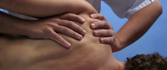
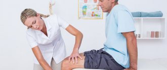
The physical examination is the second part of the patient's assessment. The doctor evaluates the patient’s gait; upon examination, the position of the limbs (free/forced), the presence of any tremors or deformities in the joints or bones are determined. The orthopedist may ask the patient to perform various actions - sit down, bend over, lift an object. Some of them may seem strange to the patient, but they are an important element of the examination, since they allow you to assess the active mobility of individual joints. In addition, passive mobility is also examined - it consists in the fact that the doctor moves the patient’s limbs in separate planes, looking for possible pathological resistance.
During the consultation with an orthopedic traumatologist, the patient’s muscle strength is also assessed. Typically, the examination is carried out in such a way that the patient sequentially performs individual movements, while the doctor tries to counteract them. Severe distortions can be seen with the naked eye, but sometimes visual examination is necessary.
fusion
Another technique in orthopedic surgery is fusion, which is used in the treatment of spinal-joint fusion. If a patient is experiencing severe arthritis pain, his/her orthopedist may suggest joint fusion surgery. This procedure brings together the two bones that make up his/her diseased joint. This causes the bones to become one solid bone, and this can reduce pain. It can also make the joint more stable and help the patient bear more weight. Joint fusion surgery can be performed on different joints such as: spine, ankles, wrists, fingers, thumbs and feet. It may take some time—sometimes many months—to heal from joint fusion surgery, and orthopedic doctors will advise patients about their recovery time before using fusion surgery.
Additional diagnostic methods
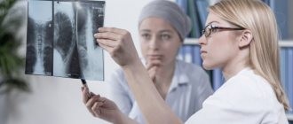
The orthopedist may order various tests. The most important thing is to check the mobility of the joints, their stability and position. In addition, the orthopedist must check the spine and the entire skeletal system. To do this, X-rays are taken, ultrasound, computed tomography and magnetic resonance imaging are performed.
An orthopedist can also order arthroscopy, a surgical procedure during which a device with a camera is inserted into the joint cavity through a puncture. There is no need to worry about the procedure: it is minimally invasive. Arthrography, an X-ray examination using a contrast agent, may also be required.
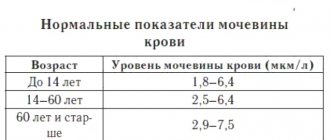
In addition, the doctor often prescribes laboratory tests. These include a general blood test, urine test, and rheumatic tests may be required.
Internal fixation and orthopedic implants
Internal fixation is an orthopedic procedure that involves the surgical placement of orthopedic implants to restore bone. This procedure uses an internal fixator, which can be made of stainless steel, titanium alloy, or cobalt-chromium alloy. These fasteners come in three main types:
- Plate and screws
- Kirchner Wires
- Intramedullary Nails Internal fixation procedures are performed in two ways: open repair internal fixation (ORIF) and closed repair internal fixation (CRIF). Open reduction here refers to open surgery to set the bones needed for some fractures. Internal fixation here refers to the fixation of screws and/or plates, intramedullary rods and other devices to enable or facilitate healing. Closed reduction internal fixation (CRIF) is a reduction without any open surgery followed by internal fixation.
Joint replacement/implant replacement
Replacement arthroplasty, or joint replacement surgery, is an orthopedic surgery procedure in which an arthritic or dysfunctional joint surface is replaced with an orthopedic prosthesis. When severe joint pain or dysfunction is not relieved by less invasive treatments, joint replacement is considered treatment by orthopedic doctors. Joint replacement is used for many joint pains such as shoulder, hip, knee, ankle and finger. The prosthetic material used in joint replacement surgeries can be metal, ceramic or alloy. Some ceramic materials commonly used in joint replacement are aluminum oxide, zirconia, silica, hydroxyapatite, titanium nitride, and silicon nitride. Orthopedic surgeons decide which material should be used.
Treatment
Orthopedic surgeons are familiar with and use technological advances such as joint replacements and the arthroscope, which allows the orthopedist to look inside the joint.
A visit to an orthopedist will begin with a personal interview and physical examination. This may be followed by diagnostic tests such as blood tests, x-rays or other tests.
Your treatment may include medical consultations, medications, casts, splints, and therapy such as exercise or surgery.
For most orthopedic conditions and injuries, there are several forms of treatment available. Your podiatrist can advise you on treatment options and help you choose the best treatment plan that will allow you to live an active and functional life.
osteotomy
In short, an osteotomy is any surgery that cuts and changes bones. This type of procedure is used by orthopedic doctors to repair a damaged joint. It is also used to shorten or lengthen deformed bone that does not fit into the joint as it should. Osteotomy is one of the orthopedic methods for relieving arthritis pain, especially in the hip and knee. This procedure can correct problems in many different bones and joints, such as: Hip, Knee, Spine, Jaw, Thumb and Chin. Due to the serious nature of this procedure, recovery can be extensive. Careful consultation with an orthopedic physician is important to ensure proper planning of non-surgical treatments during the recovery phase.
What does an orthopedist do during an appointment?
A consultation with a doctor includes taking a medical history, listening to complaints and a full visual examination.
What does the orthopedist look for first:
- posture and condition of the spine in general;
- joint mobility;
- presence of reflexes;
- gait.
If necessary, he prescribes additional diagnostics using radiography, ultrasound, scintigraphy (radioisotope scanning), histology and tissue cytology (for example, synovial fluid).
Soft tissue restoration
Soft tissue includes tissues that connect, support, or surround other structures and organs of the body, other than hard tissue such as bone. Soft tissues include tendons, ligaments, fascia, skin, fibrous tissue, fat, and synovial membranes (which are connective tissue), as well as muscles, nerves, and blood vessels. Soft tissue injury is damage to the ligaments, muscles and tendons of the hip, knee, shoulder and elbow. Soft tissue damage usually occurs from strain, sprains and repeated use of a particular body part, and can lead to swelling, pain, bruising and loss of function. To treat a soft tissue injury, orthopedic doctors use a soft tissue reconstruction procedure.
Injections and pain relief
Orthopedic injections deliver medications to a specific area of the body to help reduce or eliminate pain. Injections may not completely eliminate all symptoms of severe orthopedic pain, whether immediate or ongoing, but they can significantly help reduce pain and allow for increased comfort and function in daily life. There are four main types of orthopedic injections used by orthopedic doctors:
- Anti-inflammatory injections : This type of injection quickly relieves inflammation and is most often used in the wrist, spine, shoulder, knee, hip, elbow, or ankle.
- Viscous Supplement Injections : The injection goes directly into the joint, reducing friction during movement and reducing pain associated with osteoarthritis, improving the range of motion of the joint.
- Platelet-rich plasma (PRP) injections . PRP therapy uses injections of the patient's own platelets to speed up the healing of damaged joints, tendons and soft tissues. Specific conditions that can be treated with PRP include: osteoarthritis, tendonitis/chronic tendon injuries, back and spine conditions, nerve injuries and damage, ligament and muscle injuries, and non-healing wounds.
- Stem cell injections injections use a patient's own stem cells to treat osteoarthritis and soft tissue injuries. This type of treatment is at the forefront of non-surgical orthopedic treatment. Regenerative stem cells are collected from the patient's bone marrow in the pelvic or hip area. These cells can help rebuild soft tissue, cartilage, and bone, making them an excellent resource for accelerating the healing process and improving overall function.
Treatment methods and follow-up features
Treatment depends on the diagnosis. Orthopedic treatment options include, for example, limb immobilization, surgery, pharmacological treatment, as well as rehabilitation and physical therapy.
Physiotherapeutic procedures can be represented by an extensive list of techniques. Among them:
- Electrophoresis . The procedure is based on the use of weak electrical currents and injections of drugs into the affected area. The procedure allows you to enhance the effectiveness of pharmacotherapy by delivering medications to the diseased area, relieves swelling and pain, and improves metabolic processes.
- Medical massage . Relieves pain, helps accelerate regeneration processes and quickly alleviate symptoms.
- Exercise therapy . Helps restore joint mobility, promotes the formation of a strong muscle corset.
- Phonophoresis . The method involves delivering medications directly to the affected area. The procedure is non-invasive; pharmacological agents are introduced under the skin using ultrasound.
- Manual therapy . It has a powerful therapeutic effect, especially relevant for restoring the correct position of intervertebral discs.
Further observation in the case of chronic diseases should be continuous. The patient will be recommended moderate physical activity, proper nutrition while maintaining a normal weight, healthy sleep and rest patterns, etc. As with the treatment of other diseases, a comprehensive approach is critical.