Twisting of the intestine around the intestinal or mesenteric axis leads to intestinal obstruction. The disease is called volvulus. The intestinal walls are compressed, which leads to disturbances in the intestinal walls. Lack of supply of nutritional components and blood circulation leads to necrosis and peritonitis of the digestive tract.
The essence of pathology
In cases of volvulus, the intestinal loops twist around their axis or the mesenteric one. Transport of food is disrupted even when the baby's intestinal loop deviates 90 degrees from its normal direction. A larger angle causes compression of the arterial vessels and nerve branches passing between the layers of the mesentery. Volvulus can be observed in several areas or in one place.
As a result, a section of the intestine stops receiving nutrition, and the wall becomes necrotic. Following necrosis, vascular permeability increases, and effusion into the abdominal cavity begins. The intestinal wall ruptures and the contents exit into the abdominal cavity. This means that fecal peritonitis is developing. This creates a mortal danger for the baby. The only way to save the child is urgent surgery.
Types and forms of volvulus
The disease is classified according to localization, etiology of occurrence, and form of twisting. More often, torsion is localized in the large intestine - about 80% of cases. In the sigmoid colon - 80% of cases, in the cecum - 15%, in the transverse colon - 3%, in the splenic flexure - 2%.
Kinds:
- intussusception;
- strangulation - a severe form, occurs when the mesentery is too long, twisting around its axis; more common in school-age children;
- adhesive - formed during the formation of postoperative adhesions in the intestine at the site of surgery.
During intussusception, the outer tube of the intestine is called the vagina, and the inner tube is called the intussusception. The heads of the intussusception are the initial section of the invaginated intestine. When twisted, the loops create internal knots at 90 degrees. In severe cases, the angle of inversion is 150-10080 degrees. A rotation of up to 270 degrees indicates partial twisting, and up to 360 degrees indicates complete twisting around its axis. Repeated and single intestinal wrapping occurs.
Type of rotation:
- the intestine turns around its axis;
- volvulus with mesentery around another loop of intestine;
- volvulus of the intestine together with the mesentery around its axis.
According to etiology, it can be primary or secondary. If the cause of development is unclear, a primary volvulus is diagnosed; in the presence of provoking factors, a secondary one is diagnosed.
Why does it occur in infants?
The most common cause of intestinal volvulus, as observed by pediatric surgeons, is congenital developmental anomalies. They are more concerned with the small intestine. Determined in a newborn or under one year of age.
If the intrauterine formation of the fetus is disrupted, anatomical deviations are possible:
- excessively long mesentery (the most common cause of small intestinal volvulus);
- violation of the size of the initial and final sections of the intestine;
- internal hernia;
- formation of a cyst or tumor on the mesentery;
- functional insufficiency of the sphincter system and valves of the gastrointestinal tract;
- connection of intestinal loops by the common mesentery.
The development of anomalies is associated with:
- with a complicated pregnancy;
- forced treatment of the expectant mother with antibacterial drugs;
- malnutrition;
- hereditary disorders.
In infants, the first signs of obstruction may begin with the introduction of complementary foods or a sudden transition to artificial feeding
What matters is the unpreparedness of the underdeveloped digestive system to accept a new food substance. A reaction occurs in the form of impaired peristalsis: instead of alternating contractions of the circular and longitudinal fibers, pronounced spasms and a reverse wave are formed. Volvulus is caused by constipation in infants and stagnation of feces.
Causes
The following reasons can provoke intestinal blockage:
- prolonged constipation;
- foreign body ingress (swelling may form);
- pinching, abdominal injuries;
- regular overeating;
- hiatal hernia;
- infections, polyps;
- long mesentery;
- helminthiasis, blocking the internal space of the intestine;
- development of adhesions, tumor formations;
- paresis or intestinal spasms.
Torsion of loops in newborns occurs due to pathologies of intrauterine development. The organ defect is manifested by torsion. In infants, torsion occurs due to overfeeding with breast milk. Transferring a child under one year of age to formula milk can also provoke bloat. Early introduction of complementary foods also results in intestinal torsion.
Volvulus can lead to inflammatory processes in the abdominal cavity. Infants are more susceptible to the disease than older children. The consumption of fermented milk products and solid foods is limited. Children over three years of age are susceptible to bloat due to poor diet.
Pathological conditions that contribute to bloat in a child
At the age of 6 years, a common cause is the formation of a helminthic ball in the intestines to the point of complete obstruction. Other reasons include:
- dehydration of the body due to lack of drinking, in the heat, in cases of severe intoxication;
- abdominal injury;
- functional or pathological spasm and intestinal paresis;
- adhesions after surgery or untreated enterocolitis;
- compression of intestinal loops by a tumor formation or cyst;
- Hirschsprung's disease is a rare congenital disease, determined by the absence of nerve endings in the wall of the large intestine, in these areas peristalsis is impossible, constipation occurs; but the overlying intestine actively contracts, this creates conditions for volvulus of the sigmoid colon in newborns;
- megacolon is a congenital anomaly, expressed in a significant enlargement of the large intestine and underdevelopment of the system of nerve endings, manifested by persistent constipation from the first month of life.
Clinical manifestations
Symptoms of the disease are associated with the resulting intoxication of the body. The mechanism of its appearance is determined by blocking the access of blood to the intestinal sections. During torsion, a significant part of the intestine is compressed, and the blood supply and innervation to this and nearby areas and the mesentery are cut off. Without blood flow, nutrients and oxygen do not enter the tissues, and necrosis develops. When the intestinal lining dies, its permeability increases, which serves as an entry point for toxins and infection, causing fecal peritonitis.
The onset of the disease is characterized by general symptoms of intestinal dysfunction, which develop into symptoms of obstruction:
- Dyspeptic disorders. The first manifestations are nausea and vomiting. Rare vomiting indicates damage to the thick part, profuse vomiting indicates damage to the thin part. Vomit has a strong pungent odor.
- Stopping gas emissions within a few days.
- Stool disorders in the form of constipation due to blockage of a section of the intestine.
- Bloating or hard belly syndrome.
- Pain in the abdominal area. Severe cramping pain appears in the abdomen, the patient takes a forced position in bed.
In infants, at the onset of the disease, severe pain is replaced by periods of relief; the child can fall asleep and eat. As it worsens, weakness and moodiness appear. During the period of intoxication, body temperature rises and vomiting occurs. Before the complete cessation of bowel movements, blood elements are detected in the stool.
Intoxication is life-threatening for children and adults. The condition is dangerous due to the development of dehydration of brain tissue, systems and organs.
Symptoms of volvulus in children
Clinically, volvulus is manifested by mechanical obstruction at the site of torsion. Attentive parents can notice the initial symptoms. The pain syndrome occurs suddenly, the child screams. Older children describe the cramping nature of the pain. Pain shock may develop.
Repeated painful vomiting with an unpleasant odor. In case of small intestinal obstruction, first the food eaten, then the intestinal stool. Vomiting does not bring relief; it is more pronounced with volvulus of the small intestine. A rare gag reflex indicates a lesion in the large intestine.
Asymmetrical bloating of the abdomen due to overinflation of the adductor colon and collapse of the efferent colon, tension in the muscles of the abdominal wall. In the initial period, the passage of feces, then complete retention of stool and gases. When giving an enema, if a volvulus occurs in the small intestine, feces are released from the lower sections, and blood may be included.
A strangulated umbilical hernia creates conditions for intestinal torsion
Temperature rises to high levels with an abrupt decline. Babies often hiccup for three days, even after surgery. In a newborn baby, during volvulus, attention is paid to the cyanosis of the nasolabial triangle and the tendency to pull the knees towards the body. In the subacute course, attacks are accompanied by breaks and improvement. At the height of the illness, the baby becomes weak, lethargic, and restless. These are signs of increasing intoxication.
Bowel function
Pathological twisting occurs in the small and large intestines with the mesentery or blockage of the space by neoplasms. This is due to anatomical features.
The digestive system consists of several sections of the gastrointestinal tract, salivary glands, liver, pancreas, and gall bladder. It begins with the oral cavity, passes into the pharynx, then the esophagus. Subsequent departments:
- stomach,
- small intestine,
- colon,
- rectum.
The function of the digestive system is to process incoming nutrients into energy and distribute it further in the body. After the gastric stage of digestion, food enters the duodenum of the small intestine. The food lump (chyme) is broken down under the influence of bile.
All necessary enzymes enter the duodenum. Digestion in the large intestine ends with the formation of feces. Beneficial substances from broken down food are absorbed into the blood through the portal vein of the liver.
The organs are attached to the posterior wall of the abdominal cavity using the mesentery. It is a separate organ in the form of a thin film covering all the organs of the cavity. The mesentery unites all parts of the intestine, blood vessels, ligaments, and prevents them from twisting together. Twisting of the small and large intestines compresses the mesentery.
Dangerous complications
The goal of starting therapy within the first 3 days from the onset of volvulus symptoms is to avoid complications. When they occur, the child’s condition sharply worsens and the prognosis for a successful outcome decreases.
Dehydration
Frequent, painful vomiting contributes to the loss of a significant amount of fluid and electrolytes. There is no absorption of the liquid part of feces in the colon during volvulus, which further aggravates the state of dehydration:
- blood thickens;
- blood pressure decreases;
- the strength of heart contractions decreases.
First of all, the brain suffers (dizziness, drowsiness, loss of consciousness). If 15% of fluid is lost, the patient dies.
Intoxication
Due to the increased permeability of necrotic areas of the intestine, non-neutralized waste and toxic substances enter the bloodstream through it. The body reacts with an increase in temperature, chills, muscle pain and headaches.
Wall perforation
Perforation (perforation) of the intestinal wall occurs as a result of its thinning due to poor nutrition. Therefore, a wave of peristalsis causes rupture of the intestine that has lost its strength. The contents of the loop (waste, poisons, feces, intestinal bacteria) enter the abdominal cavity. This is how fecal peritonitis is formed. The leaves of the peritoneum have a high absorption capacity. Through them, all toxic substances will additionally enter the bloodstream and further increase intoxication.
Gangrene of the wall
Necrosis of an area of the intestine with cessation of blood supply is called gangrene. Dead tissue enters the abdominal cavity during peritonitis. Therefore, for treatment it is so important to remove (excise) non-viable tissue in a timely manner. Then rinse the abdominal cavity repeatedly with antimicrobial agents.
Sepsis
Sepsis is a pathological condition caused by pathogenic microbes entering the bloodstream. They spread throughout the body, forming ulcers in any organ. Manifests itself as severe intoxication and resistance to antibiotics.
Adhesive disease
Adhesions are the growth of scar tissue at the site of former inflammation. They begin with the loss of fibrin film. The adjacent loops then become glued together, and the scar tissue displaces and changes the normal position of the loops. When it grows, the loop may be compressed. This promotes volvulus and poses a threat to life, since the adhesions will not disappear on their own.
Removed area with internal volvulus of the small intestine and gangrene
Complications
An advanced form of obstruction is characterized by the entry of feces into the stomach, and the vomit acquires an unpleasant odor. There are symptoms of intoxication of the body with toxins and wastes. If the child's parents do not begin to worry at this moment, dangerous complications arise;
- peritonitis;
- blood poisoning;
- dehydration;
- rupture of the problem area;
- tissue necrosis;
- violation of water-salt level.
This could lead to the death of the child. Treatment of an advanced form leads to problems with the digestive tract; food is poorly digested, which results in constipation or diarrhea. Sometimes the symptoms disappear, the patient does not feel pain, and calms down. A dangerous indicator – against this background, peritonitis develops.
Diagnostics
It is difficult to hold a small child in front of the X-ray screen or give him barium to drink. The basis of diagnosis is the experience of surgeons. When palpating the abdomen, the specialist palpates the swollen soft formation of the afferent loop, an empty space in the area of the efferent intestine.
Pay attention to the asymmetrical bloating of the child’s abdomen. A digital examination reveals an empty rectum. The absence of peristalsis is judged by auscultation of the abdomen and splashing noise. It is very difficult to distinguish intestinal volvulus from appendicitis in a small child. Almost final confidence in the diagnosis comes during surgery and visual examination of the intestine.
Tests suggest inflammation: leukocytosis and ESR are increasing. In the severe stage, the hematocrit drops due to dehydration, and electrolytes decrease.
Methods of examination for volvulus
Patients are seen by a gastroenterologist. In addition to collecting anamnesis and palpating pathological areas of the intestine, the doctor prescribes additional diagnostics in the form of:
- x-ray of internal organs of the peritoneum;
- barium enemas (a radiopaque agent that helps get a picture of the condition of the intestinal tract);
- laboratory blood test;
- sigmoidoscopy (visual examination of the membrane of the rectum and sigmoid colon);
- endoscopic examination of the intestine;
- laparoscopic examination of internal organs;
- Ultrasound.
Treatment
If similar symptoms appear, you must call an ambulance. Children, together with one of their parents, are hospitalized in the surgical department and are first observed. They are trying to relieve the volvulus and pain with novocaine blockade. The meaning of conservative measures is to relieve increased tension in the adductor loop and spontaneous deployment of the node.
Saline solutions, anti-shock drugs, painkillers, and Hemodez are administered through the subclavian vein to combat intoxication. Be sure to wash the intestines with a siphon enema. The condition improves with gastric lavage and installation of a thin gas outlet tube. Antibacterial drugs are administered intravenously or intramuscularly, depending on the child’s condition.
Symptoms and treatment of intestinal diverticulosis
Surgical treatment consists of straightening the loops and securing the mesentery if the intestinal tissue is viable. If necrotic areas are present, they must be removed (bowel resection).
The undamaged ends are connected using one of the methods. The adhesions are dissected. In specialized clinics, surgery is performed using laparoscopic technology.
In severe cases of peritonitis and inflammation of the intestinal loops, the ends cannot be sutured until the inflammatory reaction has resolved. Then you have to operate step by step:
- First, the necrotic area is removed and the stoma is placed on the skin of the abdomen (feces will flow into the colostomy bag);
- after the peritonitis is cured, about 3 months later, the second stage is carried out - the ends of the intestine are connected and the stoma is removed.
For newborns, a permanent catheter is installed in the umbilical vein for long-term fluid transfusion.
Diagnostics and examination
When a patient comes in with general complaints of vomiting and bowel movements, a thorough examination of all systems is carried out. In the final stages, the doctor’s preliminary diagnosis is confirmed by instrumental research methods.
Collecting a life history is necessary to identify possible causes that provoked the pathology. The physical examination consists of palpating the abdominal area. When inverted, it is hard and swollen.
At the first stages of the examination, the patient undergoes biochemical and clinical blood tests, a general analysis of urine and feces. Signs of an inflammatory process are found in the blood in the form of an increase in the number of leukocytes and an increase in the erythrocyte sedimentation rate. With necrosis, elements of decay are revealed. In the stool there are traces of blood and elements of the intestinal mucosa.
Endoscopic methods:
- X-ray visualizes intestinal patency and obstruction of intestinal loops. The use of a coloring contrast agent allows you to determine the location of the twisted area.
- Computed tomography is a detailed scan of the intestinal walls and mesentery.
- Ultrasound scanning allows you to determine damage to intestinal tissue and assess blood flow.
Nutritional Features
After successful conservative straightening of the volvulus, the risk of recurrence remains. Parents are informed about the prognosis and are recommended to organize dietary meals for the baby. Feeding must be done 6 times a day. All dishes should be pureed and easy to digest.
Liquid porridges made from oatmeal, rice, buckwheat, jelly, and slimy soups are suitable. You can add milk to them, but not fat. It is best for infants to continue breastfeeding. Dishes made from raw vegetables, juices, and flour products should not be allowed.
What it is?
In medical practice, twisting of the intestines is called “volvulus.” The pathology has several forms of development and is divided into certain types depending on the scale of distribution and the area of damage.
The disease is one of the rare but extremely dangerous conditions. As a result of twisting of the intestine, its lumen is closed, which leads to obstruction . The pathology develops at a rapid pace and is always accompanied by certain symptoms.
Types and forms of volvulus:
- full or partial form (depending on the scale of the pathology);
- low and high view (depending on location);
- colonic or small intestinal type (depending on the location of the pathology);
- congenital or acquired form;
- dynamic and paralytic form (intestinal spasm or paresis of muscle fibers).
Why do relapses occur?
The operation was successful, the volvulus was straightened. But the patient or the baby’s parents are warned about the danger of relapse (repetition). Surgeons believe that the following play a role in the occurrence of relapses:
- Reduced scope of surgical intervention due to the severity of the patient’s condition (the loop was “untwisted”, but a long mesentery and a mobile cecum remained). Relapse can be avoided by suturing the wrapped loop to the abdominal wall.
- The formation of adhesions between the intestines, gluing together different parts of the intestine and predisposing to a new volvulus.
To prevent relapse, it is recommended:
- control the diet, avoid long breaks and overfeeding;
- prevent constipation, do not use laxatives;
- remember the importance of physical activity for a child, do joint exercises, and take more walks.
Clinical and anatomical aspects of intestinal malrotation in children
Malrotation is an abnormal position of the small and large intestine and defective formation of the mesentery as a result of incomplete or incorrect rotation of the embryonic umbilical cord loop. A common mesentery, insufficient or absent, or pathological fixation of various parts of the midgut at the stages of rotation, caused by various ante and/or postnatal disorders, can serve as an anatomical prerequisite for the occurrence of numerous types of malrotation and anomalies of intestinal fixation [3, 5]. Depending on the period and stage of intrauterine development of the fetus, variants of the anomaly appear in the absence or incomplete rotation, reverse, excessive rotation or the formation of an internal hernia. At the stages of intestinal rotation, pathological fixation in an unusual place, or a lack of physiological attachment of the mesentery or individual parts of the intestine, is possible.
Aspects of embryogenesis and clinical and anatomical characteristics of individual forms of intestinal malrotation are covered in classic manuals on pediatric surgery [1,7-10]. The data on intestinal malrotation available in the scientific literature are sporadic and in most cases are reduced only to a description of identified individual cases, however, certain anatomical variants, having differences in clinical and radiological manifestations, require differentiated treatment tactics [2, 4, 6]. Intestinal malrotation is often an accidental finding during laparotomy undertaken for acute surgical pathology of the abdominal organs. Sometimes it will be difficult for an experienced surgeon to navigate the diagnosis and identify its individual morphological variants.
The purpose of this study is to systematize the characteristic intraoperative manifestations of certain anatomical forms of intestinal malrotation in children, based on our own clinical observations and literature data.
Materials and methods of research. During 2002-2013, 107 children aged from one day to 15 years with various forms of intestinal malrotation were treated at the clinical sites of the Department of Hospital Pediatric Surgery of the Tashkent Pediatric Medical Institute. Among the patients, boys predominated - 71 (66.4%), girls - 36 (33.6%).
Of all patients with anomalies of intestinal rotation and fixation, 101 (94.4%) cases underwent surgical treatment, while 6 (5.6%) children received conservative treatment.
The age gradation of the examined children is presented in Table. 1.
The occurrence of this pathology in most cases (64.5%) is registered during the neonatal period. Of all the forms of intestinal rotation and fixation defects, the most common were volvulus of the mid and small intestine (31.8%) and Ladd syndrome (27.1%).
All patients underwent comprehensive clinical, laboratory and radiation diagnostic methods: ultrasound, radiological - survey radiography of the abdominal organs, contrast study of the gastrointestinal tract (GIT), irrigography, ultrasound examination (US) of the abdominal organs. If diagnostic difficulties were encountered, a computed tomogram (CT) scan of the abdominal organs was performed.
Table 1. Distribution of sick children with defects of intestinal rotation and fixation depending on age and form of pathology
Note: the number of unoperated children is indicated in parentheses (n=6)
Results and its discussion. An analysis of our clinical material and literature data shows that in cases of recurrent intestinal obstruction, a child’s lagging behind in physical development, the presence of paroxysmal abdominal pain, and chronic constipation, doctors should have increased alertness regarding violations of intestinal rotation and fixation.
Suspicion of such anomalies should arise when the following radiological signs are detected in the patient: a distended gas bubble of the stomach; phenomena of partial intestinal obstruction; atypical arrangement of loops of the small and large intestine, elongation of the large intestine. During ultrasound of the abdominal organs, the absence of the appendix or its detection in an atypical location, the presence of echoscopic signs of acute or chronic intestinal obstruction, and the abnormal location of internal organs should also be alarming regarding intestinal malrotation.
Regardless of the age of the children, the course of rotational disorders depends on the degree of compression or volvulus (partial, complete), both in individual parts of the small intestine and throughout the entire midgut, or on the severity of the violation of passage through the large intestine. In 87 (81.3%) patients, malrotation occurred as an acute course, in 13 (12.1%) as a chronic course, and in 7 (6.6%) as a chronically recurrent course.
In 57 (50.44%) patients, intestinal malrotation manifested itself with classic clinical and radiological signs of high intestinal obstruction: in 43 (75.4%) - partial, in 14 (24.6%) - complete.
The severity of clinical signs of high obstruction depended on the degree of compression of the duodenum by pathological cords of the peritoneum and/or abnormal location of the colon.
The early and main signs were vomiting and disturbances in the passage of intestinal contents. With obstructions above the large duodenal nipple in patients, the vomit looked like curdled milk with a significant amount of sticky mucus with a weak alkaline odor, in contrast to the sour one characteristic of gastric obstruction. With obstructions below the major duodenal nipple, vomit mixed with bile contained lumps of mucus.
In cases of low intestinal obstruction, patients experienced vomiting of the contents of the small intestine; in advanced cases, it took on a fecal character. Blood in the vomit was found in the form of streaks, vomit - coffee grounds or in the form of hematemesis. Vomiting mixed with bile and streaked with blood, and bleeding from the rectum clearly indicated volvulus.
Disturbances in the passage of intestinal contents ranged from retention of stool and gases in small portions to their complete cessation.
In newborns with malrotation with volvulus, meconium began to pass at the usual time and was released until the 4-5th day of life, then it became scanty, mixed with blood, and, finally, its release stopped. At the same time, in 4 newborns, from the very first days of life, a predominance of bloody mass in the feces was noted, which indicates the occurrence of volvulus in the prenatal period. Signs of low intestinal obstruction were observed with isolated volvulus of sections of the small intestine and mesocolicoparietal hernia in newborns. In 5 children, despite the involvement of a significant area of the small and large intestine in the volvulus, the leading clinical sign was the phenomenon of high intestinal obstruction.
Regardless of the level of obstruction, paroxysmal pain and bloating were noted, which were expressed in newborns by anxiety and irritability. Infants twisted their legs; in older age groups, obvious paroxysmal pain was noted. In 6.5% of cases, asymmetry of the abdomen was observed with swelling in the epigastric region and retraction below the navel. Tension of the muscles of the anterior abdominal wall and an increase in the child’s painful reaction were determined by palpation. The children's condition progressively worsened, with increasing manifestations of intoxication, dehydration and electrolyte disturbances. Irritability was replaced by lethargy, adynamia, decreased physiological reflexes in newborns, pale gray coloration of the skin and its marbling.
In the chronically recurrent type (7), the clinical manifestation of malrotation in the form of partial intestinal obstruction was observed in children under the age of one year (3), 3 years (2), 10 years (1), and over 10 years (1). The duration of the attack ranged from 4 hours to 2-3 days. Children periodically experienced vomiting, accompanied by abdominal pain 2-3 times a month; for some children - once every two to three months, sometimes the light period lasted from several months to several years. With this type of malrotation, the physical development of the child did not correspond to his actual age. With increasing age, body weight deficit increased. With a similar clinical picture, 4 patients were repeatedly hospitalized with various diagnoses in somatic, infectious or surgical hospitals. Despite many similar episodes, patients were not given a full comprehensive examination for a long time, which caused tactical errors.
Two of the 7 patients were operated on with various presumptive diagnoses. However, in these cases, the operations performed were ineffective. Analysis of clinical manifestations and preoperative studies does not always allow the identification of individual types among numerous forms. For surgical practice, it is important to clarify during surgery the characteristic anatomical changes in relation to certain types of anomalies of intestinal rotation and fixation, since the surgical approach may differ. Below are clinical semiotics and characteristic anatomical findings for certain types of malrotation and anomalies of intestinal fixation in children.
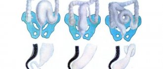
Ladd's syndrome, the most common type of intestinal malrotation, was observed in 29 (27.1%) patients, mainly in the neonatal period. Typical for this type of malrotation was the volvulus of the midgut around the common mesentery. Duodenal obstruction is caused by compression of it by cords of peritoneum connecting the duodenum with a highly located and fixed cecum (Fig. 1a).
This classic triad of components was observed in 17 of 29 patients with Ladd syndrome. In the remaining 12 cases, along with the main components of Ladd syndrome, anatomical variations were identified that arise from anomalies in the rotation of the midgut on the way from the left upper quadrant of the abdomen through the upper right to the lower right. In 7 children, different sections of the large intestine were fused with the duodenum or separate sections of the small intestine, without having a typical localization. These circumstances often caused changes in the typical anatomical structure, attachment, disruption of its syntopy and excessive expansion of the upper horizontal and descending part of the duodenum. In 5 patients, the ligament of Treitz was absent, the duodenal junction was located to the right of it, and the typical attachment of the mesentery was absent.
The main clinical sign of Ladd's syndrome was the signs of high partial intestinal obstruction, detected in all patients, regardless of the form of its course. Characteristic of Ledd's syndrome are, in some patients, the absence of trophic disorders in the intestines in the presence of volvulus or the identification of mesenteric circulation disorders from minor to intestinal necrosis of varying extent. The severity of ischemic intestinal disorders depended on the degree of disturbance of arterial inflow and venous outflow through the superior mesenteric vessels. Intestinal ischemia was manifested by symptoms of acute abdomen, intestinal bleeding, progression of multiple organ failure and determines the severity of Ladd syndrome.
Rice. 1. Schematic representations of variants of violations of rotation and fixation of the intestine (explanation in the text) a) Ledd syndrome b) right-sided mesocolicoparietal hernia c) left-sided mesocolicoparietal hernia d) incomplete rotation e) reverse rotation f) hyperrotation g) pathological fixation h) malrotation with volvulus intestines i) malrotation with volvulus of the small intestine
Among 25 operated newborns, 7 had circulatory disorders in the intestines, and 3 of them were operated on on days 1-2 of life, which indicates the occurrence of bloat even before the birth of the child. In the chronically relapsing course of Ladd syndrome, vascular and circulatory disorders of the intestine detected in children under one year of age were characterized by moderate severity. However, long-term volvulus led to venous stagnation and lymphostasis in the intestinal mesentery, which were clearly identified during surgery, obviously causing the development of secondary disorders of the gastrointestinal tract, worsening dysbiosis, impaired intestinal absorption and delayed physical development of the child.
Mesocolicoparietal hernias are the movement of the small intestine and its retroperitoneal fixation into the mesocolon within the abdominal cavity, formed in the peritoneal pockets and folds during the process of intrauterine rotation. With a right-sided hernia (Fig. 1 b), the small intestine does not make a normal turn around the superior mesenteric artery, but remains in the right upper quadrant and is enclosed on the right behind the mesocolon. The right half of the colon is rotated and fixed retroperitoneally on any part of the right flank of the abdomen. A mesocolicoparietal hernia on the left (Fig. 1 c) occurs when the small intestine rotates to the left and penetrates into the mesocolon between the inferior mesenteric vein and the retroperitoneal space. The large intestine continues to rotate to its normal position, enclosing the small intestine in a mesocolon sac, with the inferior mesenteric vein forming a rather narrow neck of the sac [8].
We observed 12 children with mesocolicoparietal hernia, among them: 5 newborns, one child was 7 months old, six were older than one year: 2, 6, 7(2), 13 and 14 years old. Four patients were female, 8 were male.
A right-sided hernia was diagnosed in 7 patients, a left-sided one in 5. In 9 patients, part of the intestine was found in the hernial sac, in 3, complete penetration of the small intestinal loops was noted. In newborns with mesocolicoparietal hernia, clinical signs of acute intestinal obstruction were observed; in other patients, signs of chronically recurrent intestinal obstruction were periodically observed and resolved independently. In 7 cases, mesocolicoparietal hernia was an independent disease; in 5 patients, along with internal hernia, concomitant pathology was found. In two out of 5 newborns, mesocolicoparietal hernia was one of the manifestations of multiple gastrointestinal malformations: in one it was combined with necrosis and perforation of the atretic terminal portion of the ileum, which ended up in the hernial sac. In another child, in the hernial sac, along with loops of the small intestine, there was an atretic section of the transverse colon, complicated by perforation of the adductor segment. An abdominal cyst in 1 child and in 1 patient Hirschsprung's disease were also associated with a mesocolicoparietal hernia, determined, as in other cases, during surgery.
Incomplete rotation - counterclockwise rotation stops when turning 180º. In this case, the fixed cecum lies under the liver in the area of the right hypochondrium. Rotation of the duodenum also does not occur; the duodenojejunal flexure is located to the right of the midline and in relation to the superior mesenteric vessels (Fig. 1 d). Narrowing of the root of the common mesentery, containing mesenteric vessels and attachments in the form of a narrow stalk, creates the prerequisites for the occurrence of volvulus, which was observed in 12 (11.2%) patients. Moderate signs of recurrent intestinal obstruction were characteristic. Among all operated children with this pathology, there was no complication of volvulus in the form of necrosis, however, in three cases, venous stagnation and mild lymphostasis in the intestinal mesentery were noted. The absence of critical volvulus with the development of intestinal necrosis in these cases can be explained by partial fixation of the cecum in the right hypochondrium and the presence of a relatively larger area of fixation (but incomplete) of the mesenteric root.
Lack of rotation occurs when there is a delay in further rotation of the midgut after its repeated return to the abdominal cavity from the coelom of the umbilical peduncle. With this type of rotation disorder, in the presence of a common mesentery of the stomach, the duodenum and the entire large intestine lie on the left, and the small intestine on the right. It is appropriate to note the difficulties of preoperative verification of this anatomical variant of malrotation and its combination with congenital intestinal obstruction. Data from a survey radiography of the abdominal cavity and contrast studies of the gastrointestinal tract in the form of the location of the large intestine on the left and the small intestine on the right are the most common radiological findings, which allows us to evaluate it as a synistrous position of the large intestine. Only during surgery for congenital intestinal obstruction, the above characteristic changes for the absence of intestinal rotation were discovered in 3 newborns.
Reverse rotation in 3 (2.8%) patients occurred in cases where the last 180º rotation of the intestine occurs in the opposite direction, that is, clockwise (with physiological rotation - counterclockwise), when the postarterial segment of the intestine enters the abdominal cavity first , while the rudiment of the right half of the colon pushes its distal parts to the left, and as a result, the area from which the transverse colon is formed appears under the superior mesenteric artery (Fig. 1 e). Further formation of the defect is aggravated by improper fixation of other parts of the large intestine to the posterior abdominal wall or mesentery of the small intestine. Defects in fixation, disproportionate growth of individual sections of the intestine, and often combined anomalies of the gastrointestinal tract create favorable conditions for the occurrence of volvulus. Options for reverse rotation may be the right-sided arrangement of the entire colon or the left-sided arrangement of the liver and all parts of the colon, observed with a mirror arrangement of the abdominal organs [1,4].
Hyperrotation occurs when part of the midgut rotates 360º or more. In this case, the cecum and ascending colon are located in the left upper quadrant of the abdomen medial to the splenic flexure. The mesentery is quite developed and is common to the small and large intestines. The ileum enters the cecum from the medial side, and the ileocecal angle has a rotated appearance. The ascending colon, at some distance, located along the descending colon and forming a loop, takes an obliquely ascending direction, passes into the right flank of the abdomen, and continues in the form of a normally located transverse colon and distal sections of the colon (Fig. 1 e).
With pathological fixation, in 14 (13.1%) patients, against the background of completion of intestinal rotation, or in cases of malrotation, fusion of various parts of the small and or large intestine or unusual adhesion of intestinal loops to the parietal peritoneum, in which there is no volvulus, are noted (Fig. 1 g). The clinical picture of pathological fixation was distinguished by the paucity of clinical symptoms, expressed in moderate pain syndrome with symptoms of partial intestinal obstruction in newborns, or signs of colonic stasis in children of older age groups. Emptying the bowel with enemas or while taking laxatives led to the disappearance of abdominal pain and a decrease in the severity of symptoms of intestinal obstruction. Plain radiographs showed signs of partial intestinal obstruction - the presence of fluid levels in the dilated stomach, upper intestines and decreased pneumotization, up to the complete absence of gas in the underlying intestinal loops. Contrast studies of the gastrointestinal tract conducted in newborns also revealed signs of partial low intestinal obstruction. Irrigograms failed to identify specific signs, however, elongation of individual parts or the entire colon was present in the majority of those examined, as with other types of rotational disorders. Radiological signs and clinical findings are non-pathognomonic. The diagnosis of pathological fixation in all our cases was an intraoperative finding.
Malrotation with midgut volvulus in 16 (15.0%) patients was characterized by the presence of a non-rotated midgut with the entire colon located on one side (on the left), and in rare cases on the right. Incorrectly developed segments of the duodenum and the initial part of the small intestine are located laterally in relation to the superior mesenteric vessels, there is no duodenojejunal flexure and the usual fixation of the duodenum to the posterior abdominal wall. The relationship between duodenum and the pancreas and liver also changes. The large intestine, located together with the small intestine, having a common mesentery, most often was not fixed (Fig. 1h). Midgut volvulus was observed in 16 (15.0%) children with malrotations. The pathology was predominantly diagnosed in newborns - 12 (75.0%) out of 16, in 2 to 3 months of age and in 2 over one year of age. Among newborn children, clinical and radiological signs of high (56.3%) and low (43.7%) intestinal obstruction were noted. 12 (75.0%) had signs of complete obstruction, which appeared in early neonatal age, and 4 (25.0%) had partial obstruction.
Midgut volvulus in 10 (62.5%) cases was an isolated nosological form, in 6 (37.5%) patients it was combined with other anomalies of the gastrointestinal tract: atresia of various parts of the intestine - 4, mesenteric defect - 1, lymphoid cyst - 1. Among 16 operated children with midgut volvulus, 2 (12.5%) had local areas of necrosis of individual segments of the small or large intestine. In the presence of necrosis within the midgut, symptoms of an acute abdomen were prevalent in the clinical picture.
Malrotation with small bowel volvulus occurred in 18 (16.8%) patients. The volvulus mainly involves part of the small intestine with the normal location and fixation of the large intestine (Fig. 1 i).
The pathology, as a rule, was accompanied by a violation of trophism to necrosis of varying extent and intestinal perforation with the development of peritonitis. Therefore, in case of malrotation with volvulus of the small intestine, all patients showed clinical and radiological signs of complete intestinal obstruction and peritonitis, 4 of them had perforation. In the clinical and radiological picture in 13 (72.2%) patients, signs of complete low intestinal obstruction predominated, at the same time, in 2 observations, data characteristic of high intestinal obstruction were revealed, which was due to concomitant anomalies in the development of the gastrointestinal tract or compression of the initial part of the small intestine separate adhesions or pathological formations that create a conglomerate around the wrapped section of the small intestine. Volvulus of the small intestine in 13 (72.2%) cases occurred in the presence of a number of predisposing anatomical causes: anomalies in the development of mesenteric vessels, causing disruption of intestinal trophism (7); small intestinal atresia (4); membranes of the small intestine (1); combined anomalies of the gastrointestinal tract (ileal atresia, mesenteric cyst) (1).
Conclusion. Thus, rotation disorders and anomalies of intestinal fixation are represented by numerous anatomical forms and varying frequencies of occurrence. Among them, volvulus of the middle and small intestine predominates, Ladd syndrome, mesocolicoparietal hernias and pathological fixation occur with equal frequency and were observed in 12 (11.2%) and 14 (13.1%) patients, respectively. Various forms of intestinal rotation and fixation defects are predominantly detected among newborns and manifest themselves in the form of an acute course. As the age of children increases, the frequency and type of rotational disorders decrease with a tendency to increase anomalies of colon fixation, characterized by a chronically recurrent course and progressive colostasis. The clinical picture of malrotation is dominated by signs of high partial or complete intestinal obstruction, depending on the degree of obstruction, the nature of the complications encountered and concomitant pathology of the gastrointestinal tract. Based on clinical and radiological data before surgery, it is difficult to determine the exact anatomical shape. The final diagnosis is often made during surgery. In case of malrotation, regardless of the type of rotation disorder, during surgery through a thorough revision and in the process of eliminating existing anatomical rotation disorders and pathological fixations, it is necessary to evaluate the possibility of giving the small, large intestine and mesentery a look corresponding to the physiological position, and carry out surgical fixation as a complete rotations. In this case, the right half of the colon is mixed along the abdominal wall to the iliac region with the typical localization of the small intestine and the ilocecal angle only if the mesentery is of sufficient length and width without tension on the mesenteric vessels. In cases where it is impossible to achieve this goal due to underdevelopment of the mesentery or the relative shortness of the mesenteric vessels, regardless of the type of malrotation, the intestine should be given the position as in the absence of rotation.
What to do when the baby grows?
As children grow older, anatomical anomalies become less dangerous or are completely eliminated. The consistency of the intestinal sphincters is improved. The size of the intestines becomes proportional. The body adapts quite well to life with its own characteristics.
But a threat can arise when chronic diseases appear during adolescence. Therefore, when raising a child, you need to pay attention to proper nutrition, avoiding any alcohol, soda, or fast food. The disease spares neither children nor the elderly. Seeking medical help gives you a chance not only to stay healthy, but also to survive.
What danger does the disease pose?
Ignoring or untimely provision of medical care is fraught with the appearance of complicated conditions:
- general poisoning (intoxication) of the victim’s body;
- development of a state of dehydration;
- the appearance of peritonitis - a dangerous inflammation of the peritoneum;
- gangrene develops in the affected area of the intestine;
- perforation of the intestine.
What is not recommended to do?
Intestinal volvulus in a child should under no circumstances be treated at home. It is not possible to eliminate pathology only with folk remedies or medications. To restore the normal state of the intestines, doctors use special techniques that do not always fall into the category of surgical procedures. If symptoms of the disease appear or a pathology diagnosis is made, the child should be provided with qualified medical care.
If there is a volvulus in a child, it is not recommended to:
- ignore the symptoms of pathology;
- self-medicate;
- use folk remedies as the main method of therapy;
- do not follow the recommendations of a specialist;
- interrupt the course of therapy at the first relief of the condition;
- Uncontrollably give drugs to a child without a specialist’s prescription.
Symptoms and clinical picture
The first symptoms of volvulus are always associated with a violation of the digestive system .
In the early stages of the development of the disease, only an experienced specialist can diagnose the pathology.
The intensity of symptoms depends on the degree of intestinal twisting and the individual characteristics of the child’s body. Some signs may indicate pathology in advance .
For example, the pungent smell of vomit indicates the accumulation of feces in the intestines and a violation of its patency.
Symptoms of volvulus may include the following conditions:
- sudden increase in body temperature;
- pain in the abdominal area;
- acrid smell of vomit;
- attacks of nausea and vomiting;
- no gas formation;
- constipation;
- blood impurities in stool;
- decreased blood pressure;
- abdominal consolidation;
- deterioration of the general condition of the body.
Recommendations for the treatment of intestinal dolichosigma in a child can be found on our website.
Treatment methods
Treatment of volvulus in most cases is carried out in a hospital setting . Therapy can be carried out using conservative or radical methods.
If a cleansing procedure using a siphon enema does not eliminate the accumulation of feces in the intestines, then surgery becomes the only method to solve the problem.
Drug treatment is not able to relieve volvulus. The drugs are used only at the rehabilitation stage.
The main methods of treating volvulus in children are the following procedures:
- Blowing out the sunken intestine (the method is effective only for mild cases of the disease).
- Laparotomy surgery (the method is used for early diagnosis of pathology).
- In the presence of tumors that provoked intestinal volvulus, the formations are cut out and the damaged sections of the intestine are connected.
- Toxic substances are removed from the child’s body using droppers.
- Adhesions are eliminated using a tumor removal algorithm.
- Dissection of the peritoneum and manual reduction of the intestines.