Patau syndrome is a chromosomal disorder caused by the presence of an extra copy of chromosome 13 (trisomy 13).
The structure of Patau syndrome includes multiple defects of the nervous system (microcephaly, holoprosencephaly), eyes (microphthalmia, cataracts), musculoskeletal system (polydactyly, cleft lip and palate, omphalocele), heart, urogenital system, etc.
At its core, Patau syndrome is a pathological condition of the human genome in which an extra thirteenth chromosome is detected. In this regard, the disease received the scientific name trisomy 13. The frequency of this pathology reaches 1:7000, but usually this figure fluctuates around 1:10000-1:14000. The sex ratio of patients with trisomy 13 is approximately 1:1.
A characteristic feature of newborns with this disease is true prenatal hypoplasia in 25-30%, which manifests itself during normal pregnancy (without minor prematurity).
Genetics
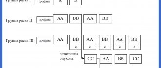
Basically, there are two genetic forms of trisomy on the thirteenth chromosome:
- Simple trisomy - three chromosomes of the same type exist freely and realize their genetic potential.
- Robertsonian translocation - two chromosomes remain free, and the third merges with long arms with another acrocentric chromosome (for example, with 14 or 21). Since acrocentric chromosomes in short arms have only rRNA genes that are duplicated many times in the karyotype, their functionality is preserved in most cases.
In any case, the karyotype of a patient with Patau syndrome is determined by the formula 47 XX (XY) 13+. Sometimes cases of non-Robertsonian translocations, isochromosomal and mosaic forms of trisomy 13 are recorded.
The pathological picture and symptoms do not differ in the genetic varieties of Patau syndrome. In 3/4 of patients, an extra 13th chromosome is found, the remaining quarter of patients suffer from the involvement of the 13th pair of chromosomes in a translocation, which in 75% of cases occurs due to a denovo mutation (new, independently occurring) - the so-called “genetic error”. Studies of the heritability of Patau syndrome indicate that the remaining 25% of trisomy 13 cases are due to the transmission of a translocalized thirteenth chromosome from one parent. At the same time, the risk of recurrence of the case in the next child (recurrent risk) is 14%.
Recommendations
- ^ a b
"Trisomy 13: a brief description".
GOV.UK.
_ - "Prevalence and incidence of Patau syndrome". Disease Center - Patau Syndrome
. Adviware Pty Ltd. 2008-02-04. Retrieved 2008-02-17. the average maternal age for this anomaly is about 31 years - About.com>Patau Syndrome (Trisomy 13) From Chrissy Danielsson. Updated June 10, 2009
- H. Bruce Ostler (2004). Diseases of the eyes and skin: color atlas
. Lippincott Williams and Wilkins. paragraph 72. ISBN 978-0-7817-4999-2. Retrieved April 13, 2010. - "Trisomy 13: MedlinePlus Medical Encyclopedia." Retrieved 2010-04-12.
- Callahan, Tamara L., and Aaron B. Caughey. Obstetrics and gynecology drawings. Baltimore, MD: Lippincott Williams & Wilkins, 2013.
- Janvier, Annie; Farlow, Wilfond (1 August 2012). "The experiences of families with children with trisomy 13 and 18 on social networks." Pediatrics
.
130
(2):293–298. Doi:10.1542/ped.2012-0151. PMID 22826570. - "Patau Syndrome". StatPearls
. StatPearls. 2021. - "Patau syndrome (trisomy 13)." Retrieved July 3, 2014.
- Nelson, Catherine E.; Rosella, Laura C.; Mahant, Sanjay; Guttmann, Astrid (2016). "Survival and surgical interventions for children with trisomy 13 and 18." JAMA
.
316
(4):420–8. doi:10.1001/jama.2016.9819. PMID 27458947. Retrieved December 3, 2021. - sind / 1024
in Who named it? - Patau K., Smith D.W., Terman E., Inhorn S.L., Wagner H.P. (1960). "Multiple congenital anomaly caused by an additional autosome." Lancet
.
1
(7128): 790–3. Doi:10.1016/S0140-6736(60)90676-0. PMID 14430807. - “National Cytogenetic Registry for Down Syndrome Annual Reports 2008/09.” Archived from the original on April 15, 2010.
Causes
The causes of this disease are not yet fully understood. It is believed that the main reasons that lead to the development of this syndrome are mutations at various levels of child development (ovum, embryo, fetus level), as well as heredity.
Reasons that contribute to the development of Patau syndrome:
- during the process of division of the germ cell, chromosome nondisjunction is possible;
- Robertsonian translocation, in which the embryo receives an additional copy of genes;
- various anomalies that form at the level of genetic information in the cell;
- various mutational changes;
- mutations of the fertilized egg;
- mutations of fetal cells.
The following risk factors contribute to the development of this disease:
- poor environmental situation in the area of residence;
- children born from marriages between close relatives;
- the presence of hereditary diseases in all previous generations of parents;
- old age of the mother (this disease is diagnosed in children who were born to women after 45 years of age).
This syndrome is characterized by spontaneity and cannot be prevented.
Risk factors
The main risk factors are age (especially significant for Down syndrome), as well as exposure to radiation and certain heavy metals. It should be borne in mind that even without risk factors, the fetus can have pathology.
As can be seen from the graph, the dependence of the risk on age is most significant for Down syndrome, and less significant for the other two trisomies.
Symptoms
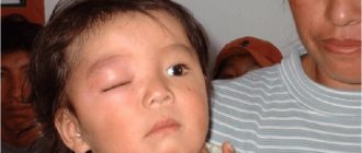
Patau syndrome (see photo) entails multiple disorders of both internal organs and systems, so the symptoms of the disease are divided depending on the location of the pathology.
Symptoms of this disorder related to the nervous system:
- reduced brain volumes;
- underdevelopment or complete absence of some components of the brain, for example, the cerebellum;
- delay in mental, physical and mental formation of personality.
Signs of this disorder in terms of structural changes in the skull and skeleton are:
- the baby’s body weight is critically low – less than two kilograms;
- deformation of the skull – the child’s head is much smaller than the body;
- the location of the ears is much lower than in a healthy child;
- pronounced bilateral clefts on the palate and upper lip;
- palpebral fissures are significantly narrowed;
- in some cases there is an increase in the number of fingers on the upper and lower extremities;
- short neck;
- skin imperfections in the back of the head;
- irregular foot shape.
Manifestation of such a karyotype disorder in the formation of the genital organs:
- underdevelopment – reduction in penis size;
- detection of a testicle in the abdominal cavity;
- duplication of the genital organs in girls, in particular the uterus or vagina.
Signs of Patau syndrome, characterizing anomalies in the structure of vital organs:
- structural pathologies of the heart;
- changes in the normal volumes of large and medium-sized vessels;
- underdevelopment or duality of the ureter;
- neoplasms on the kidneys in the form of cysts;
- umbilical cord deformities, such as hernia;
- changes in the natural position of all organs.
Symptoms of Patau disease from damage to the organs of vision and hearing:
- possible lack of one eyeball;
- defects of the membranes of the eye and lens;
- complete deafness caused by irregularly shaped ears;
- congenital cataract.
Due to the presence of multiple congenital defects in children, the prognosis for them is quite sad - on average, 90% of infants will not live their first year of life completely. Only a sixth of children live to be five years old, and only three percent of the total live to be ten years old. In most cases, babies with Patau syndrome die inside the womb.
Edwards syndrome
Edwards syndrome is characterized by trisomy 18 and a complex of multiple malformations.
In one case out of 10, mosaicism is observed, that is, not all cells of the body have an extra chromosome. Partial trisomy is also possible with the attachment of part of chromosome 18 to another chromosome.
During pregnancy, low fetal weight, polyhydramnios, a small placenta and the presence of one placental artery are observed.
Newborns have a change in the shape of the skull, a small mouth and jaw, facial dysmorphism, eye defects and low deformed ears. Numerical anomalies of the fingers and toes, and foot deformity (“rocker foot”) are also observed.
Among the defects of internal organs, the most common are heart and vascular defects. All have cerebellar hypoplasia.
Edwards syndrome is characterized by mental retardation and developmental delays.
Most children die in the first months of life.
Diagnostics
All chromosomal diseases are diagnosed according to the same principle. At the first stage of screening, ultrasound and determination of biochemical markers (PAPP-A, beta-hCG) are performed. During prenatal diagnosis, the data makes it possible to determine the degree of risk of having a child with a chromosomal abnormality. Further invasive diagnosis of Patau syndrome with a cytogenetic diagnosis in case of a high probability of the disease is carried out as follows:
- at 8-12 weeks of pregnancy - chorionic villus biopsy;
- at 14-18 weeks of pregnancy - amniocentesis;
- after 20 weeks of pregnancy - cordocentesis.
The material obtained as a result of research is studied using the QF-PCR method or karyotyping with differential staining. Chromosome 13 is detected and based on the ultrasound results, the doctor focuses on the clinical signs not of the mother, but of the child. Confirmation of Patau disease can only be done with the help of prenatal diagnostics, which is responsible for determining the baby's chromosome set. If there is a high probability of a hereditary disease, the baby is given a thorough examination, which includes:
- neurosonography;
- Ultrasound of the kidneys, abdominal cavity;
- echocardiography and so on.
Changes in internal organs
Unfortunately, the presence of an additional chromosome does not only manifest itself in appearance; the whole body suffers. Therefore, if external signs are detected, the child is prescribed a full examination of the internal organs.
central nervous system
In such children, the brain is smaller and entire sections may be missing. Or they are underdeveloped. As a rule, the corpus callosum and cerebellum are affected by this disease. With such an anomaly, the child is affected by a severe form of idiocy. He is unable to speak, think, or understand what is happening around him. Such patients even find it difficult to control their movements. Therefore, they may never learn to walk, eat, and do other everyday things. This pathology does not allow a person to feel and express their emotions. There is no sensitivity to external stimuli, such as bright lights or loud sounds.
The cardiovascular system
With Patau syndrome, abnormalities of the cardiac septum, abnormal structure of large vessels, heart disease, and patent ductus arteriosus may occur.
Genitourinary system
Kidneys with this syndrome can be affected by polycystic disease, horseshoe kidney, hydronephrosis. The ureter is shortened. In boys, the external genital organs are underdeveloped and the testicle does not descend into the scrotum. Girls have two uteruses, a double vagina, hypertrophy of the external genitalia.
Sense organs
Underdevelopment of the brain leads to abnormalities in the formation and functionality of the organs of vision and hearing. Complete deafness may be present. The birth of children with this syndrome was noted, in which one eye was defective or completely absent. In addition, other painful signs appear: cataracts, retinal dysplasia and others.
Gastrointestinal tract
Cysts on the pancreas, Meckel's diverticulum, incomplete intestinal rotation.
Other manifestations
Pathology can cause a change in the location of organs, their defects and underdevelopment. In addition, an umbilical hernia is observed at birth. The skin may also have pathological changes, for example, the absence of hair or skin on an area of the body or folds. One of the dangerous signs of the disease is leukemia - blood cancer.
Attention! An established diagnosis does not yet indicate the presence of all of the above changes in a child. Sometimes the pathology is limited to some external signs and mental retardation.
Treatment
Patau syndrome cannot be cured. Pathology leads to disturbances in the development and structure of internal organs. Doctors can try to relieve the child's condition. For this, surgery is performed. Most often, with Patau syndrome, the following is performed:
- Facial plastic surgery. Many patients with this disease experience cleft lips. Doctors are eliminating this deficiency.
- Removal of an additional uterus. A similar action is performed in relation to other similar pathologies, if they occur.
- Surgeries are performed on internal organs. Usually treatment is carried out in relation to the kidneys, ureters and heart. This makes caring for the child easier.
Additionally, symptomatic treatment is carried out. Doctors eliminate associated symptoms and strengthen the child’s immunity. Actions must be performed in order to avoid inflammation of the organs. Children with Patau syndrome are always underdeveloped. This applies to physical and mental development. Such patients are unlikely to be able to live a full life. The child will not be independent. This is why doctors recommend terminating a pregnancy before 22 weeks.
Forecast
If a woman nevertheless chooses to continue the pregnancy, or if abortion is already contraindicated due to timing, then, with the exception of rare cases, she will have to refuse termination. In addition, serious attention should be focused on exactly how to give birth.
A fetus with Patau syndrome in most cases dies antenatally (during pregnancy) or is born dead. This is explained by the significant severity of congenital anomalies of internal organs. Babies with this pathology, who remain alive after birth, are completely disabled throughout their lives and suffer from idiocy.
If the cause of the disease was a random factor, then subsequent pregnancies in this woman can be successful.
Prevention
There are no uniform and effective measures that would protect against the development of trisomy 13. The risk of developing the pathology can be reduced by following some recommendations: carefully monitor the absence of blood ties between future parents, give birth to children under 40 years of age, move from areas where there is increased radiological contamination .
Patau syndrome is a genetic disorder that cannot be treated and is accompanied by external defects and pathologies of physical and mental development. Children with this diagnosis extremely rarely live to be one year old, but even in this case they need round-the-clock careful care, constant health monitoring and regular examinations by doctors.
Historical facts
Trisomy 13 was first described in 1657 by Erasmus Bartholin. And in 1960, Dr. Klaus Patau revealed the chromosomal nature of this disease. It was named in his honor. The syndrome has been described in Pacific Island tribes. It is also believed that the anomaly was caused by radiation contamination resulting from nuclear weapons testing.
Approximately one in 7–10 thousand babies is born with this syndrome. As a rule, it occurs equally in both boys and girls.