Author
— Natalya Reznik.
Pelvic floor muscle dysfunction is a common problem affecting approximately the world's population. It is not life-threatening, but it significantly complicates it. In this case, you need to contact specialists. They provide potential clients with information sufficient to independently make a primary diagnosis and take the necessary measures.
How does it work?
The position of our body is supported by a cylinder-like muscular frame (core). Its walls are made up of the transverse, oblique and rectus abdominis, multifidus (deep dorsal muscles) and erector spinae muscles. The cylinder is closed from above by the diaphragm, and from below by the pelvic floor muscles. It is a three-layer muscle plate stretched like a hammock from the pubic bone in front to the tailbone in the back. The thickness of the plate is about 1 cm. If all the pelvic floor muscles contracted individually, they would pull in different directions. However, they work together to support the bladder and intestines, the prostate in men and the uterus in women, and therefore partly control the functioning of these organs. In addition, the pelvic floor muscles strengthen the rectal sphincter and urethra (Fig. 1).
Contractions of the pelvic floor muscles can be regulated by volition, but usually they contract unconsciously, in coordination with the work of the deep muscles of the abdomen and back and the diaphragm and help control pressure in the abdominal cavity during physical activity. In an ideal situation, intra-abdominal pressure is automatically regulated. For example, when lifting a weight, the core muscles work in ensemble: the pelvic floor muscles rise, the abdominal and back muscles retract to support the spine, and breathing is not difficult. If any of the core muscles, including the pelvic floor muscles, are weakened or damaged, automatic coordination is impaired. Then, in situations that increase intra-abdominal pressure, there is a possibility of overload of the pelvic floor, it weakens and the pressure decreases. If this happens repeatedly, the stress on the pelvic organs increases over time, which can lead to loss of bladder or bowel control or pelvic organ prolapse.
To work as part of the core, the pelvic floor muscles must be flexible, meaning they can not only contract and hold tension, but also relax. Constant tension can cause the muscles to lose flexibility and become very stiff, and pelvic floor muscle stiffness is usually associated with weakness, which can lead to urinary incontinence, pelvic pain, pain with intercourse and difficulty urinating.
Symptoms of pelvic floor muscle dysfunction
- Uncontrollable leakage of urine when exercising, laughing, coughing or sneezing (stress incontinence).
- The need to go to the toilet frequently or urgently (urge incontinence).
- Incontinence of gases and intestinal contents.
- Difficulty emptying your bladder or bowels.
- Prolapse of internal organs. In women, it is felt as a bulge in the vagina or a feeling of discomfort or heaviness. In men, there is a bulge in the rectum or a desire to empty the bladder, although there is no objective need for this.
- Pain in the bladder, bowel, or back near the pelvic floor when doing pelvic floor exercises or during sexual intercourse.
Experts emphasize that the above problems are not necessarily associated with weakness of the pelvic floor muscles, so their cause should certainly be found out. Diagnosis is carried out using electromyography, ultrasound or MRI.
Diagnostics
When conducting a clinical examination, true muscle weakness with a decrease in strength is distinguished from increased fatigue as a subjective sign. The anamnesis is used to find out information about the intensity and rate of development of the symptom, and the presence of additional signs. During a neurological examination, the doctor determines muscle strength (in points), range of active and passive movements, and reflexes. To determine the cause of muscle weakness, additional studies are prescribed:
- Blood analysis.
If an infection is suspected, a hemogram gives an idea of the leukocyte formula and ESR. Biochemical analysis allows us to identify electrolyte and hormonal disorders, the presence of antibodies to the pathogen. Toxicological examination shows the content of toxic substances in the blood. - Lumbar puncture.
Cerebrospinal fluid is taken for examination to exclude subarachnoid hemorrhages, infectious and inflammatory diseases of the brain and membranes. CSF pressure is assessed in case of volumetric processes (tumors, abscesses, hematomas). - X-ray.
X-ray of the spine is the first test prescribed for spinal injuries. Standard photographs do not show fractures of the odontoid process or the lower cervical segment, which requires special radiographs and the use of other imaging techniques. - Tomography.
In case of damage to the central nervous system, MRI is considered the main method of neuroimaging. The method is highly informative for pathology of the posterior cranial fossa, inflammation of the meninges, and myelopathy. CT scan of the brain is more suitable for patients with basal skull fractures in the acute period of stroke. - Myelography.
This is an X-ray examination of the central canal of the spinal cord. The procedure is performed for herniated intervertebral discs, spinal injuries, and tumors. The technique shows any obstacles to normal liquor dynamics. - Electroneuromyography.
Damage to nerve trunks and fibers requires a functional study with analysis of impulse conduction to the muscles, which allows one to evaluate the speed of signal transmission, the location of damage, and the ability of muscles to contract.
If muscle weakness is accompanied by systemic disorders, the examination plan includes ultrasound of the kidneys and adrenal glands, thyroid and parathyroid glands. For vascular disorders, ultrasound and angiography are performed. The search for a malignant tumor may require radioisotope scintigraphy. Given the variety of causes, a neurologist has to carry out a thorough differential diagnosis with the involvement of related specialists.
Physiotherapy is used in the complex treatment of paresis.
Who is at risk?
Pelvic floor muscle dysfunction occurs when the muscles are stretched, weakened, or too tight. Some people have weak pelvic floor muscles from childhood, but more often problems develop as they age. The risk group includes
- pregnant women and those who have already given birth,
- women going through or past menopause,
- women who have undergone gynecological surgery, such as hysterectomy,
- men who have had their prostate gland removed
- high-level athletes such as gymnasts, runners or trampoline jumpers.
Additional risk factors include:
- back pain,
- pelvic trauma or radiation therapy,
- constant constipation (you have to strain to empty your bowels),
- chronic cough or sneezing (asthma, smoking or hay fever),
- overweight or obesity,
- Regular lifting of weights at work or in the gym.
Let's look at these cases in more detail.
Pregnancy and childbirth
affect the physical shape of the pelvic floor muscles. The growing fetus puts pressure on the pelvic floor muscles. During pregnancy, the hormone relaxin is released, which makes tissue more elastic, allowing the muscles and ligaments of the pelvic floor to stretch during childbirth. Sometimes they remain stretched for a long time. The action of relaxin and fetal pressure prevents the muscles from holding the pelvic organs in the correct position.
Women at greatest risk during pregnancy and childbirth are those who:
- gave birth repeatedly
- gave birth using instruments: forceps or vacuum extractor,
- the second stage of labor lasted more than an hour,
- during childbirth there was a rupture of the perineum,
- the baby weighed more than 4 kg.
During menopause
The female body undergoes significant changes. All muscles weaken, including the pelvic floor muscles. Because they support the pelvic organs, problems in this area can occur, particularly difficulty with bladder or bowel control.
Menopause is often accompanied by weight gain, which also weakens the pelvic floor muscles.
The situation is also aggravated by:
- less elastic bladder
- anal trauma as a result of childbirth,
- chronic diseases (diabetes or asthma).
Gynecological or pelvic surgery
Treatments such as hysterectomy or radiation therapy can disrupt the shape of the pelvic floor muscles and cause incontinence.
After prostate surgery
Incontinence also often develops. Most men regain full bladder control within 6 to 12 months, and exercise makes a significant difference.
Professional athletes and just big fitness enthusiasts
often experience constant strong pressure on the pelvic floor. As a rule, these are gymnasts, trampoline jumpers and runners, fans of games in which you have to hit the ground a lot, such as basketball or netball. Athletes who lift weights or do high-intensity interval training are also at risk.
Urinary incontinence may be common when performing these types of exercises, but this is not normal.
Why does muscle weakness occur?
Causes of weakness in the limbs
When considering the origin of skeletal muscle weakness, it is worth separating the concept of paresis associated with damage to nerve pathways and symptoms caused by defects in synaptic transmission and muscle damage. Often we are talking about blockade or increased destruction of acetylcholine, primary or secondary muscle pathology, reflex syndromes. Among the most well-known causes of weakness are the following:
- Myasthenia.
- Myasthenic syndromes:
Lambert-Eaton associated with slow closure of ion channels, familial infantile myasthenia. - Hereditary paroxysmal myoplegia:
hyper-, hypo- and normokalemic, Andersen-Tawil syndrome. - Infections:
botulism, tick-borne encephalitis. - Endocrine pathology:
hyper- and hypothyroidism, Conn's syndrome, Addison's disease. - Myopathies:
inflammatory (acute myositis, polymyositis, dermatomyositis), metabolic (including storage diseases), mitochondrial. - Muscular dystrophies:
progressive (Duchenne, Becker, Emery-Dreyfus), non-progressive (myotubular myopathy). - Diseases of the musculoskeletal system:
arthrosis, tendonitis, injuries. - Vascular pathology:
obliterating endarteritis, varicose veins of the lower extremities, Takayasu's disease. - Poisoning:
organophosphorus compounds, carbon monoxide, cyanides, aromatic hydrocarbons (toluene, benzene), plants (hemlock), nicotine, cocaine. - Taking medications:
D-penicillamine, antibiotics, antitumor drugs, statins, cortisone, colchicine, chloroquine.
Weakness in the muscles of the whole body is provoked by various systemic diseases and intoxications. It is noted in acute and chronic infections, autoimmune pathology, and malignant tumors. Low motor activity in older people, with immobilization, prolonged bed rest are common causes of widespread limb weakness.
Causes of facial muscle weakness
The facial muscles are innervated by the facial nerve (VII pair). Any pathology that disrupts the conduction of impulses along motor fibers - from the corticonuclear pathway to the peripheral part - can cause muscle weakness. It is also worth considering the direct damage to the muscles themselves. The list of probable pathologies includes the following conditions:
- Neuritis of the facial nerve (Bell's palsy).
- Facioscapulohumeral myodystrophy of Landouzy-Dejerine.
- Degenerative diseases:
progressive bulbar palsy, syringobulbia. - Tumors:
base of the skull (Garcin's syndrome), cerebellopontine angle, temporal bone and middle ear. - Hemorrhages and infarctions of the pons area.
- Congenital anomalies:
Chiari malformation, Klippel-Feil syndrome. - Infections:
herpetic ganglionitis of the geniculate ganglion (Ramsay-Hunt syndrome), polioencephalitis, Lyme disease, syphilis. - Meningitis:
bacterial, tuberculous, fungal. - Systemic pathology:
sarcoidosis, periarteritis nodosa, Behcet's disease. - Consequence of injuries, facial surgery, installation of a cochlear implant.
- The effect of chemotherapy drugs.
The facial nerve is often damaged by inflammatory processes in the ear and parotid salivary glands. Compression or rupture of its fibers is observed in TBI with a fracture of the base of the skull. The risk of facial nerve paresis increases in older people, with a long history of smoking, and the presence of concomitant pathologies (diabetes mellitus, arterial hypertension).
Causes of paresis
The upper or first neuron of the motor pathway begins in the motor cortex of the precentral gyrus, goes as part of the pyramidal tract through the internal capsule and trunk, ending in the anterior horns of the spinal cord. There, the impulse is transmitted to the second motor neuron, the axons of which make up the roots and peripheral nerves. Damage to these structures at any length is accompanied by paresis, which is typical for various pathologies of the nervous system:
- Strokes:
hemorrhagic, ischemic, subarachnoid hemorrhage. - Tumors and traumatic brain injuries.
- Demyelinating diseases:
leukodystrophies, multiple sclerosis, Devic's opticomyelitis. - Motor neuron diseases:
amyotrophic lateral sclerosis, spinocerebellar and bulbospinal atrophy. - Cerebral palsy.
- Neurodegenerative processes:
Wilson-Konovalov's disease, Shtrumpel's disease, Refsum's disease. - Myelopathy:
compression, spinal cord ischemia, transverse myelitis. - Damage to peripheral nerves:
polyneuropathies (metabolic, toxic, hereditary), tunnel syndromes, plexopathies. - Acute polyradiculoneuritis:
diphtheria, Guillain-Barré syndrome, ascending Landry's palsy. - Infections:
polio, meningococcal meningoencephalitis, rabies. - Vertebrogenic pathology:
intervertebral hernia, osteochondrosis, scoliosis. - Intoxication:
nerve poisons, salts of heavy metals (thallium, lead, arsenic).
Disturbances in spinal conduction are often caused by spinal cord injuries, which entail compression, ischemic damage, and swelling of the nervous tissue. There are also direct wounds - gunshots, bone fragments from fractures. Typically, the cervical spine is susceptible to such damage; the thoracic and lumbar spine are affected much less frequently. In case of concussion of brain tissue, the changes are transient in nature, in other cases they are more persistent.
Metastatic tumors (lung, breast or prostate cancer) are a common cause of compression of the spinal cord; lymphoma, multiple myeloma, and epidural hematomas are less common. If weakness appears in the arms or legs, tuberculous spondylitis, rheumatoid arthritis with subluxation of the atlantoaxial joint, and spinal vascular malformations are excluded.
What to do?
Pelvic floor muscle dysfunction. There are surgical methods, but we will not discuss them. There is electrical stimulation, in which an electrode is placed on the vagina or rectum. However, the ability of electrical stimulation to increase skeletal muscle strength remains questionable, and side effects are common with this procedure. In most cases, patients complain of soreness, local irritation, pain and psychological discomfort.
Drug therapy does not strengthen the muscles, but can reduce resting pressure in the urethra by increasing the tone of its smooth or striated muscles. Alpha adrenergic agonists commonly used are ephedrine, norepinephrine, or phenylpropanolamine; imipramine, clebuterol, duloxetine, estrogen. Clinical studies confirming the effect of these drugs are few.
The urethra is sensitive to the effects of estrogen, data on its effectiveness is conflicting, and use is fraught with side effects, such as an increased risk of coronary heart disease and cancer.
For incontinence, experts recommend bladder training—scheduled urination. Their essence is that the patient urinates according to an individual plan, at certain intervals, and restrains untimely urges by contracting the sphincter. Gradually, the intervals between urination increase. It is believed that this restraint will increase the activity of the pelvic floor muscles. This method has been practiced since the 1960s, mainly for urge incontinence, but its effectiveness is questionable.
The first choice method is exercises to strengthen the pelvic floor muscles.
How to train your pelvic floor muscles?
If the muscles are weakened, they need to be strengthened. The muscles contract by force of will and try to hold them in this position for some time. The purpose of the exercises is to increase the tone and cross-sectional area of the pelvic floor muscles and the stiffness of the connective tissue and thus lift the pelvic floor. The exercises were first proposed in 1948 by American gynecologist Arnold Kegel. However, they are quite effective, and patients performing them very rarely feel pain or discomfort. The frequency, intensity and duration of exercise may vary and there is no standard protocol.
The key to success is to train exactly the muscles you need. You can't see them, but you can feel them. There are several ways to do this.
Feel the muscles - stop the flow
You need to try to stop or slow down the flow of urine in the toilet. This is not an exercise, but just a way to feel the pelvic floor muscles and determine which muscles control the bladder.
Feel the muscles: visualization for women
Another method is to mentally stop urination and at the same time hold it in the body. This can be done lying, sitting or standing, with your feet shoulder-width apart.
- Relax the muscles of your thighs, bottom and abdomen.
- Squeeze the muscles around your anterior passage as if you were trying to stop the flow of urine.
- Squeeze the muscles around the vagina and pull it up into the pelvis.
- Squeeze the muscles around your anus as if trying to hold back gas.
- The muscles around the anterior and anal passages should contract and draw into the pelvis.
- Feel the muscles that contract as you perform this complex, then relax them.
Feel the muscles: visualization for men
Men are asked to stand in front of a mirror without clothes and tense their pelvic floor muscles. If done correctly, they will see the base of the penis retract and the scrotum rise. The anus will also retract, but that is not the purpose of the exercise. After relaxing the muscles, you should feel like you have let go.
Contracting muscles correctly
During exercises, only the pelvic floor muscles should work. The lower part of the abdominal wall will tighten and flatten, this is not a problem, since this part of the abdomen works together with the pelvic floor muscles. The muscles above the navel should be completely relaxed, including the diaphragm.
Try to gently tighten just your pelvic floor muscles so that they rise and contract while breathing freely. See if you can do it. After contracting, it is important to relax the muscles. This will allow them to recover and prepare for the next contraction.
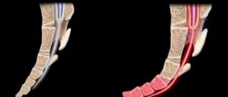
People often strain their external muscles out of zeal, usually the abdominal muscles, buttocks and hip adductors. However, the joint contraction of these muscles along with the pelvic floor muscles does not support the internal organs. You only need to tense the internal muscles. Exercising incorrectly can be harmful (Fig. 2).
If you don't feel your pelvic muscles contracting, change your position and try again. For example, if you are sitting, try lying down or standing up. If this does not help, contact a specialist. Research shows that more than 30% of women even after they are explained in detail how to do it.
Training mode
Having learned how to properly contract the pelvic floor muscles, you can begin training. Try to hold the muscles contracted for up to 10 seconds before relaxing. Don't forget to breathe while doing this. Repeat the exercise up to 10 times, but only as long as you can do it correctly. Exercises can be repeated several times during the day. They can be done lying down, sitting or standing with your legs apart, but the muscles of the thighs, buttocks and abdomen should be relaxed.
As a rule, to achieve this, the exercise must be performed for at least 6-8 weeks, and preferably 6 months. By themselves they can be. A good addition to this daily independent activity is a weekly lesson with an instructor. Exercises are performed standing, sitting, lying or kneeling. The pelvic floor muscles are contracted as strongly as possible and held in this position for 6 - 8 seconds. After each long contraction, do 3-4 quick ones. In each pose, perform 8-12 long contractions and a corresponding number of fast ones. In this case, all contractions must be performed with the same intensity.
Sometimes people forget to do pelvic floor exercises, so it's best to associate them with some regular activity, such as eating or brushing your teeth. This is a great way to integrate exercise into your regular routine.
Women who have recently given birth are advised to reduce the load on their pelvic floor muscles in the first few months to allow them to recover.
Men who have had prostate surgery should begin exercise as soon as their urinary catheter is removed (it can irritate the bladder and cause discomfort during exercise).
Pelvic floor exercises and other physical activities
No matter how strong and strong a person is, if his pelvic floor function is impaired, it is necessary to restore it. There is no need to give up regular sports activities, but for all types of training: cardio, endurance training or strength training, the number of repetitions, sets and frequency of training should depend on the reaction of the pelvic floor. If necessary, you need to reduce the intensity, impact, load, number of repetitions or duration of the workout, and then, as your pelvic floor functions improve, gradually return to the previous regime.
It is better to coordinate the training program with specialists, because people are different, and what suits one person may not suit another. But there are some general rules:
- Avoid high-impact or high-intensity exercises that reduce pressure on the pelvic floor.
- Don't lift heavy objects unless absolutely necessary.
- Before lifting weights, tighten your pelvic floor muscles.
There are two ways to assess the load on the pelvic floor. It is large if you feel heaviness or leak urine during the exercise, and if you are unable to tense your pelvic floor muscles while doing the exercise.
It's unrealistic to constantly think about your pelvic floor muscles during an hour-long workout, but it's helpful to pay attention to them regularly. If you can't engage and contract your muscles while doing a squat, biceps curl, or biking up a hill, you may need to cut your workout short or choose something easier. If your pelvic floor isn't ready for running, you can go for a walk in the hills. If five squats are tiring, do three. Over time you will progress.
How to treat muscle weakness?
The course of treatment is prescribed after diagnosis. Therapy depends on what disease has been identified. It can be based on medications, physiotherapeutic methods, surgical treatment methods, and can also be a combination of several techniques.
References
- Nikitin S. S. et al. Clinical recommendations for the provision of medical care to patients with Pompe disease // Neuromuscular diseases - 2021. - T. 6. - No. 1.
- Vorobyova O. V. Chronic fatigue syndrome (from symptom to diagnosis) // Difficult Patient - 2010. - T. 8. - No. 10.
- Agafonov B.V., Kotov S.V., Sidorova O.P. Myasthenia and congenital myasthenic syndromes - 2013.
- Antelava O. A. et al. Features of the onset and course of antisynthetase syndrome as the most severe subtype of polymyositis/dermatomyositis // RMZh - 2009. - T. 17. - No. 21. - P. 1443-1447.
- Muravyova G. B., Devlikamova F. I. Neuromuscular complications in diseases of the thyroid gland // Practical Medicine - 2013. - No. 1. P. 66.
- Rivier F. et al. Congenital muscular dystrophies: classification and diagnosis // Neuromuscular diseases – 2014. – No. 1. – P. 7-18.
- Belozerov Yu. M. et al. Progressive muscular dystrophy of Emery-Dreyfuss //Almanac of Clinical Medicine – 2001. – No. 4. – P.66-71.
GZEA.PD.18.09.0435g


