Osteochondroma (osteocartilaginous exostosis)
A benign exophytic formation, consisting of a bone base, covered with cartilage tissue on top and located on the surface of the bone.
The formation can be single or generalized in the form of multiple exostotic chondrodysplasia. According to the ICD for oncology (ICD-O3), osteochondroma is coded as 9210/0; according to ICD-10, MEC is coded as Q78.6.
Treatment of osteophytes
Treatment of osteophytes of the knee joint, spine or any other location always begins with treatment of the underlying disease due to which they appeared. The fight against the bone outgrowths itself is carried out in several directions:
- drug therapy to relieve pain and inflammation, restore tissue trophism and prevent further joint destruction (anti-inflammatory drugs, angio- and chondroprotectors);
- therapeutic exercises (relieve muscle spasms and reduce the load on sore joints, improve metabolism in tissues);
- physiotherapeutic treatment (the doctor chooses the method taking into account the location and type of osteophytes);
- maintaining a healthy lifestyle and daily activity (patients should give up bad habits, walk as much as possible, swim, choose active types of recreation);
- massage and self-massage, as well as the use of orthoses (collars, corsets, walking aids);
- following a non-strict diet (foods rich in calcium and magnesium are recommended, such as fish, seafood, nuts and seeds, spinach and other greens, legumes and dairy products - in the absence of contraindications due to metabolic diseases). Patients should avoid sweet, smoked and highly salty foods, as well as any semi-finished products (except homemade preparations) and fast food. Overweight people need to lose weight to successfully treat osteophytes.
Although osteophytes cannot always be completely eliminated with conservative treatment alone, it can reduce their size and completely stop the progression of the pathology. In advanced cases, surgical removal of osteophytes and part or all of the joint is performed.
.
Physiotherapy
Taking courses of physiotherapy helps stop the growth of osteophytes, support joints and significantly alleviate the patient’s condition. To combat the problem, methods such as:
- shock wave therapy
; - electrophoresis
(especially with medications - glucocorticoids, lithium, sulfur, zinc); - phonophoresis with analgesics
; - diadynamic therapy
; - galvanization with caripain
; - massotherapy;
- balneotherapy
(radon, turpentine baths),
mud therapy
; - kinesiotherapy and therapeutic exercises.
Most physiotherapeutic procedures are contraindicated in the presence of cancer and cardiovascular diseases.
Drug treatment
Local drug treatment of osteophytes is carried out in the early stages of the disease and consists of rubbing in warming, anti-inflammatory, locally irritating compounds (ointments, gels, creams and balms). Patients can also do baths and compresses. In all other cases, you need to take pills, and sometimes even injections.
The following groups of drugs are used to treat osteophytes in the spine and joints:
- Non-steroidal anti-inflammatory drugs.
- Glucocorticoids.
- Chondroprotectors (Artracam)
are the only group of drugs that can prevent the destruction of cartilage and joint tissue, as well as start the processes of its regeneration. Other medications are usually used for symptomatic relief. - Muscle relaxants.
- Vitamin and mineral preparations.
In case of severe pain, when it is not possible to urgently consult a doctor, temporary use of simple analgesics is allowed (taking into account their side effects).
Surgery
Surgical treatment of osteophytes of the spine and joints is carried out at the 4th stage of the disease, in which the patient suffers from chronic pain that is not amenable to drug therapy. Such cases include late stages of spondylosis with ring-shaped osteophytes and other severe conditions. A timely operation allows you to save the nervous tissue from irreversible changes and eliminate pain
, and in some cases, even return a certain range of movements.
Minimally invasive operations with a short recovery period are performed in situations where bone spurs pose a serious threat to blood flow and nerve endings and entail severe symptoms, as well as interfere with the patient’s movement and are accompanied by severe pain.
Surgical treatment of spinal osteophytes is carried out in the case of massive growths that disrupt the functions of tissues in the places of occurrence.
In case of degenerative joint diseases, not only the osteophyte is removed, but also excess bone tissue. In the case of knee, elbow and some other joints, joint replacement is indicated.
Please note that gentle arthroscopic operations do not relieve the patient of responsibility for his joints
: after removal of osteophytes, new ones may grow in their place if the patient is not concerned about his health.
Take care of yourself and be happy!
Clinic
Osteochondroma is the most common skeletal tumor. It is detected mainly in children and young adults. Exostoses are most often localized in the metaphyses and growth zones of long tubular bones (femur, tibia, humerus), but can also develop in flat bones (pelvis, scapula, ribs); lesions of the spine are rare. With multiple chondrodysplasia, exostoses affect several segments and have different shapes and sizes. Osteochondromas do not appear simultaneously; their growth and the appearance of new exostoses continue until the growth zones close. It is also possible that osteochondral exostoses will continue to grow against the background of hormonal changes in the body in women.
Clinical manifestations of exostotic chondrodysplasia depend on the location, size, and relationship with surrounding structures. At the beginning of the disease, the process is asymptomatic and the first sign is the identification of a palpable formation (dense, immobile, painless). Pain occurs when there is compression of the surrounding soft tissues, involvement of the neurovascular bundle, a fracture of the base of the osteochondroma, or malignancy. Often, during periods of active skeletal growth, bone deformations are observed in the form of curvature or shortening of a segment. If the pelvic bones, ribs or spine are affected, dysfunction of the pelvic organs, lungs or neurological symptoms may occur.
What treatments are used?
- Resection (or removal) of osteochondroma(s). Indications for this operation are long-term pain and neuropathy. Osteochondroma, which is the cause of deformation. Thus, osteochondromas located in the interosseous space on the forearm that are not identified and not removed in a timely manner can cause retarded growth of the ulna, which leads to deformation of the radius and, accordingly, ulnar clubhand. Also, the forearm loses its rotational function;
- Hemiepiphysiodesis with figure-of-eight plates and screws (controlled growth). An extremely effective method in the treatment of lower limb deformities in growing children. In some cases, the method is used repeatedly as the child grows. When using controlled growth, the child does not lose the ability to move for a single day;
- Corrective osteotomies (crossing the bone with immediate elimination of deformity) with internal fixation (plates locked with intramedullary rods). This option of surgical treatment is indicated in cases where the patient did not undergo controlled growth in a timely manner or it was impossible to use it;
- Osteotomies (bone transection) in combination with transosseous osteosynthesis and gradual lengthening of the bone(s) correction of deformity. The method is indicated in cases of shortening of the bone(s), its deformation. For example, with ulnar clubhand, lengthening of the ulna and correction of deformation of the radius are often indicated, which can only be done using the method of transosseous osteosynthesis.
For more information about the treatment methods used, see the “Treatment Methods” section.
In each case, the decision on the choice of surgical treatment method and its necessity is made by the surgeon individually.
Example 1: Photographs and radiographs of an 11-year-old boy with MEHD, who simultaneously underwent treatment of left femoral deformity using hemiepiphysiodesis, resection of osteochondromas of the forearm bones, and lengthening of the ulna using transosseous osteosynthesis.
Example 2: Photographs and radiographs of a 7-year-old boy with MEHD, for which deformity of both legs and the right thigh was simultaneously treated with hemiepiphysiodesis, resection of osteochondromas of the forearm bones and lengthening of the ulna using transosseous osteosynthesis.
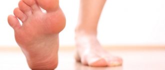
Marginal bony exostoses of the calcaneus
Bone exostosis of the calcaneus is better known as a heel spur. This is a neoplasm that begins to develop from tendon and fascial tissue. The primary factor in the inflammatory reaction is a violation of the integrity of individual fibers of the plantar fascia. It begins to gradually thicken. Plantar fasciitis develops.
This pathology entails constant injury to the synovial cartilage tissue in the heel bone area. The cartilage begins to grow and marginal bone exostoses form, causing severe suffering to the patient when walking.
It is possible to cure bone exostoses on the heels without surgery only in the initial stages. Therefore, if you experience pain in the heels that develops after physical activity (for example, after walking long distances), you should immediately consult an orthopedist.
The doctor will order an x-ray of the heel bone. It will show the formation of marginal bone exostoses along the surface of the heel bone. These formations injure the surrounding soft tissues when walking. An inflammatory process occurs, which stimulates further growth of cartilage and bone tissue.
Causes of exostosis
The causes of the formation of exostosis can be an inflammatory process, bruise, pinching, abnormalities of the periosteum and cartilage, infectious diseases such as syphilis, insufficiency of the functions of the endocrine system or its individual glands. Exostosis is presented, in general, as a persistent formation, however, there are cases when the process of formation of exostosis decreases over time and the exostosis disappears forever.
Often, slowly increasing and not causing pain, exostosis is not marked by clinical symptoms, remaining invisible to both the patient and the doctor. Exostosis is detected by X-ray examination, or by palpation of seals that are already visible during examination.
A large number of scientific works are devoted to elucidating the causes of exostosis; their attention is directed to the study of heredity in this disease . However, even the presence in certain cases of family exostoses, which are inherited, does not yet provide any basis for explaining the occurrence of this disease.
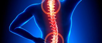
Bone exostoses of the spine and ribs
Cartilaginous and bone exostoses of the spine are a consequence of a long-term degenerative dystrophic process in the area of the intervertebral discs and endplates of the vertebral bodies. The degeneration process begins with a violation of the diffuse nutrition of cartilage tissue. It is able to receive nutrition only when the muscular frame of the back is actively working. If a person leads a sedentary lifestyle, muscles lose elasticity and performance. They cannot transport blood and nutrients dissolved in it to the cartilage tissue of the intervertebral discs.
As a result, the process of dehydration of the fibrous ring begins. It loses its shock-absorbing ability and becomes covered with a network of cracks. Calcium salts are deposited in them.
As a compensatory reaction, chondrosis and sclerosis of the endplates of the vertebral bodies begin. They increase in volume and become deformed. As a result of strengthening the blood supply point, the process of nutrition of bone tissue is enhanced. Deformation of the vertebral bodies and bases begins.
Bone exostoses of the ribs are formed as a result of traumatic exposure and curvature of the spinal column. These tight junctions fix the deformation of the chest, limit the vital capacity of the lungs and can lead to severe oxygen deficiency in the structures of the brain and vital internal organs (heart, liver, spleen).
Diagnosis is made using x-rays. For treatment, conservative methods of influence can be used, such as manual therapy, physiotherapy, shock wave therapy, etc. Surgical intervention for this localization of exostoses is used relatively rarely.