Arthritis of the hip joint (coxitis) is an articular pathology that is characterized by activation of the inflammatory process in the cavity of the corresponding joint of various origins. The etiological basis is inflammatory lesions of the hip joint of infectious, autoimmune, traumatic origin, etc. The disease can affect one or both hip joints. Inflammation initially begins in the synovial membrane, and in the final stages it spreads to cartilaginous, ligamentous, and bone tissues. The course of coxitis is quite variable: acute, subacute, and chronic variants are observed.
Diagnosis by X-ray.
Arthritis of any etiology in the largest, functionally significant joint brings a lot of suffering and trials, provoking excruciating pain, stiffness, and impaired mobility. A disease that lasts a long time and/or with frequent relapses, in most cases leads to persistent chronicity of the process, serious complications, in particular to the destruction of all structural elements of the joint, gross deformation of the bone joint. The most severe type, which with inadequate treatment literally leads to disability within 2 years from the onset of symptoms, is rheumatoid arthritis (RA). RA is a most insidious systemic disease, from which it is impossible to completely recover.
Arthritis pathologies of the hip joint have become so firmly established in modern society that they can rightfully be called the epidemic of the century. According to official data, hip arthritis is diagnosed in people of all ages: the elderly, young, teenagers and even children. Statistics show that women suffer from the disease 2-2.5 times more often than men. The peak incidence falls in the age range of 30-55 years. The average age of patients is 47 years. As for the statistics of the total number of patients with this diagnosis, official data is so far only announced for rheumatoid arthritis. Namely: today in Russia, 2 million people suffer from RA of all types and localizations, of which approximately 35% of patients have an unfavorable focus in the hip joints.
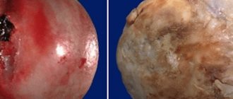
Removed femoral head affected by arthritis.
Experts emphasize that there are approximately the same number of registered patients with rheumoatritis of the hip joint and other forms of coxitis as those who do not go to medical institutions for diagnosis and treatment. Moreover, in just 10 years, the number of people with arthritis of the hip joints has almost doubled. An incredibly rapid growth trend in incidence is predicted for the next decade, and experts do not rule out even worse results.
Doctors urge you not to try to treat arthritic pathogenesis on your own. This is a complex, multifactorial inflammation of the most massive and loaded joint of the musculoskeletal system, difficult to treat. Self-medication will not bring any good, but will only worsen the problem. Forecasts directly depend on the timeliness of visiting a doctor, correct diagnosis and a competent, qualified approach to treatment, based on the principle of individuality. Otherwise, the risks of significant or complete loss of movement in the hip joint, serious health problems (infectious-toxic shock, sepsis in advanced infectious forms, severe cardiac and pulmonary damage in RA, etc.), and in some cases even death, are too high.
Minimally invasive endoprosthetics in the Czech Republic: doctors, rehabilitation, terms and prices.
Find out more
Causes and types of disease
The causes of the inflammatory reaction in the tissues of the hip joint can be due to infection, allergens, and systemic diseases. In addition, injuries, physical overload, and even simple hypothermia can lead to the development of inflammatory lesions in the musculoskeletal area of the pelvis and femur. Sometimes articular tissues become inflamed due to previously performed surgical interventions of any content within the hip region. According to doctors, cancer, blood pathologies, and hereditary factors play a significant role in the appearance of a lesion.
In medicine, there are more than 150 different formulations of diagnoses of arthritic diseases. But in general, arthritis of the hip joints is usually divided into 3 predominant pathogenetic forms: rheumatoid, infectious, reactive. More details about each in the table.
| Form of hip | Frequent provocateurs | Peculiarities | Diagnoses |
| Rheumatoid | The etiology has not been established. But, presumably, RA can be triggered by: viruses (eg, Epstein-Barr, herpes simplex, hepatitis B), bacteria (usually the respiratory system), tobacco toxins, injuries, allergies, stress, metabolic pathologies, abuse of steroid therapy, heredity, etc. | Autoimmune inflammation of synovial joints of the erosive-destructive type. This is a severe chronic pathology caused by the abnormal production of aggressive antibodies aimed at destroying the articular apparatus. Specific autoimmune aggressors affect not only the TB joints specifically (in turn or two at once), but also other types of joints, sometimes even involving internal organs. Monoarthritis of the rheumatoid type is extremely rare, except at the very onset of the disease. The inflammatory process is initiated by antibodies such as RF and ACCP, which are the leading biomarkers in the diagnosis of rheumatoid arthritis of the hip joint. | Seropositive, seronegative RA, Still's disease, juvenile RA, etc. |
| Infectious | Streptococcus Staphylococcus Gonococcus Mycobacterium Brucella Haemophilus influenzae Pseudomonas aeruginosa | The pathogenic environment penetrates directly into the joint cavity by lymphogenous or hematogenous route. It can migrate to the hip joint from any infected area of the body. Harmful microorganisms settle and become active in the hip joint, which is why a local focus of inflammation occurs. It is also possible to introduce infection through injection/surgical means or from the external environment during an open injury. Important: pathological microorganisms are always cultured during an intra-articular tank examination of synovial fluid. | Purulent septic, tuberculous, brucellosis arthritis, etc. |
| Reactive "sterile" | Chlamydia Ureaplasma Mycoplasma Yersinia infection Salmonella Campylobacter Clostridium Shigella | An immunoinflammatory reaction in response to an acute or persistent genitourinary or intestinal infection. The infectious focus that induces arthritis is located outside the joint - in the urogenital tract or gastrointestinal tract. That is, the infection is not detected in the joint media. Inflammation occurs due to a genetic factor: carriage of the HLA-B27 antigen. When this infection + HLA-B27 is combined, the immune system, along with the destruction of the main pathogen, begins to attack tissue cells of a healthy joint, mistakenly perceiving them as foreign agents. When diagnosed, HLA-B27 is detected in the blood, while rheumatoid factor is absent. | Reiter's disease, urogenic arthritis, postenterocolitic, etc. |
It cannot be said that separate groups include gouty, psoriatic and post-traumatic arthritis, the root causes of which have nothing in common with infections or rheumatism. However, it is, of course, impossible to completely exclude the detection of associated rheumatoid pathology at the time of diagnosis. Gouty and psoriatic types are accompanied by joint inflammation due to complex and, unfortunately, incurable diseases of a systemic nature: gout and psoriasis. Post-traumatic type is an inflammatory reaction of the joint that occurs after closed injuries or physical overload.
- Arthritis due to gout
. Gouty arthritis is caused by a violation of purine metabolism in the body, which causes an increase in the concentration of uric acid in the blood. For this reason, urates (crystals) of uric acid are deposited in the cavities of the hip joints, which provoke local inflammation and have a destructive effect on the articular cartilage and periarticular tissues. The etiological basis of gouty arthritis is a systemic metabolic failure. - Arthritis with psoriasis
. This is an inflammatory process in the hip joint, occurring against the background of a severe skin disease, the mechanism of development of which still remains a mystery to specialists. An immunological factor is involved in the development of joint inflammation due to psoriasis. It is characterized by an imbalance of pro-inflammatory and anti-inflammatory cytokines, which produces a destructive-inflammatory effect on the osteochondral segment. - Post-traumatic arthritis of the hip joint
. This arthritic disease often occurs, for example, after an injury with hemorrhage into the joint cavity, after dislocations/subluxations of the hip, severe local contusion with trauma to the capsule or ligamentous-muscular system. The lesion can also develop after systematic heavy physical activity, which leads to microdamage to the articular cartilage. Under the influence of these factors, the synovial membranes and periarticular tissues become inflamed, as a result of which pathological fluid (effusion) accumulates in the joints.
With timely (early) and proper treatment, getting rid of post-traumatic arthritis forever is quite possible. This is the only form that has the best prognostic indicators. However, untreated injuries and their complications in the form of synovitis can develop into a serious, chronic arthritic pathology, which in turn can be complicated by irreversible deforming coxarthrosis.
General information about the disease
Arthritis of the hip joint (coxitis) is a polyetiological disease in the development of which many factors take part. There are several clinical forms of the disease, during which there are both similar and different symptoms. ICD-10 code M00 – M99.
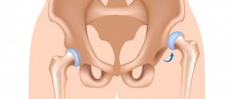
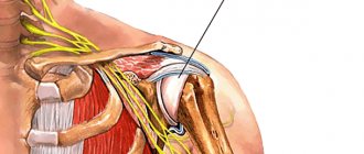
The hip joint (HJ) is formed by the acetabulum of the pelvic bone and the head of the femur. The spherical shape makes it mobile; a ligament fits from the head of the femur to the acetabulum, holding the articular parts in a normal position. Externally, the hip joint is also strengthened by ligaments and a thick layer of soft tissue. This is the leading supporting joint, located deep in the tissues, so it is not so easy to identify a slow inflammatory process with mild symptoms in it.
Arthritis of the hip joint develops at any age, since the disease can have different origins. The most relevant clinical forms are lesions in childhood and adolescence, as well as tuberculous arthritis.
Symptoms of hip arthritis
The standard symptoms of arthritis affecting the hip part of the musculoskeletal system are local signs of inflammation, and these are:
- pain syndrome, aches in the corresponding area (pain most often occurs during or after a long period of rest, for example, in the middle of a night's sleep or in the morning after waking up);
- swelling of the soft tissues near the TB joint as a result of the formation of inflammatory effusion in the joint;
- sensation of “hot” skin covering the sore joint;
- a feeling of squeezing and bursting in the hip joint;
- redness of the skin in the inflamed area (not always);
- limited range of motion, stiffness.
Pain is the main symptom.
Since the basis of the disease is severe autoimmune and infectious processes within the body, the clinical picture is often complemented and aggravated by symptoms of general intoxication:
- increased body temperature;
- chills and loss of strength;
- headache;
- dizziness;
- loss of appetite, nausea;
- nervousness;
- anemia;
- bad dream.
The intensity of symptoms is influenced by the nature of the pathological process: acute, subacute or chronic. The acute course is characterized by the sudden onset of an arthritic disease with pronounced painful symptoms. For a subacute course, a gradual increase in signs of inflammation over 1-2 weeks is typical. Acute and subacute arthritis can become chronic, when periods of remission periodically alternate with periods of relapse.
A chronic variant of the course can occur from the very beginning (for example, with RA). Moreover, such arthritis can develop for a long time and not particularly bother, and after a few years begin to persistently annoy with attacks of inflammation, which, as the duration of the disease increases, becomes more and more protracted and painful. If a chronic disease is not properly controlled, it will inevitably lead to the destruction of the articular ends with their further deformation, fusion with each other, resulting in immobility of the hip joint, muscle atrophy and disability of the patient.
General clinical recommendations
To avoid relapses of hip arthritis and forget about pain, patients are recommended to:
- lead an active healthy lifestyle;
- Healthy food;
- get rid of excess weight and bad habits - smoking and alcohol abuse;
- do exercise therapy every day; Swimming is especially beneficial;
- avoid heavy physical activity, hypothermia and stress;
- carefully treat all acute and chronic diseases;
- take courses of anti-relapse therapy several times a year as prescribed by a doctor.
How not to get sick
The hip joints should be especially taken care of by persons with a family history and who have relatives with a similar pathology. To do this, you need to avoid any factors that provoke the disease: infections, hypothermia, stress. It is also necessary to move more, engage in feasible sports to strengthen the muscles of the back and lower extremities, and also maintain normal body weight.
What to eat
Meals should be varied and regular. It is worth limiting: spicy seasonings, fried, fatty, smoked foods, alcohol, sweets - all this can provoke inflammatory processes in the joints.
Diagnosis of hip arthritis
It will not be difficult for the doctor to tell that the patient has arthritis during the initial listening to complaints, palpation and visual examination of the problem area. But this is absolutely not enough to recommend therapeutic measures to the patient. It is alarming that with good rheumatologists, who have the highest competence in differentiating the forms of arthritic pathologies among the huge range of possible ones, there is tension in domestic medical institutions. In fact, patients today are too often diagnosed with unidentified arthritis, which often results in the wrong treatment being prescribed, further exacerbating the joint problem.
Diagnosis in the picture.
Therefore, in the interests of the patient, it is important to find a highly qualified doctor who will competently conduct an examination using effective diagnostic tools recommended by modern rheumatology. As a result, it will accurately establish a diagnosis, determining its etiology, severity and, above all, exclude or confirm the existence of RA - the most aggressive type of disease, known for its worst prognosis. And, of course, he will competently develop the concept of effective treatment.
As you correctly understood, a correctly made diagnosis at an early stage of arthritis manifestations, within the minimum allotted time from the patient’s first visit to the doctor, is of clinical value. This is called the “window of maximum opportunity”: when an adequate therapy program can radically change the course and outcome of the pathology for the better. To verify the true diagnosis in the presence of inflammatory arthropathy, specialists use the following diagnostic tools:
- a list of classification features of specific nosological forms;
- instrumental methods for examining the hip joint (axial x-ray, MRI, ultrasound);
- laboratory research methods:
- general and biochemical blood test;
- urine and stool tests;
- immunological (for RF and ACCP) and immunogenetic tests (for HLA);
- blood examination to identify infection (ELISA, PCR methods);
- puncture collection of synovial fluid for organoleptic, cytological and bacteriological examination;
- synovial membrane biopsy;
- arthroscopic examination of the joint cavity.
The first place in diagnosis, of course, is given to laboratory examination methods, which significantly increase the likelihood of making a correct diagnosis. It should be emphasized that in addition to all the listed diagnostic techniques, a thorough study of the entire history of a person’s illnesses, analysis of diseases, conditions, lifestyle, etc. that preceded arthritis also play an important role.
In the diagnostic and treatment process, in addition to the rheumatologist and orthopedist, in most cases it is necessary to involve doctors of other specialties: gastroenterologist, urologist, dermatologist, infectious disease specialist, ENT specialist, pulmonologist, allergist, immunologist, cardiologist, etc.
Modern drug therapy for rheumatoid arthritis
Rheumatoid arthritis is a disease that has been the focus of attention of rheumatologists around the world for decades. This is due to the great medical and social significance of this disease. Its prevalence reaches 0.5–2% of the total population in industrialized countries [1, 2]. Patients with rheumatoid arthritis experience a decrease in life expectancy compared to the general population by 3–7 years [3]. It is difficult to overestimate the colossal damage caused by this disease to society due to the early disability of patients, which, in the absence of timely active therapy, can occur in the first 5 years from the onset of the disease.
Rheumatoid arthritis is a chronic inflammatory disease of unknown etiology, which is characterized by damage to peripheral synovial joints and periarticular tissues, accompanied by autoimmune disorders and can lead to destruction of articular cartilage and bone, as well as systemic inflammatory changes.
The pathogenesis of the disease is very complex and largely insufficiently studied. Despite this, by now some key points in the development of rheumatoid inflammation are well known, which determine the main methods of therapeutic treatment for it (Fig. 1). The development of chronic inflammation in this case is associated with the activation and proliferation of immunocompetent cells (macrophages, T- and B-lymphocytes), which is accompanied by the release of cellular mediators - cytokines, growth factors, adhesion molecules, as well as the synthesis of autoantibodies (for example, anticitrullinated antibodies) and the formation immune complexes (rheumatoid factors). These processes lead to the formation of new capillary vessels (angiogenesis) and the proliferation of connective tissue in the synovial membrane, to the activation of cyclooxygenase-2 (COX-2) with an increase in the synthesis of prostaglandins and the development of an inflammatory reaction, to the release of proteolytic enzymes, activation of osteoclasts, and as a result - to the destruction of normal joint tissues and the occurrence of deformities.
Treatment of rheumatoid arthritis
Treatment includes:
- drug therapy;
- non-drug therapy methods;
- orthopedic treatment, rehabilitation.
Based on the pathogenesis of the disease, it becomes obvious that it is possible to effectively influence the development of the disease at two levels:
- suppressing excessive activity of the immune system;
- blocking the production of inflammatory mediators, primarily prostaglandins.
Since, in addition to inflammation itself, activation of the immune system is accompanied by many other pathological processes, the effect at the first level is significantly deeper and more effective than at the second. Drug immunosuppression is the mainstay of treatment for rheumatoid arthritis. Immunosuppressive drugs used to treat this disease include disease-modifying anti-inflammatory drugs (DMARDs), biological agents, and glucocorticosteroids. At the second level, non-steroidal anti-inflammatory drugs (NSAIDs) and glucocorticosteroids act.
In general, immunosuppressive therapy is accompanied by a slower development of the clinical effect (in a broad framework - from several days in the case of biological therapy to several months in the case of some DMARDs), which at the same time can be very pronounced (up to the development of clinical remission) and persistent, and is also characterized by inhibition of joint destruction.
Anti-inflammatory therapy itself (NSAIDs) can produce a clinical effect (pain relief, reduction of stiffness) very quickly - within 1-2 hours, however, with the help of such treatment it is almost impossible to completely stop the symptoms of active rheumatoid arthritis and, apparently, it has no effect at all on the development of destructive processes in tissues.
Glucocorticosteroids have both immunosuppressive and direct anti-inflammatory effects, so clinical improvement can develop quickly (within a few hours when administered intravenously or intra-articularly). There is evidence of suppression of the progression of the erosive process in joints during long-term therapy with low doses of glucocorticosteroids and their positive effect on the functional status of the patient. At the same time, it is well known from practice that prescribing only glucocorticosteroids, without other immunosuppressive drugs (DMARDs), rarely provides an opportunity to effectively control the course of the disease.
Non-drug methods of treating rheumatoid arthritis (physiotherapy, balneotherapy, diet therapy, acupuncture, etc.) are additional methods that can slightly improve the patient’s well-being and functional status, but do not relieve symptoms and significantly influence joint destruction.
Orthopedic treatment, including orthotics and surgical correction of joint deformities, as well as rehabilitation measures (physical therapy, etc.) are of particular importance, mainly in the later stages of the disease, to maintain functional ability and improve the patient’s quality of life.
The main goals of treatment for RA are [2, 6]:
- relief of disease symptoms, achievement of clinical remission or at least low disease activity;
- inhibition of the progression of structural changes in joints and corresponding functional disorders;
- improving the quality of life of patients, maintaining their ability to work.
It must be kept in mind that treatment goals may vary significantly depending on the duration of the disease. At an early stage of the disease, i.e., with a disease duration of 6–12 months, achieving clinical remission is a very realistic goal, as well as inhibiting the development of erosions in the joints. With the help of modern methods of active drug therapy, it is possible to achieve remission in 40–50% of patients [4, 5], and the absence of the appearance of new erosions according to radiography [7] and magnetic resonance imaging [8] has also been shown in a significant number of patients with a follow-up duration of 1 -2 years.
With long-term rheumatoid arthritis, especially with insufficiently active therapy in the first years of the disease, achieving complete remission is theoretically also possible, but the likelihood of this is much lower. The same can be said about the possibility of stopping the progression of destruction in joints that have already been significantly destroyed over several years of illness. Therefore, with advanced rheumatoid arthritis, the role of rehabilitation measures and orthopedic surgery increases. In addition, in the later stages of the disease, long-term basic maintenance therapy can be used for secondary prevention of complications of the disease, such as systemic manifestations (vasculitis, etc.), secondary amyloidosis.
Basic therapy for rheumatoid arthritis. DMARDs (synonyms: disease-modifying antirheumatic drugs, slow-acting drugs) are the main component of the treatment of rheumatoid arthritis and, in the absence of contraindications, should be prescribed to every patient with this diagnosis [9]. It is especially important to prescribe DMARDs as quickly as possible (immediately after diagnosis) at an early stage, when there is a limited period of time (several months from the onset of symptoms) to achieve the best long-term results - the so-called “therapeutic window” [10].
Classic DMARDs have the following properties.
- The ability to suppress the activity and proliferation of immunocompetent cells (immunosuppression), as well as the proliferation of synoviocytes and fibroblasts, which is accompanied by a pronounced decrease in the clinical and laboratory activity of RA.
- Persistence of the clinical effect, including its persistence after discontinuation of the drug.
- The ability to delay the development of the erosive process in joints.
- Ability to induce clinical remission.
- Slow development of a clinically significant effect (usually within 1–3 months from the start of treatment).
DMARDs differ significantly in their mechanism of action and features of use. The main parameters characterizing DMARDs are presented in Table 1.
DMARDs can be roughly divided into first-line and second-line drugs. First-line drugs have the best balance of effectiveness (reliably suppress both clinical symptoms and the progression of the erosive process in the joints) and tolerability, and therefore are prescribed to most patients.
First-line DMARDs include the following.
- Methotrexate is the “gold standard” for the treatment of rheumatoid arthritis. Recommended doses of 7.5–25 mg per week are individualized by gradual increases of 2.5 mg every 2–4 weeks until a good clinical response is achieved or intolerance occurs. The drug is given orally (weekly for two consecutive days in 3-4 divided doses every 12 hours). In case of unsatisfactory tolerability of methotrexate when taken orally due to dyspepsia and other complaints associated with the gastrointestinal tract (GIT), the drug can be prescribed parenterally (one IM or IV injection per week).
- Leflunomide (Arava). Standard treatment regimen: 100 mg orally per day for 3 days, then 20 mg/day continuously. If there is a risk of intolerance to the drug (old age, liver disease, etc.), treatment can be started with a dose of 20 mg/day. It is comparable in effectiveness to methotrexate and has slightly better tolerability. There is evidence of higher effectiveness of leflunomide in relation to the quality of life of patients, especially in early rheumatoid arthritis. The cost of treatment with leflunomide is quite high, so it is often prescribed when there are contraindications to the use of methotrexate, its ineffectiveness or intolerance, but it can also be used as the first basic drug.
- Sulfasalazine. In clinical trials, it was not inferior in effectiveness to other DMARDs, but clinical practice shows that sulfasalazine usually provides sufficient control over the course of the disease in cases of moderate and low activity of rheumatoid arthritis.
Second-line DMARDs are used much less frequently due to lower clinical efficacy and/or greater toxicity. They are prescribed, as a rule, when first-line DMARDs are ineffective or intolerable.
DMARDs can cause significant improvement (good clinical response) in approximately 60% of patients. Due to the slow development of the clinical effect, the prescription of DMARDs for periods of less than 6 months is not recommended. The duration of treatment is determined individually; the typical duration of a “course” of treatment with one drug (in case of a satisfactory response to therapy) is 2–3 years or more. Most clinical recommendations suggest indefinite use of maintenance dosages of DMARDs to maintain the achieved improvement.
If monotherapy with any basic drug is insufficiently effective, a combination basic therapy regimen, i.e., a combination of two or three DMARDs, can be chosen. The following combinations have proven themselves to be the most effective:
- methotrexate + leflunomide;
- methotrexate + cyclosporine;
- methotrexate + sulfasalazine;
- methotrexate + sulfasalazine + hydroxychloroquine.
In combination regimens, drugs are usually used in moderate dosages. A number of clinical studies have demonstrated the superiority of combination basic therapy over monotherapy, but the higher effectiveness of combination regimens is not considered strictly proven. Combination DMARDs are associated with a moderate increase in side effects.
Biological drugs in the treatment of rheumatoid arthritis. The term biological drugs (from English biologics) is used in relation to drugs produced using biotechnology and carrying out targeted (“point”) blocking of key moments of inflammation using antibodies or soluble receptors to cytokines, as well as other biologically active molecules. Thus, biological products have nothing to do with “dietary supplements.” Due to the large number of “target molecules” that can potentially suppress immune inflammation, a number of drugs from this group have been developed and several more are undergoing clinical trials.
The main biological drugs registered in the world for the treatment of rheumatoid arthritis include:
- infliximab, adalimumab, etanercept (act on tumor necrosis factor (TNF-α);
- rituximab (acts on CD 20 (B lymphocytes));
- anakinra (acts on interleukin-1);
- abatasept (affects CD 80, CD 86, CD 28).
Biological drugs are characterized by a pronounced clinical effect and reliably proven inhibition of joint destruction. These signs allow biological drugs to be classified as DMARDs. At the same time, a feature of the group is the rapid (often within a few days) development of significant improvement, which combines biological therapy with intensive care methods. A characteristic feature of biological agents is the potentiation of the effect in combination with DMARDs, primarily methotrexate. Due to its high effectiveness in rheumatoid arthritis, including in patients resistant to conventional therapy, biological therapy has now moved to second place (after DMARDs) in the treatment of this disease.
The negative aspects of biological therapy include:
- inhibition of anti-infective and (potentially) anti-tumor immunity;
- the risk of developing allergic reactions and inducing autoimmune syndromes due to the fact that biological drugs are proteins in their chemical structure;
- high cost of treatment.
Biological therapies are indicated if treatment with DMARDs (such as methotrexate) is not adequate due to lack of effectiveness or poor tolerability.
One of the most important target molecules is TNF-a, which has many pro-inflammatory biological effects and contributes to the persistence of the inflammatory process in the synovium, destruction of cartilage and bone tissue through a direct effect on synovial fibroblasts, chondrocytes and osteoclasts. TNF-α blockers are the most widely used biological agents in the world.
A drug from this group, infliximab (Remicade), which is a chimeric monoclonal antibody to TNF-α, has been registered in Russia. The drug is usually prescribed in combination with methotrexate. In patients with insufficient effectiveness of therapy with medium and high doses of methotrexate, infliximab significantly improves the response to treatment and functional indicators, and also leads to a significant inhibition of the progression of joint space narrowing and the development of the erosive process.
The indication for the use of infliximab in combination with methotrexate is the ineffectiveness of one or more DMARDs used at full dose (primarily methotrexate), with the persistence of high inflammatory activity (five or more swollen joints, erythrocyte sedimentation rate (ESR) more than 30 mm/h, C-reactive protein (CRP) more than 20 mg/l). In early rheumatoid arthritis with high inflammatory activity and a rapid increase in structural disorders in the joints, combination therapy with methotrexate and infliximab can be prescribed immediately.
Before prescribing infliximab, a screening examination for tuberculosis (chest x-ray, tuberculin test) is required. Recommended regimen: initial dose of 3 mg/kg of the patient’s body weight intravenously, then 3 mg/kg of body weight after 2, 6 and 8 weeks, then 3 mg/kg of body weight every 8 weeks, if the dose is insufficiently effective may increase up to 10 mg/kg body weight. The duration of treatment is determined individually, usually at least 1 year. After discontinuation of infliximab, maintenance therapy with methotrexate is continued. It should be kept in mind that re-administration of infliximab after completion of treatment with this drug is associated with an increased likelihood of delayed-type hypersensitivity reactions.
The second drug registered in our country for biological therapy is rituximab (Mabthera). The action of rituximab is aimed at suppressing B lymphocytes, which are not only the key cells responsible for the synthesis of autoantibodies, but also perform important regulatory functions in the early stages of immune reactions. The drug has pronounced clinical efficacy, including in patients who do not respond sufficiently to infliximab therapy.
For the treatment of rheumatoid arthritis, the drug is used at a dose of 2000 mg per course (two infusions of 1000 mg, each with an interval of 2 weeks). Rituximab is administered intravenously slowly; infusion in a hospital setting is recommended with the possibility of precise control over the rate of administration. To prevent infusion reactions, it is advisable to pre-administer methylprednisolone 100 mg. If necessary, a second course of rituximab infusions can be performed after 6–12 months.
According to European clinical guidelines, it is advisable to prescribe rituximab in cases of ineffectiveness or impossibility of infliximab therapy. The possibility of using rituximab as the first biological drug is currently the subject of research.
Glucocorticosteroids. Glucocorticosteroids have a multifaceted anti-inflammatory effect due to the blockade of the synthesis of proinflammatory cytokines and prostaglandins, as well as inhibition of proliferation due to their effect on the genetic apparatus of cells. Glucocorticosteroids have a rapid and pronounced dose-dependent effect on the clinical and laboratory manifestations of inflammation. The use of glucocorticosteroids is fraught with the development of undesirable reactions, the frequency of which also increases with increasing doses of the drug (steroid osteoporosis, drug-induced Itsenko-Cushing syndrome, damage to the gastrointestinal mucosa). These drugs alone in most cases cannot provide complete control over the course of rheumatoid arthritis and must be prescribed together with DMARDs.
Glucocorticosteroids for this disease are used systemically and locally. For systemic use, the main method of treatment is the administration of low doses orally (prednisolone - up to 10 mg/day, methylprednisolone - up to 8 mg/day) for a long period with high inflammatory activity, polyarticular lesions, and insufficient effectiveness of DMARDs.
Medium and high doses of glucocorticosteroids orally (15 mg/day or more, usually 30-40 mg/day in terms of prednisolone), as well as pulse therapy with glucocorticosteroids - intravenous administration of high doses of methylprednisolone (250-1000 mg) or dexamethasone (40-1000 mg). 120 mg) can be used to treat severe systemic manifestations of rheumatoid arthritis (effusive serositis, hemolytic anemia, cutaneous vasculitis, fever, etc.), as well as some special forms of the disease. The duration of treatment is determined by the time required to relieve symptoms and is usually 4–6 weeks, after which a gradual stepwise dose reduction is carried out with a transition to treatment with low doses of glucocorticosteroids.
Glucocorticosteroids in medium and high doses, pulse therapy, apparently, do not have an independent effect on the course of rheumatoid arthritis and the development of the erosive process in the joints.
For local therapy, drugs are used in microcrystalline form, prescribed in the form of intra-articular and periarticular injections: betamethasone, triamsinolone, methylprednisolone, hydrocortisone.
Glucocorticosteroids for local use have a pronounced anti-inflammatory effect, mainly at the injection site, and in some cases - a systemic effect. Recommended daily doses are: 7 mg for betamethasone, 40 mg for triamsinolone and methylprednisolone, 125 mg for hydrocortisone. This dose (in total) can be used for intra-articular injection into one large (knee) joint, two medium-sized joints (elbows, ankles, etc.), 4-5 small joints (metacarpophalangeal, etc.), or for periarticular administration of the drug at 3–4 points.
The effect after a single injection usually occurs within 1–3 days and lasts for 2–4 weeks if well tolerated.
In this regard, it is not advisable to prescribe repeated injections of glucocorticosteroids into one joint earlier than after 3-4 weeks. Carrying out a course of several intra-articular injections into the same joint has no therapeutic meaning and is fraught with complications (local osteoporosis, increased destruction of cartilage, osteonecrosis, suppuration). Due to the increased risk of osteonecrosis, intra-articular injection of glucocorticosteroids into the hip joint is generally not recommended.
Glucocorticosteroids for local use are prescribed as an additional method for relieving exacerbations of rheumatoid arthritis and cannot serve as a replacement for systemic therapy.
NSAIDs. The importance of NSAIDs in the treatment of rheumatoid arthritis has decreased significantly in recent years due to the emergence of new effective pathogenetic therapy regimens. The anti-inflammatory effect of NSAIDs is achieved by suppressing the activity of COX, or selectively COX-2, and thereby reducing the synthesis of prostaglandins. Thus, NSAIDs act on the final link of rheumatoid inflammation.
The effect of NSAIDs in rheumatoid arthritis is to reduce the severity of symptoms of the disease (pain, stiffness, swelling of the joints). NSAIDs have an analgesic, anti-inflammatory, and antipyretic effect, but have little effect on laboratory parameters of inflammation. In the vast majority of cases, NSAIDs are not able to significantly change the course of the disease. Their prescription as the only antirheumatic drug for a definite diagnosis of rheumatoid arthritis is currently considered a mistake. However, NSAIDs are the mainstay of symptomatic therapy for this disease and in most cases are prescribed in combination with DMARDs.
Along with the therapeutic effect, all NSAIDs, including selective ones (COX-2 inhibitors), can cause erosive and ulcerative damage to the gastrointestinal tract (primarily its upper parts - “NSAID gastropathy”) with possible complications (bleeding, perforation, etc.), as well as nephrotoxic and other adverse reactions.
The main characteristics that must be considered when prescribing NSAIDs are as follows.
- There are no significant differences between NSAIDs in terms of effectiveness (for most drugs, the effect is proportional to the dose up to the maximum recommended).
- There are significant differences in tolerability between different NSAIDs, especially with regard to gastrointestinal involvement.
- The incidence of adverse effects is usually proportional to the dose of NSAIDs.
- In patients with an increased risk of developing NSAID-associated gastrointestinal lesions, the risk can be reduced by concurrent administration of proton pump blockers and misoprostol.
There is individual sensitivity to various NSAIDs in terms of both effectiveness and tolerability of treatment. Doses of NSAIDs for rheumatoid arthritis correspond to standard doses. The duration of NSAID treatment is determined individually and depends on the patient’s need for symptomatic therapy. If there is a good response to DMARD therapy, the NSAID drug may be discontinued.
The most commonly used NSAIDs for rheumatoid arthritis include:
- diclofenac (50–150 mg/day);
- nimesulide (200–400 mg/day);
- celecoxib (200–400 mg/day);
- meloxicam (7.5–15 mg/day);
- ibuprofen (800–2400 mg/day);
- lornoxicam (8–12 mg/day).
Selective NSAIDs, while not significantly different in effectiveness from non-selective ones, are less likely to cause NSAID gastropathy and serious adverse reactions from the gastrointestinal tract, although they do not exclude the development of these complications. A number of clinical studies have demonstrated an increased likelihood of developing severe vascular pathology (myocardial infarction, stroke) in patients receiving drugs from the coxib group, and therefore the possibility of treatment with celecoxib should be discussed with particular caution in patients with coronary artery disease and other serious cardiovascular pathologies.
Additional drug treatments. As a symptomatic analgesic (or an additional analgesic if NSAIDs are insufficiently effective), paracetamol (acetaminophen) can be used at a dose of 500–1500 mg/day, which has relatively low toxicity. For local symptomatic therapy, NSAIDs are used in the form of gels and ointments, as well as dimethyl sulfoxide in the form of a 30–50% aqueous solution in the form of applications. In the presence of osteoporosis, appropriate treatment with calcium, vitamin D3, bisphosphonates, and calcitonin is indicated.
General principles of management of patients with RA
A patient diagnosed with rheumatoid arthritis should be prescribed a drug from the DMARD group, which, with a good clinical effect, can be used as the only method of therapy [9]. Other medications are used as needed.
The patient should be informed about the nature of his disease, course, prognosis, the need for long-term complex treatment, as well as possible adverse reactions and a treatment monitoring scheme, unfavorable combinations with other drugs (in particular, alcohol), possible activation of foci of chronic infection during treatment , the advisability of temporarily discontinuing immunosuppressive drugs in the event of acute infectious diseases, the need for contraception during treatment.
Therapy for rheumatoid arthritis should be prescribed by a rheumatologist and carried out under his supervision. Treatment with biological drugs can only be carried out under the supervision of a rheumatologist who has sufficient knowledge and experience to carry it out. Therapy is long-term and involves periodic monitoring of disease activity and assessment of response to therapy. A simplified algorithm is presented in Figure 2.
Monitoring disease activity and response to therapy includes assessment of joint status indicators (number of painful and swollen joints, etc.), acute-phase blood parameters (ESR, CRP), assessment of pain and disease activity using a visual analogue scale, assessment of the patient’s functional activity in daily activities with using the Russian version of the Health Questionnaire (HAQ). There are internationally recognized methods for quantifying response to treatment using the DAS (Disease Activity Score) recommended by the European League Against Rheumatism (EULAR) and the American College of Rheumatology (ACR) criteria [1]. In addition, the safety of the therapy administered to the patient should be monitored (in accordance with both the formulary and existing clinical guidelines). Due to the fact that the erosive process can develop even with low inflammatory activity, in addition to assessing the activity of the disease and response to therapy, radiography of the joints is mandatory. The progression of destructive changes in the joints is assessed by standard radiography of the hands and feet using the radiological classification of the stages of rheumatoid arthritis, quantitative methods using the Sharp and Larsen indices. In order to monitor the patient’s condition, examinations are recommended to be carried out at certain intervals (Table 2).
Treatment of treatment-resistant RA
It is advisable to consider a patient resistant to treatment if there is ineffectiveness (lack of 20% improvement in the main indicators) of at least two standard DMARDs in sufficiently high doses (methotrexate - 15-20 mg/week, sulfasalazine - 2000 mg/day, leflunomide - 20 mg/day) . Failure can be primary or secondary (occurring after a period of satisfactory response to therapy or when the drug is re-prescribed). There are the following ways to overcome resistance to therapy:
- prescription of biological drugs (infliximab, rituximab);
- prescription of glucocorticosteroids;
- use of combination basic therapy;
- use of second-line DMARDs (cyclosporine, etc.).
From the point of view of long-term results in relation to functional impairment, quality of life and its duration, the optimal treatment strategy for rheumatoid arthritis is long-term treatment with DMARDs with a systematic change in the regimen of their use as needed [11].
Literature
- Clinical recommendations. Rheumatology/ sub. ed. E. L. Nasonova. M.: GEOTAR-Media, 2006. 288 p.
- Emery P., Suarez-Almazor M. Rheumatoid Arthritis // Clin Evid. 2003; 10: 1454–1476.
- Nasonov E. L., Karateev D. E., Chichasova N. V., Chemeris N. A. Modern standards of pharmacotherapy for rheumatoid arthritis // Clinical pharmacology and therapy. 2005. T. 14. No. 1. P. 72–75.
- Balabanova R. M., Karateev D. E., Kashevarov R. Yu., Luchikhina E. L. Leflunomide (Arava) for early rheumatoid arthritis // Scientific and practical rheumatology. 2005. No. 5. pp. 31–34.
- Goekoop-Ruiterman YP, de Vries-Bouwstra JK, Allaart CF et al. Clinical and radiographic outcomes of four different treatment strategies in patients with early rheumatoid arthritis (the BeSt study): a randomized, controlled trial // Arthritis Rheum. 2005; v. 52; 11: 3381–90.
- Smolen et al. Therapeutic strategies in early rheumatoid arthritis //Best Practice & Research Clinical Rheumatology. 2005; 19; 1: 163–177.
- Breeveld F.C., Emery P., Keystone E. et al. Infliximab in active early rheumatoid arthritis // Ann Rheum Dis 2004; 63: 149–155.
- Quinn MA, Conaghan PG, O'Connor PJ et al. Very early treatment with infliximab in addition to methotrexate in early, poor-prognosis rheumatoid arthritis reduces magnetic resonance imaging evidence of synovitis and damage, with sustained benefit after infliximab withdrawal: results from a twelve-month randomized, double-blind, placebo-controlled trial // Arthritis Rheum. 2005; 52; 1:27–35.
- American College of Rheumatology Subcommittee on Rheumatoid Arthritis Guidelines. Guidelines for the management of rheumatoid arthritis. 2002 Update // Arthritis Rheum. 2002; 46: 328–346.
- Quinn MA, Emery P. Window of opportunity in early rheumatoid arthritis: possibility of altering the disease process with early intervention// Clin Exp Rheumatol. 2003; 21; Suppl 31:154–157.
- Karateev D. E. Retrospective assessment of long-term basic therapy in patients with rheumatoid arthritis // Scientific and practical rheumatology. 2003. No. 3. pp. 32–36.
D. E. Karateev , Doctor of Medical Sciences Institute of Rheumatology RAMS, Moscow
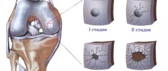
1-2-3 stages
The severity of the disease is characterized by 3 stages. The first is the stage of synovitis, the second is productive-destructive, the third is deforming and ankylosing.
- The first stage is initial and low active in relation to the structures of the articular apparatus. Its characteristic signs are inflammation and thickening of the synovial membrane of the hip joint, accumulation of inflammatory exudate in the cavity. Vital movements may be slightly difficult due to pain and swelling due to synovitis. In general, the integrity and shape of the structures are preserved.
- The second stage is arthritis with moderate activity and severity. At this stage, destruction processes begin to progress. That is, there is ulceration and loss of the hyaline cartilage that covers the surfaces of the femoral head and acetabulum. X-rays reveal a narrowing of the joint space, and periarticular osteoporosis may be detected. The range of motion is noticeably reduced, and stiffness is pronounced. By the middle of this stage, articular cartilage has been massively lost, and the ends of the articular bones are practically exposed.
- In the third phase - highly active and severe - deformation of the bone elements occurs at a rapid pace, their partial or absolute fusion in an intact position. Ankylosis is a “classic” of late rheumatoid arthritis. The joint space is critically narrowed or completely blocked. The patient is unable to move normally and independently perform even basic daily tasks. At this stage, surgery is recommended.
Disease in dynamics.
Clinical manifestation of arthrosis of the hip joint
At the stage of the inception of the clinical picture of the pathology, the first symptoms of arthrosis of the hip joint are painful sensations in the pelvic area that occur after physical stress. The pain can radiate to the groin area and knee, becoming especially pronounced when walking. After a few seconds or minutes, the intensity of the pain decreases, and after a while it increases again. Subsequently, painful sensations are observed at rest, especially at night. In the absence of conservative treatment, the patient soon develops the chroma typical of arthrosis of the hip joint and the need to use a cane or even a walker.
Principles of treatment
Based on the fact that the disease has different roots of origin, the same treatment regimen does not exist for everyone. Therapeutic methods are developed only individually, taking into account all clinical criteria of the pathological process, as well as the age and concomitant diseases in the patient’s medical history. Treatment should only be recommended by a highly specialized specialist! With a competent approach, in many cases it is possible to completely eliminate the pathology. If we are talking about a chronic disease, then significantly reduce the severity of its course, productively slow down or prevent the destruction of functionally significant structures.
It is of primary importance during an inflammatory exacerbation to ensure the leg is as completely immobilized as possible in an advantageous position. Remember that if arthritis manifests itself, it is contraindicated to load or try to work out the problem area. Attention! Physical rehabilitation (physical therapy for the affected limb, massage, physiotherapy) is possible only after the acute phase of coxitis has subsided.
Drug treatment
The only medical method common to all types of arthritic diseases is the use of drugs from a number of NSAIDs in the form of tablets, injections (IM), and ointments. They effectively suppress the severity of pain, alleviate other local symptoms of inflammation, and reduce body temperature. Usually, doctors recommend using anti-inflammatory medications such as Ibuprofen, Xefocam, Nise, Diclofenac for severe painful symptoms. Dimexide (50%) is widely used against arthritic inflammation in the form of compresses applied to the sore joint.
If analgesics and NSAIDs are low or ineffective, a specialist may individually consider the possibility of using glucocorticosteroids (Prednisolone, Diprospan, Hydrocortisone).
Depending on the cause of the disease, patients are prescribed immunosuppressants and antibiotics that have pronounced antibacterial activity against the identified pathogen.
- Among antibacterial agents, drugs from the group of macrolides, fluoroquinolones, tetracyclines, or others may be recommended. Antibiotic therapy is indicated if an infectious agent is involved in the pathogenesis; it is carried out over a long course, usually for at least 1 month.
- In case of severe autoimmune pathologies, a unique therapy is prescribed using immunosuppressants, for example, based on sulfasalazine or cytostatics. Immunosuppressants are dangerous due to many side effects, therefore, against the background of their use, it is mandatory to perform special control hematological and liver studies.
In addition, in the treatment of arthritis, doctors often turn to metabolites and vitamins (preference to group B), as well as antitoxic drugs to cleanse the body of waste and toxins. We must not forget that in gout and psoriasis, stable remission of coxitis is possible when compensation for the underlying disease is achieved.
Physiotherapy
Physiotherapy is started at the stage of remission or after maximum reduction of the inflammatory picture. This category of treatment is aimed at improving blood circulation, metabolism and nutrition of joint tissues, increasing the endurance of the problem area to adverse factors. Physiotherapeutic tactics can prevent the appearance of pain and swelling in the future, reduce the likelihood of relapses, restore mobility and regenerate weakened hip muscles. The following physiotherapy sessions (locally) produce an effective therapeutic effect:
- phonophoresis with anti-inflammatory drugs;
- ultrasound therapy;
- laser treatment;
- UV irradiation;
- electromagnetic therapy.
For reactive forms, a popular procedure is plasmapheresis, which allows one to productively neutralize pro-inflammatory cytokines, reduce the amount of autoantibodies, and remove harmful toxins and waste from the body. However, in case of purulent processes, this method of blood purification is contraindicated.
Gymnastics
Physiotherapy exercises are an important part of treatment, but they are used only when painful manifestations subside. While the patient is forced to rest in bed, he is shown general strengthening exercises, breathing exercises in combination with muscle relaxation techniques. A little later, lighter swinging movements and techniques for lightly swinging the leg are allowed. It is imperative to monitor fairly frequent changes in body position while in bed (from back to stomach, to healthy side, etc.) in order to prevent disturbances in the respiratory system and inhibition of circulatory functions.
Subsequently, passive and passive-active exercises are introduced, which a person first performs with the help of unloading equipment, for example, through special planes on rollers, sliding platforms, etc. As you recover, special training is included without unloading the affected leg: squats, limb abduction with hold, walking on steps, resistance/weight training exercises, etc.
Therapeutic gymnastics is gradually expanding and diversifying with active exercises with a ball, on gymnastic walls, hurdle apparatus for stepping over obstacles of various shapes and heights, on an exercise bike, balance beams to develop coordination, and in the pool. A complex of physical therapy is compiled and adjusted as needed exclusively by the specialists leading the patient - a rehabilitation specialist, a physical therapy methodologist, a rheumatologist/arthrologist.
Approach to treating the disease at the Paramita clinic, Moscow
Patients with suspected hip arthritis are always carefully examined by specialists at our clinic using modern laboratory and instrumental methods, including MRI. During the examination, the main and concomitant diseases are identified and only after that individually selected treatment is prescribed.
Our doctors are trained in all existing methods of treating this disease. The Western techniques they use allow them to act directly on the source of inflammation, while the Eastern ones – on the body as a whole, restoring the proper functioning of all organs and systems, including the hip joint.
This approach to treatment allows you to quickly eliminate the pain syndrome and stimulate the patient’s desire for recovery. Then the main course of treatment is carried out, aimed at suppressing inflammation and progression of the disease. The final stage is the restoration of lost joint function. Treatment allows patients to forget about pain and lead a normal life. More information about treatment methods for hip arthritis can be found on our website.
We combine proven techniques of the East and innovative methods of Western medicine.
Read more about our unique method of treating arthritis
Surgery for hip arthritis
Minimally invasive endoprosthetics in the Czech Republic: doctors, rehabilitation, terms and prices.
Find out more
Among the surgical treatment technologies today, endoprosthetics and arthroscopic synovectomy predominate. Hip replacement is primarily indicated for people with rheumatoid arthritis, preferably in the phase before the joint “locks up.” And this is stage 2 of RA. If bone ankylosis has occurred, endoprosthesis replacement surgery is also possible, although it will involve the greatest technical difficulties (separation of ankylosis and determination of the direction of femoral neck osteotomy), but quite surmountable for experienced specialists. Installation of an endoprosthesis in place of a native joint affected by severe arthritic arthrosis is the only way to return the patient’s quality of life with good functional results. Various conservative methods are ineffective for severely advanced forms of coxitis.
As for synovectomy, it is performed when the inflammatory process with the accumulation of pathological fluid persists for a long time and is not amenable to drug treatment. Chronic synovitis is treated using an arthroscopic technique, which is minimally invasive (the procedure is done through small punctures). The essence of this operation is partial or complete excision of the synovial membrane of the hip joint. Along with the removal of this tissue, the pathological cells located on it, which produced a large amount of complement and immunoglobulins responsible for tissue inflammation, are also removed.
Note that with the help of arthroscopy, in case of acute purulent pathogenesis, a puncture and washing of the articular cavity can be performed, followed by the introduction of antibiotics or antiseptics into it
Frequently asked questions about the disease
Which doctor should I contact?
It’s better to start with a therapist, he will advise who to contact. A surgeon treats purulent processes, a rheumatologist - everything else, except for tuberculosis, you need to contact a phthisiatrician.
Can juvenile arthritis be cured without surgery?
With adequate treatment, you can get rid of relapses of the disease and its progression. Modern methods of conservative treatment allow patients to forget about exacerbations forever during maintenance treatment. Surgery is a last resort, but sometimes it is still necessary.
Literature:
- Yablokova E. A. Clinical features and impaired mineralization of bone tissue in children with inflammatory bowel diseases. Diss. Ph.D. honey. Sci. M., 2006. 185.
- Dzyuba G.G., Reznik L.B., Erofeev S.A., Stasenko I.V. Modern methods of treating surgical infection of the hip joint // Modern problems of science and education. – 2021. – No. 5.
- D'Incà R., Podswiadek M., Ferronato A., Punzi L., Salvagnini M., Sturniolo GC Articular manifestation in inflammatory bowel disease patients. A prospective study // Dig Liver Dis. 2009, Mar 9.
- Rodriguez VE, Costas PJ, Vazquez M., Alvarez G., Perez-Kraft G., Climent C., Nazario CM Prevalence of spondyloarthropathy in Puerto Rican patients with inflammatory bowel disease/Ethn Dis. 2008, Spring; 18(2 Suppl 2):S2–225–9.
Themes
Arthritis, Joints, Pain, Treatment without surgery Date of publication: 12/09/2020 Date of update: 03/12/2021
Reader rating
Rating: 5 / 5 (2)
Anti-arthritic prevention measures
To avoid the recurrence of arthritis after successful therapy, as well as to prevent the disease if a person has not yet encountered it, please familiarize yourself with the general principles of prevention and adhere to them. Recommended rules for carrying out preventive control are as follows:
- avoid hypothermia of the body and joints in particular, dress appropriately in cold weather;
- maintain the correct daily routine - balanced and normal frequency of meals, reasonable hours for rest and work, daily exercise and healthy physical activity;
- adding foods containing B vitamins to your diet if there are not enough of them on your table;
- wash vegetables and fruits before eating, and cook foods that require mandatory heat treatment until fully cooked;
- drink enough clean water daily (2-2.5 liters);
- avoid stress and nervous situations;
- watch your weight, high body weight is an enemy for bones and joints;
- promptly and efficiently treat any bacterial, infectious and viral diseases, including even common colds and caries;
- completely give up smoking, eliminate alcohol or minimize its consumption as much as possible (a little bit and only on holidays);
- do not sit for a long time in a monotonous position, eradicate habits such as crossing “legs over legs” and tucking your limbs in a bent position under you;
- maintain general and intimate hygiene;
- be vigilant about casual sex (increased risk of catching an STI), so if you are not sure about your partner, use a condom for the entire session, and then be examined for sexually transmitted infections;
- at the first unpleasant sensation localized in the joint, immediately go to the doctor (self-medication will not be without consequences);
- carry out all treatment measures and prevention of all existing chronic diseases in a timely manner, according to the instructions of a specialized doctor.
Trochanteritis: signs, symptoms and treatment
In this disease, inflammation directly affects the hip part of the skeleton, the greater trochanter of the hip joint (trochanter). This pathology is otherwise called trochanteric bursitis. Painful symptoms with trochanteric bursitis are often similar to signs of hip coxarthrosis. With trochanteritis, the inflammatory process covers the tendons and muscle ligaments located on the surface of the greater trochanter of the femur.
Most often, only one hip trochanter is affected. Moreover, trochanteritis, in comparison with arthrosis of the hip joint, is not accompanied by restrictions in motor activity.
Depending on the location of the inflammatory process, trochanteritis can be:
- aseptic form - inflammation covers the synovial membrane, but pathogens are not involved in the process;
- septic flow - an infectious, bacterial, viral or fungal pathogen spreads throughout the entire pelvic area. The purulent-inflammatory process also affects other surfaces in the vital organs;
- The tuberculosis form is a fairly rare disease, mostly diagnosed in childhood. A distinctive feature is the spread of inflammation both to the greater trochanter and to the tissue adjacent to the bones of the thigh.
Possible factors in the formation of inflammation:
- excess body weight;
- the presence of anatomical disorders in the pelvis and legs;
- hypothermia;
- problems with the endocrine system;
- excessive physical activity.
Most often, trochanteritis is diagnosed in women 25-30 years of age or during menopause due to hormonal imbalances.
Regardless of the age category and gender of the patient, pain symptoms characteristic of trochanteritis can appear against the background of problems with calcium metabolism and after severe damage to bones and joints by osteoporosis.
The doctor selects the treatment, taking into account the origin of the inflammatory process occurring in the greater trochanter. If the cause of the development of the pathology lies in infection of the body, antibiotics are indicated. The aseptic form of the disease involves treatment with non-steroidal anti-inflammatory drugs. If tuberculous trochanteritis is detected, the doctor prescribes anti-tuberculosis medications.
Thanks to intensive treatment, literally after 2 weeks a person can be completely cured. The main condition is to contact a specialist in time.
Pelvic pain in women
The joint connecting the pelvic and femur bones bears the heaviest load in a woman’s body, so its wear and tear is also extremely high. A common cause of its damage is a fracture of the femoral neck - an injury that most often affects women over 60 years of age. At this age, many of them develop osteoporosis after menopause. Bones become fragile, and any fall from height can lead to a severe fracture. In old age, its treatment usually requires surgery.
Another disease that often affects women is coxarthrosis, which is characterized by the gradual destruction of interarticular cartilage tissue. One of the typical manifestations is pain in the hip joint when walking, which subsides with rest. In women, the disease begins during menopause and progresses steadily, sometimes leading to complete inability to move independently.
Possible complications of post-traumatic arthritis
Failure to consult a doctor in a timely manner, inaccurate diagnostic measures, as well as refusal or deviation from the prescribed treatment plan can cause the development of unwanted complications
, among which:
Causes of post-traumatic arthritis
Before considering the symptoms of post-traumatic arthritis, it is important to understand the causes of its occurrence and development.
Key reason
post-traumatic arthritis - damage to joint tissues (muscle, connective, bone), as well as the vessels and nerves inside them. Cartilage tissue rarely suffers from injury, however, inflammatory foci located near it provoke an acceleration of its wear.
Damage to joint tissues can occur due to reasons such as:
- injuries received at home, at work or during sports training;
- damage received as a result of an accident and other unusual situations;
- surgeries and other medical events.
In addition, the causes of post-traumatic arthritis are not always serious injuries. Pathology can develop due to lack of treatment for even mild bruises.
What is post-traumatic arthritis?
Arthritis
– this is a group of diseases involving damage to the joint of a degenerative-dystrophic type. Pathology can be an independent disease or a symptom of another.
Post-traumatic arthritis
– a type of arthritis that involves the development of degenerative-dystrophic processes against the background of a previous injury, in a situation where dysfunction of the damaged part of the body is observed.
Athletes and elderly people are considered to be particularly susceptible to the disease.
In the absence of timely diagnosis and necessary treatment, the pathology leads to irreversible disability of the patient.
